
 Worksheets
Patho - CVS Preview
Total questions: 224
Worksheet time: 2hrs 28mins
What are arrhythmias?
Normal heart rates produced through the SA node
Any abnormalities in pattern of cardiac activities
Flow of blood through the ventricles.
Normal patterns of cardiac activity
What is the purpose of the heart valves?
To minimize heart arrhythmias
To protect the chordae tendiae
To allow blood to move from the ventricles to the atria
To prevent any backflow of blood between atrium and ventricles
What blood vessel branches from pulmonary trunk?
Pulmonary arteries
Pulmonary valve
Pulmonary veins
Superior vena cava
What happens in ventricular systole
atria contracts
ventricles relax
ventricles contract
both ventricles and atria relax
What happens during ventricular diastole
atria relax
ventricles relax
ventricles contract
both contract
What happens in atrial systole
atria relax
ventricles contract
atria contract
atria and ventricles contract
What happens during atrial diastole
atria relax
ventricles relax
atria contract
neither
What are intercalated discs
neighboring cells that each cardiac muscle are bound to help transmit action potentials to networks of cells
support cells for conduction transmission
aid in the transmission of the SA node conduction pathway
provide cardiac muscle with structure
What is the role of myocardium during contraction?
contains the purkinje fibers for the signal transduction
contains the connective tissue for the heart to expand
muscle that regulates the heart rate pulse
the muscle that produces a twisting and squeezing action during a heart contraction
What is known as the "pacemaker"
AV node
Purkinje fibers
SA node
Bundle of His
Which blood vessel delivers oxygenated blood to the left atrium?
Pulmonary arteries
Pulmonary veins
Superior vena cava
Inferior vena cava
What are the 4 chambers of the heart?
bicuspid, mitral, pulmonary, aortic
right atrium, left atrium, right ventricle, left ventricle
left atrium, bicuspid, left ventricle, aortic
right atrium, tricuspid, right ventricle, pulmonary
Which blood vessel returns systemic blood to the heart?
pulmonary veins
coronary arteries
superior/inferior vena cava
common carotid artery
What is the superior portion of the heart called?
apex
head
base
cardio head
What is the distal end of the heart called?
apex
inferior head
base
cardiac notch
The heart consists of what type of muscle?
skeletal
connective
cardiac
smooth
What is the role of the interventricular septum
separate the left and right atrium
separate the left and right ventricles
to connect the left and right ventricles
to prevent back flow of blow
Where is the heart located within the body?
chest
thorax
pleural cavity
mediastinum
What is a fast HR called?
bradycardia
tachycardia
hypercardia
hypocardia
What is a slow HR called?
hypocardia
tachycardia
hypercardia
bradycardia
What best describes capillaries?
transports blood away from the hear
functions at the blood reservoir
vessels that permit exchange of gases, nutrients, and waste between blood and tissue
they are located between the pulmonary trunk and pulmonary veins
When is blood pressure the strongest in arteries?
atrial systole
ventricular diastole
ventricular systole (contraction)
atrial contraction
What are the branches of the aorta?
brachiocephalic trunk, left common carotid, left subclavian
left and right brachiocephalic
left common carotid, left subclavian, right common carotid, right subclavian
left and right common carotid
What is a normal BP reading?
80/120
140/95
95/140
120/80
How does vasodilation affect BP?
steady BP
increases BP
decreases BP
accelerate BP
How does vasoconstriction affect BP?
increases BP
decreases BP
slows BP
steady the BP
What organ helps veins return blood to the heart?
cardiac muscle
smooth muscle
skeletal muscle
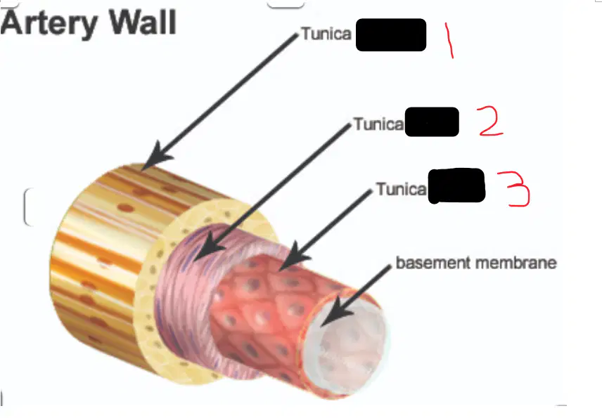
Identify the layers of the blood vessel
1: tunica interna 2: tunica media 3: tunica externa
1: tunica externa 2: tunica media 3: tunica interna
1: tunica media 2: tunica interna 3: tunica externa
1: tunica externa 2: tunica interna 3: tunica media
What is the difference in structure between veins and arteries
arteries have thin muscle layers and large lumen, veins have thick muscle layer and small lumen
arteries have thicker muscle layer and smaller lumen, veins have thin muscle layer and large lumen
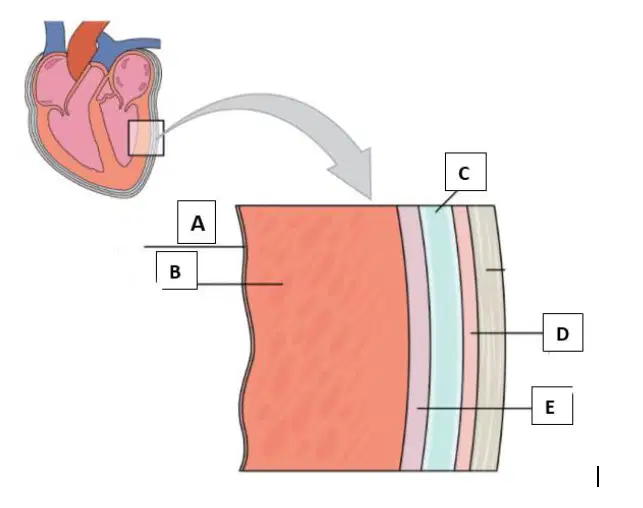
Identify letter B
myocardium
endocardium
parietal pericardium
visceral pericardium
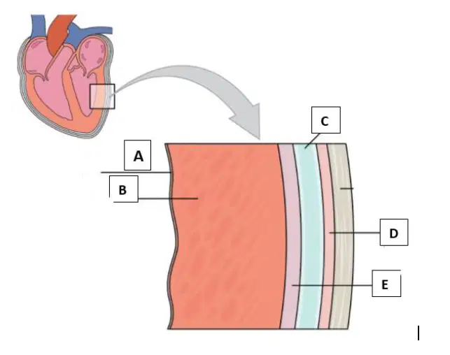
Identify letter E
endocardium
myocardium
visceral pericardium
parietal pericardium
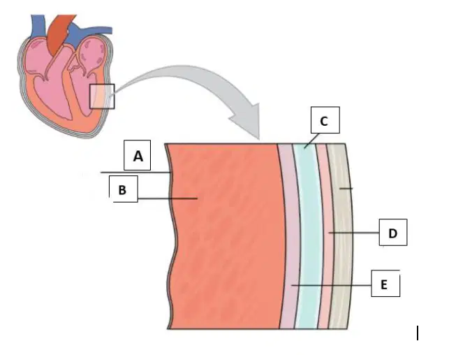
Identify letter D
myocardium
Parietal pericardium
visceral pericardium
endocardium
Type O blood is known as the
universal donor
universal recipient
universal blood type
universal receiver
Where are antigens located?
red blood cells
plasma
antibodies
capillaries
Group O can donate to
all blood types
O only
AB only
AO or BO genotypes only
Type AB is known as the
universal recipient
universal donor
universal blood type
universal transfusor
Where are antibodies located?
plasma
red blood cells
white blood cells
arteries
Choose 3 functions of blood
transport oxygen & nutrients to lungs & tissue
clot blood
carry cells & antibodies to fight infection
transport oxygen & nutrients to heart and lungs
bring waste products to pancreas and kidneys
Main components of blood
red blood cells, white blood cells, platelets, plasma
red blood cells, white blood cells, plasma
blood cells and platelets
platelets, plasma, arteries, capillaries
Blood is considered to be positive or negative based on the presence of what?
Rh factor
A and B antigens
antibodies
platelets

What is the special protein found in red blood cells that makes blood red?
Phagocytes
Hemocyanin
Antibodies
Hemoglobin
Which type of blood cells protect the body against disease-causing organisms?
White blood cells
Red blood cells
Platelets
Plasma
What is the process in which white blood cells engulf foreign pathogens called?
Hemoglobin synthesis
Blood clotting
Antibody production
Phagocytosis
What is the function of antibodies produced by white blood cells?
Carrying oxygen
Stopping bleeding
Preventing infection
Transporting nutrients
Which component of blood helps in clotting and stopping bleeding?
Red blood cells
Platelets
Plasma
White blood cells
Mature erythrocytes
are the least numerous formed element in the body
are cylindrically shaped cells
are produced in yellow bone marrow
do not have a nucleus
The most common type of leukocyte, which functions to phagocytize microorganisms and other substances?
lymphocyte
macrophage
neutrophil
thrombocyte
Platelets are a part of blood that help in the:
formation of hemoglobin
formation of a clot
release of anitbodies
absorption of amino acids

Which type of blood cell is involved in allergic reactions and parasitic infections?
Lymphocytes
Eosinophils
Platelets
Monocytes
Where are Red Blood Cells (RBCs) produced?
Liver
Spleen
Red bone marrow
Lymph nodes
Which of the following types of white blood cells (WBCs) are classified as granulocytes?
Neutrophils, Eosinophils, Basophils
Monocytes, Lymphocytes
Neutrophils, Monocytes, Lymphocytes
Eosinophils, Monocytes, Basophils
Which of the following types of white blood cells (WBCs) do not contain granules in their cytoplasm?
Neutrophils, Eosinophils, Basophils
Monocytes, Lymphocytes
Neutrophils, Monocytes, Lymphocytes
Eosinophils, Monocytes, Basophils
Where are blood platelets formed?
Liver
Red bone marrow
Spleen
Lymph nodes
What is the primary function of blood platelets?
Transport oxygen
Fight infections
Control loss of blood by coagulation
Regulate blood pressure
Which of the following is a function of blood related to regulation?
Transporting oxygen to cells
Regulating body temperature
Fighting infections
Clotting blood
A condition that develops when your blood lacks enough healthy red blood cells or hemoglobin
Lymphatic System
Not Anemia
Platelets
Anemia
Immature red blood cells have a nucleus.
True
False
What is the rarest blood type in the U.S.?
A
B
AB
O
What is the function of plasma?
transports/carries food, minerals, blood cells and waste
carries oxygen
controls bleeding by clotting blood
fights off pathogens, germs, bacteria and viruses
What disorder is characterized by an overproduction of WBCs?
Hemophilia
Sickle cell anemia
Leukemia
Hypovolemia
What disorder results in abnormally shaped RBCs that cant carry oxygen through the body efficiently?
Hemophilia
Sickle cell anemia
Leukemia
Hypovolemia
What disorder results when clotting factor proteins are missing so your platelets will not activate?
Hemophilia
Sickle cell anemia
Leukemia
Hypovolemia
When would you expect WBC numbers to increase?
During an active infection
During an active/internal bleed
When the level of oxygen has decreased
When the fluid level in the body is below normal
When would you expect platelet numbers to increase?
During an active infection
During an active/internal bleed
When the level of oxygen has decreased
When the fluid level in the body is below normal
Which of the following is a platelet?
leukocyte
erythrocyte
thrombocyte
Veins that empty blood from the lungs into the upper left heart chamber
Inferior Vena Cava
Superior Vena Cava
Pulmonary Veins
Aortic veins
Type of blood vessels taking blood to the heart
Venules
Veins
Capillaries
Arteries
Which valve sits between the left atrium and left ventricle of the heart?
Pulmonary
Bicuspid
Aortic
Tricuspid
Which valve sits between the lower right chamber and arteries leading to the lungs
Pulmonary
Bicuspid
Aortic
Tricuspid
Which valve sits between the lower left chamber and large artery leading to the body
Pulmonary
Bicuspid
Aortic
Tricuspid
Which chamber receives blood from the two largest veins in the body?
Right atrium
Right ventricle
Left atrium
Left ventricle
The blood enters on the left side of the heart (from the lungs) through the ______________ and enters the left atrium. It then passes through the _____________to enter the left ventricle.
pulmonary artery, mitral valve
pulmonary vein, tricuspid valve
pulmonary artery, aortic valve
pulmonary vein, bicuspid valve
Blood flows in the following sequence in the heart: Superior vena cava/Inferior vena cava, right atrium, tricuspid valve, right ventricle, pulmonary semi-lunar valve, pulmonary artery, lungs, pulmonary vein, left atrium, bicuspid valve, left ventricle, aortic semi-lunar valve, aorta
True
False
The function of the valves is to...
Allow for the contraction of the heart.
Prevent back-flow.
Oxygenate the blood.
Supply the myocardium with oxygen.

There are 4 of these in the heart, prevents backflow
valves
The left side of the heart
separates oxygenated and deoxygenated blood before bringing it to the lungs
pumps only deoxygenated blood
pumps oxygenated blood only
brings deoxygenated blood from the lungs to the body where it is oxygenated
Largest artery in the body taking oxygenated blood to the body from the lower left chamber
Pulmonary
Radial
Aorta
Femoral
Which valve sits between the lower left chamber and large artery leading to the body
Pulmonary
Bicuspid
Aortic
Tricuspid
Which valve sits between the lower right chamber and arteries leading to the lungs
Pulmonary
Bicuspid
Aortic
Tricuspid
Blood flows in the following sequence in the heart: Superior vena cava/Inferior vena cava, right atrium, tricuspid valve, right ventricle, pulmonary semi-lunar valve, pulmonary artery, lungs, pulmonary vein, left atrium, bicuspid valve, left ventricle, aortic semi-lunar valve, aorta
True
False
Which chambers of the heart are responsible for receiving blood?
atrium
ventricles
pulmonary artery
pulmonary veins
Which of these carries deoxygenated blood away from the heart?
pulmonary artery
pulmonary vein
aorta
vena cava
Which of these carries oxygenated blood away from the heart?
pulmonary artery
pulmonary vein
vena cava
aorta

Structure 1 in the picture is the _______
right atrium
left atrium
right ventricle
left ventricle

Structure 2 in the picture is the _______
right atrium
left atrium
right ventricle
left ventricle

Structure 5 in the picture is the ______
pulmonary artery
pulmonary vein
aorta
vena cava

Structure 7 in the picture is the _______
pulmonary artery
pulmonary vein
inferior vena cava
superior vena cava

Structure A in the picture is the _______
tricuspid valve
biscuspid valve
aortic valve
pulmonary valve

Structure B is the ______
tricuspid valve
pulmonary valve
aortic valve
mitral valve

Structure C in the picture is the ______
tricuspid valve
bicuspid valve
pulmonary valve
aortic valve

Structure D in the picture is the ______
bicuspid valve
tricuspid valve
aortic valve
pulmonary valve

Which valves are atrioventricular valves? ⭐Choose all that apply⭐.
bicuspid valve
pulmonary valve
tricuspid valve
mitral valve
aortic valve
Which valves are atrioventricular valves? ⭐Choose all that apply⭐.
bicuspid valve
pulmonary valve
tricuspid valve
mitral valve
aortic valve
Which valves are semilunar valves? ⭐Choose all that apply⭐.
bicuspid valve
pulmonary valve
tricuspid valve
mitral valve
aortic valve
Arteries carry blood....
towards the heart
away from the heart
Veins carry blood....
towards the heart
away from the heart
The blood coming from the body enters the heart via the....
Pulmonary Artery
Pulmonary Vein
Aorta
Vena Cava
From the vena cava it enters the ....
right atrium
left atrium
right ventricle
left ventricle
From the right atrium it goes through the....
pulmonary valve
semi lunar valve
tricuspid valve
bicuspid valve
After travelling through the tricuspid valve it enters the ....
right atrium
left atrium
right ventricle
left ventricle
From the right ventricle it goes through the pulmonary valve and heads through the....
Pulmonary Artery
Pulmonary Vein
Aorta
Vena Cava
Once it has left the right side of the heart the blood heads to the ....
body
working muscles
lungs
capillaries
At the lung the blood becomes
deoxygenated
oxygenated
From the lungs the blood reenters the heart via the...
Pulmonary Artery
Pulmonary Vein
Aorta
Vena Cava
After travelling through the bicuspid valve the oxygenated blood enters the...
right atrium
left atrium
right ventricle
left ventricle
From the left atrium the blood goes through the ....
tricuspid valve
bicuspid valve
From the left ventricle the blood travels through the semi-lunar valve into the ....
Pulmonary Artery
Pulmonary Vein
Aorta
Vena Cava
From the aorta the blood goes to the ...
body
lungs
brain
heart
The blood going to the body is ....
oxygenated
deoxygenated
What is the main job of blood while flowing through the body?
All of the above
Fighting infections
Removing carbon dioxide and waste products
Delivering oxygen and nutrients
What are the two main jobs of blood while flowing through the body?
Removing carbon dioxide and waste products, transporting hormones
Delivering oxygen and nutrients, removing carbon dioxide and waste products
Fighting infections, transporting hormones
Delivering oxygen and nutrients, fighting infections
Where are the electrical signals for the conduction process created?
SA Node
AV Node
Purkinje Fibres
Bundles of His
What does depolarization result in?
relaxation
contraction
repulsion
attraction
What does repolarization result in?
relaxation
contraction
attraction
repulsion

The last place electrical impulses move through the heart is located where?
the AV Node
the Purkinje fibers
the SA Node
the AV Bundle
Depolarization of a heart chamber such as the left atrium means the left atrium has
contracted
relaxed

The P wave on an ECG printout represents
contraction of the ventricles
relaxation of the ventricles
contraction of the atria
relaxation of the atria

The T wave on an ECG printout represents
contraction of the ventricles
relaxation of the ventricles
contraction of the atria
relaxation of the atria
1.atria contract
2. AV node
3. Bundle of His
4.Purkinje fibers
5. SA node
6. ventricles contract
What is another word for AV bundle?
Bundle of HIS
AV node
Pacemaker
Purkinje Fibers
What is the contraction phase of a hearbeat called?
diastole
sinoatrial
systole
atrioventricular
Which structures sends the electrical stimulus to the purkinje fibres?
SA Node
Bundles of His
AV Node
Purkinje Fibres
Which structure allows a delay in the signal between the atria and ventricles, allowing ventricles to fill with blood before contracting.
SA Node
AV Node
Purkinje Fibres
Bundles of His
Which structure is referred to as the Pace maker of the Heart?
SA Node
AV Node
Purkinje Fibres
Bundles of His
True or False: Depolarization of the heart muscle is when the muscle contracts and repolarization is when the heart muscle rests.
True
False
What is the correct sequence of electrical conduction of the heart?
AV node, internodal pathways, SA node, Bundle of his, right and left bundle branch, Purkinje fibers.
SA node, internodal pathways, AV node, Purkinje fibers, right and left bundle branches, Bundle of his.
SA node, internodal pathways, AV node, Bundle of his, right and left bundle branches, Purkinje fibers.
None of the options are correct.
Which is located within the walls of the ventricles?
Purkinje fibers
SA node
AV node
AV bundle and branches

The Purkinje fibers are indicated by ________.
A
B
C
D
E
The sinoatrial (SA) node is indicated by ________.
A
B
C
D
E
The atrioventricular bundle (bundle of His) is represented by ________.
A
B
C
D
E
The atrioventricular (AV) node is indicated by ________.
A
B
C
D
E
The sounds made by the heart are due the contractions of the atria and ventricles.
True
False
What is the delivery of blood to and from the heart itself called?
coronary circulation
pulmonary circulation
systemic circulation
cardiac circulation

What part of the hearts conduction system is shown in the diagram?
SA node
AV Node
AV bundle
Purkinje Fibers

What part of the hearts conduction system is shown in the diagram?
SA node
AV Node
AV bundle
Purkinje Fibers

What part of the hearts conduction system is shown in the diagram?
SA node
AV Node
AV bundle
Purkinje Fibers

What part of the hearts conduction system is shown in the diagram?
SA node
AV Node
AV bundle
Purkinje Fibers
What part of the hearts electrical conduction system "receives" the impulse from the nervous system telling it to contract?
SA Node
AV Node
AV Bundle
Purkinje Fibers
What part of the hearts electrical conduction system "delays" the impulse from the atria to the ventricles?
SA Node
AV Node
AV Bundle
Purkinje Fibers

What is the name of the circled area of the ECG in the diagram?
P Wave
QRS Complex
T Wave
Cardiac Cycle

What is the name of the circled area of the ECG in the diagram?
P Wave
QRS Complex
T Wave
Cardiac Cycle

What is the name of the circled area of the ECG in the diagram?
P Wave
QRS Complex
T Wave
Cardiac Cycle

The normal pattern of impulse conduction through the heart is:
AV-SA-AVB (Bundle of His)-Purkinje
AV-SA-Purkinje-AVB (Bundle of His)
SA-AV-AVB (Bundle of His)-Purkinje
SA-AV-Purkinje-AVB (Bundle of His)

In the diagram below, the P wave represents ___________, the QRS segment represents ___________ and the T wave represents _____________.
atrial depolarization, ventricular depolarization, ventricular repolarization
ventricular repolarization, atrial depolarization, ventricular depolarization
ventricular depolarization, ventricular repolarization, atrial depolarization
What does the QRS wave of the electrocardiogram (ECG) represent?
atrial depolarization
atrial repolarization
ventricular depolarization
ventricular repolarization
A patient has a normal heartbeat of 72 beats per minute. What area of the conduction system should the nurse consider as functioning normally in this patient?
Bundle of His
Sinoatrial node
Purkinje’s fibers
Atrioventricular node
What is the name of the circulation between the heart and lungs?
Systemic
Pulmonary
Arterial
Diastolic
What part of the hearts electrical conduction system is found in the ventricle muscle walls and is what causes them to contract?
SA Node
AV Node
AV Bundle
Purkinje Fibers
Which structure divides into the left and right bundle branches in the heart?
Atrioventricular bundle (Bundle of His)
Sinoatrial (SA) node
Atrioventricular (AV) node
Purkinje fibers
1.atria contract
2. AV node
3. Bundle of His
4.Purkinje fibers
5. SA node
6. ventricles contract
What is another word for AV bundle?
Bundle of HIS
AV node
Pacemaker
Purkinje Fibers
What is the contraction phase of a hearbeat called?
diastole
sinoatrial
systole
atrioventricular
Which structure is referred to as the Pace maker of the Heart?
SA Node
AV Node
Purkinje Fibres
Bundles of His
What is the delivery of blood to and from the heart itself called?
coronary circulation
pulmonary circulation
systemic circulation
cardiac circulation
What is the name of the circled area of the ECG in the diagram?
P Wave
QRS Complex
T Wave
Cardiac Cycle
The normal pattern of impulse conduction through the heart is:
AV-SA-AVB (Bundle of His)-Purkinje
AV-SA-Purkinje-AVB (Bundle of His)
SA-AV-AVB (Bundle of His)-Purkinje
SA-AV-Purkinje-AVB (Bundle of His)
The nurse is assessing the client’s electrocardiography (ECG). What does the P wave on the ECG tracing represent?
Contraction of the atria
Contraction of the ventricles
Depolarization of the atria
Depolarization of the ventricles
A patient has a normal heartbeat of 72 beats per minute. What area of the conduction system should the nurse consider as functioning normally in this patient?
Bundle of His
Sinoatrial node
Purkinje’s fibers
Atrioventricular node
What is the name of the circulation between the heart and lungs?
Systemic
Pulmonary
Arterial
Diastolic

What is one complete contraction of the heart as represented in the ECG diagram called?
Coronary Cycle
Cardiac Output
ECG Cycle
Cardiac Cycle
Which side of the heart carries poorly oxygenated (deoxygenated) blood?
Right
Left
Type of blood vessels taking blood to the heart
Venules
Veins
Capillaries
Arteries
Which valve sits between the left atrium and left ventricle of the heart?
Pulmonary
Bicuspid
Aortic
Tricuspid
Which valve sits between the lower right chamber and arteries leading to the lungs
Pulmonary
Bicuspid
Aortic
Tricuspid
Which valve sits between the lower left chamber and large artery leading to the body
Pulmonary
Bicuspid
Aortic
Tricuspid
Which chamber receives blood from the two largest veins in the body?
Right atrium
Right ventricle
Left atrium
Left ventricle
Which chamber receives blood from the upper right chamber and sends blood to the lungs?
Right atrium
Right ventricle
Left atrium
Left ventricle
The blood enters on the left side of the heart (from the lungs) through the ______________ and enters the left atrium. It then passes through the _____________to enter the left ventricle.
pulmonary artery, mitral valve
pulmonary vein, tricuspid valve
pulmonary artery, aortic valve
pulmonary vein, bicuspid valve
Blood flows in the following sequence in the heart: Superior vena cava/Inferior vena cava, right atrium, tricuspid valve, right ventricle, pulmonary semi-lunar valve, pulmonary artery, lungs, pulmonary vein, left atrium, bicuspid valve, left ventricle, aortic semi-lunar valve, aorta
True
False
On the right side of the heart, blood flows from the right ventricle to the?
Tricuspid valve to the pulmonary vein
Pulmonic valve to the pulmonary artery
Tricuspid valve to pulmonary artery
Inferior vena cava to the aorta

Place the pathway of blood through the heart in the correct sequence.
L-K-H-B-G-E-C-F-A-I-E-D
I-J-D-C-F-A-H-B-G-E-K-L
A-B-J-C-D-E-F-K-L-G-H-I
I-J-E-D-C-F-A-B-G-H-K-L
The function of the valves is to...
Allow for the contraction of the heart.
Prevent back-flow.
Oxygenate the blood.
Supply the myocardium with oxygen.


True or False: The mitral valve is located on the left side of the heart.
True
False

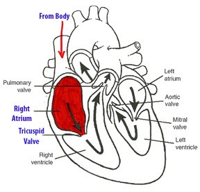


Which blood vessel takes blood AWAY from the heart?
Arteries
Veins

This is where arteries and veins meet and exchange gases
veins
capillaries
Which valve sits between the lower right chamber and arteries leading to the lungs
Pulmonary
Bicuspid
Aortic
Tricuspid
Which valves are atrioventricular valves? ⭐Choose all that apply⭐.
bicuspid valve
pulmonary valve
tricuspid valve
mitral valve
aortic valve
Which valves are semilunar valves? ⭐Choose all that apply⭐.
bicuspid valve
pulmonary valve
tricuspid valve
mitral valve
aortic valve
Veins carry blood....
towards the heart
away from the heart
The blood coming from the body enters the heart via the....
Pulmonary Artery
Pulmonary Vein
Aorta
Vena Cava
From the vena cava it enters the ....
right atrium
left atrium
right ventricle
left ventricle
From the lungs the blood reenters the heart via the...
Pulmonary Artery
Pulmonary Vein
Aorta
Vena Cava
After travelling through the bicuspid valve the oxygenated blood enters the...
right atrium
left atrium
right ventricle
left ventricle
From the aorta the blood goes to the ...
body
lungs
brain
heart
What part of the hearts electrical conduction system is found in the ventricle muscle walls and is what causes them to contract?
SA Node
AV Node
AV Bundle
Purkinje Fibers
Which of the following lines the inside of the heart chambers and makes up heart valves?
endocardium
myocardium
pericardium
epicardium
When electrical stimulation hits _____________________ the heart contracts.
myocardium
endocardium
pericardium
epicardium
Diastole is associated with
contraction of the heart
relaxation of the heart
myocardial infarction
angina
Which of the following is the measure of the number of times the heart beats in a minute?
blood pressure
heart rate
electrocardiogram (EKG)
biopsy
The right side of the heart contains _____ blood and the left side of the heart contains _____ blood.
oxygenated; deoxygenated
deoxygenated; oxygenated
deoxygenated; deoxygenated
oxygenated; oxygenated
What is the primary purpose of coronary circulation?
To supply blood to the heart muscle.
To remove waste products from the brain.
To transport oxygen to the lungs.
To regulate blood pressure in the arteries.

