
 Worksheets
Muscular System Review
Total questions: 76
Worksheet time: 55mins

Letter A represent which structure?
Endomysium
Epimysium
Fascicle
Muscle Fiber
Perimysium
Letter H represent which structure?
Endomysium
Epimysium
Fascicle
Muscle Fiber
Perimysium
Letter G represent which structure?
Endomysium
Epimysium
Fascicle
Muscle Fiber
Perimysium
Letter F represent which structure?
Endomysium
Epimysium
Fascicle
Muscle Fiber
Perimysium
What is the main function of Smooth Muscle?
Attached to bones and help with movement
Make up walls of organs and blood vessels
Make up heart
What are a type of connective tissue that connects muscles to bones?
Tendons
Cartilage
Ligaments
Joints
This type of muscle tissue is found in the heart.
Cardiac
Skeletal
Smooth
Blood vessels
All of these muscles show striations of light and dark bands when viewed under the microscope EXCEPT:
Cardiac
Skeletal
Smooth
Which type of muscle tissue connects bones to other bones, allowing for movement of our joints?
cardiac
smooth
skeletal
sympathetic
What is endomysium?
An epithelial tissue that encloses muscle fibers
A connective tissue that encloses muscle fibers
An epithelial tissue that encloses myofilaments
A connective tissue that encloses myofilaments
What is perimysium?
A fibrous membrane that wraps around blood vessels
A fibrous membrane that wraps around ONE muscle fibers
A fibrous membrane that wraps around bunders of muscle fibers.
A fibrous membrane that wraps around several fascicle
Movement of a limb laterally, away from the midline of the body.
Adduction
Abduction
Flexion
Extension
Hyperextension
Movement of a limb medially, towards the midline of the body.
Adduction
Abduction
Flexion
Extension
Hyperextension
Movement of a limb anteriorly; a forward motion; decreases angle between two bones.
Adduction
Abduction
Flexion
Extension
Hyperextension
Movement of a limb posteriorly to return body to neutral position; a backwards motion; increases angle between two bones.
Adduction
Abduction
Flexion
Extension
Hyperextension
Movement of a joint 360 degrees in a complete circle.
Supination
Pronation
Rotation
Circumduction


Which movement is shown in the picture?
eversion
rotation
extension
flexion
Straightening a joint or increasing the angle between two bones
Flexion
Extension
Hyperextension
Abduction
Bending a joint or decreasing the angle between two bones
Flexion
Extension
Hyperextension
Adduction
The meeting place between 2 or more bones is known as a _________.
Diarthrosis
Joint (or articulation)
Synovial
Junction
___________ connect bones to bones.
Joints
Tendons
Ligaments
Patellas
The more movable the joint, the _________ stable it is.
more
less
no change
equally
Synarthrosis
an immovably fixed joint between bones connected by fibrous tissue
a slightly movable articulation
joint that can move freely in various planes
type of structure in the body formed between two bones in which the articular, or free, surfaces of the bones are flat or nearly flat, enabling the bones to slide over each other
Amphiarthrosis
an immovably fixed joint between bones connected by fibrous tissue
a slightly movable articulation
joint that can move freely in various planes
type of structure in the body formed between two bones in which the articular, or free, surfaces of the bones are flat or nearly flat, enabling the bones to slide over each other
Diarthrosis
an immovably fixed joint between bones connected by fibrous tissue
a slightly movable articulation
joint that can move freely in various planes
type of structure in the body formed between two bones in which the articular, or free, surfaces of the bones are flat or nearly flat, enabling the bones to slide over each other

The movement shown is a person moving his leg outward. This movement is known as:
abduction
adduction
rotation
flexion


Name this movement
adduction
abduction
flexion
extension

Name this movement
extension
flexion
adduction
abduction
Which myofilament is thin?
Collagen
Elastin
Actin
Myosin
What is the smallest unit of a muscle to contract?
Sarcomere
Myofibril
Muscle fiber
Myofilaments

The part of the sarcomere that C (Blue) refers to is the...
Actin
Myosin
Troponin
Tropomyosin
Actin
Myosin
Troponin
Tropomyosin
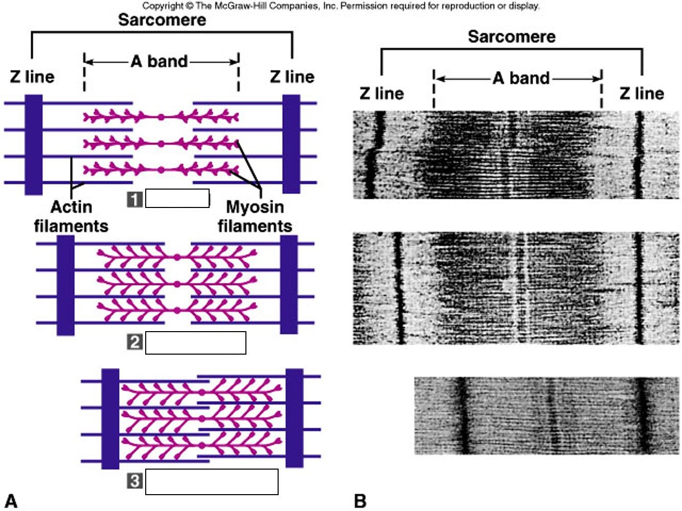
Relaxed
Contracting
Fully Contracted
None of the above
Relaxed
Contracting
Fully Contracted
None of the above
It disappears
gets closer together
It gets bigger
None of these
Sarcomere
Osteon
Myofilament
Muscle fiber
Sarcomeres
Sarcoplasm
Actin
Myosin
ATPase
Sarcoplasmic reticulum
Actin
Myosin

How may sarcomeres are shown above?
6
5
4
3

What is the area labeled 8
M line
Z line
H zone
A band
What molecule prevents actin-myosin interaction?
Troponin
Nebulin
Titin
Tropomyosin

What is the area labeled D?
Z line
H zone
I band
A band

What is the area labeled C
A band
I band
H zone
Zone of overlap
In a contracted muscle fiber, identify what occurs in the zone of overlap
Increase
Stays the same
Decrease
What is the myosin anchored to?
M-line
Z-line
Actin
Thin filament
Myofilaments are made of which 2 proteins?
Fascicles
Myosin
Myofibrils
Actin
A fascicle surrounds:
muscles
muscle fibers
myofibrils
sarcomeres
A muscle fiber contains?
fascicles
muscles
myofibrils
What is released from the sarcoplasmic reticulum?
Inorganic phosphate
ADP
Calcium
ATP
What is the name of the neurotransmitter released at a neuromuscular junction?
Calcium
Acetylcholine
Dopamine
Troponin
Which of the following statements correctly explains the sliding filament model?
Actin head attaches to a binding site on a myosin filament, forming a cross bridge.
Myosin head attaches to a binding site on an actin filament, forming a neuromuscular junction.
Actin head attaches to a binding site on a myosin filament, forming a neuromuscular junction.
Myosin head attaches to a binding site on an actin filament, forming a cross bridge.
Layers of Connective tissue on the outside of a muscle
Epimysium
Perimysium
Endomysium
Fascicle
Surrounds a single Muscle Fiber
Endomysium
Perimysium
Epimysium
Sarcoplasmic Reticulum
Molecule that provides Energy
ATP
Calcium
Acetylcholine
Actin
Found surrounding Fascicles
Endomysium
Perimysium
Epimysium
Sarcolemma
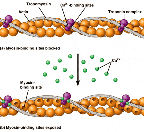
___are regulatory proteins that cover the myosin binding site on the actin.
troponin-tropomyosin
sarcomere
sarcolemma
sarcoplasmic reticulum
What substance provides the energy for muscle contraction?
ATP
Glucose
Lactic acid
Glycogen
What happens to the muscle when the concentration of calcium ions decreases?
The muscle contracts more forcefully.
The muscle relaxes.
The muscle becomes paralyzed.
The muscle begins to twitch uncontrollably.
What happens to the muscle when the concentration of ATP decreases?
The muscle contracts more forcefully.
The muscle relaxes.
The muscle becomes paralyzed.
The muscle begins to fatigue.
Please select the correct order
1. Muscle cell releases calcium
2. Neurotransmitter releases acetylcholine
3. Sodium & Potassium pump opens
4. Acteylcholine signals cell membrane gates to open
1. Neurotransmitter releases acetylcholine
2. Acteylcholine signals cell membrane gates to open
3. Sodium & Potassium pump opens
4. Muscle cell releases calcium
1. Sodium & Potassium pump opens
2. Acteylcholine signals cell membrane gates to open
3. Muscle cell releases calcium
4. Neurotransmitter releases acetylcholine
1. Neurotransmitter releases acetylcholine
2. Acteylcholine signals cell membrane gates to open
3. Muscle cell releases calcium
4.Sodium & Potassium pump opens
When ATP binds to the myosin head the myosin head then _____________.
completes a power stroke
attaches to actin
rotates
detaches from actin

