
 Worksheets
Introduction To the Wrist & Hand
Total questions: 35
Worksheet time: 24mins
Which statement best describes the wrist in relation to the forearm and hand?
It attaches fingers directly to the radius
It separates radius from the ulna
It connects proximal hand to the elbow
It connects distal forearm to the hand
How many carpal bones form the wrist?
Twelve bones total
Ten bones total
Eight bones total
Six bones total
Match each carpal bone to its correct row location.
Scaphoid
Proximal row
Lunate
Proximal row
Capitate
Distal row
Hamate
Distal row
Which set lists ONLY proximal row carpal bones from radial to ulnar side?
Scaphoid, Capitate, Lunate, Hamate
Scaphoid, Lunate, Triquetral, Pisiform
Pisiform, Trapezium, Triquetral, Radius
Trapezium, Trapezoid, Capitate, Hamate
Which bones make up the distal carpal row from radial to ulnar side?
Trapezium, Trapezoid, Capitate, Hamate
Scaphoid, Lunate, Triquetral, Pisiform
Trapezoid, Capitate, Hamate, Pisiform
Radius, Ulna, Capitate, Hamate
How many metacarpal bones are in one hand?
Seven per hand
Five per hand
Four per hand
Six per hand
Phalanges in the fingers are organized into which segments?
Basal, central, terminal
Proximal, middle, distal
Primary, secondary, tertiary
Radial, median, ulnar
What is the primary function of wrist ligaments?
To cushion impact during running
To transport nutrients to cartilage
To bind carpal, radius, ulna, metacarpals
To generate muscle contraction power
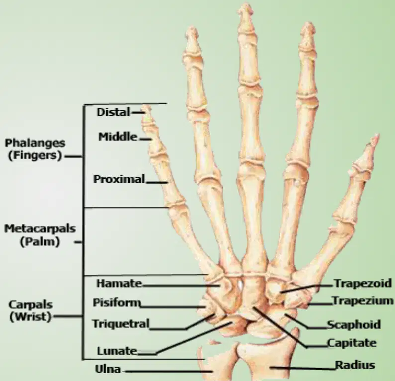
In the labeled hand diagram, which carpal is located proximal and lateral adjacent to the radius?
Pisiform on palmar ulnar
Hamate at distal ulnar
Lunate near distal ulna side
Scaphoid near radial styloid
A PA wrist radiograph highlights a small pea-shaped carpal on the palmar ulnar side. Which bone is most likely seen?
Triquetral dorsal to pisiform
Scaphoid near radial styloid
Pisiform on palmar ulnar
Trapezium under first metacarpal
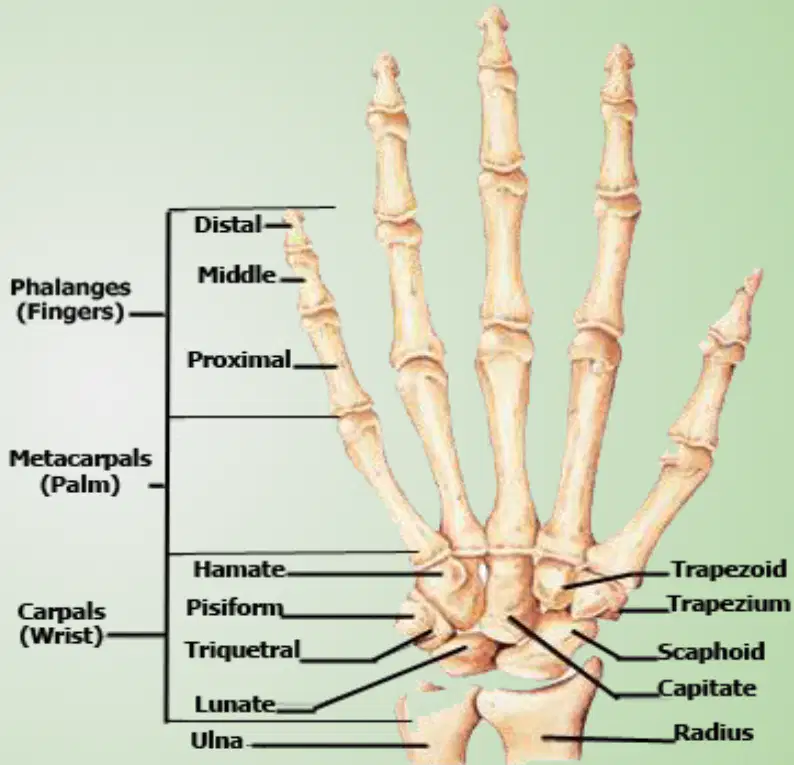
Which bones articulate directly with the metacarpals in the distal carpal row? Select all that apply.
Capitate articulating with third
Trapezoid articulating with second
Trapezium articulating with first
Lunate articulating with fourth
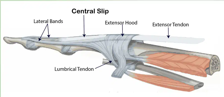
In the tendon diagrams, which structure spreads over the dorsum of the finger to distribute extensor forces?
Central slip to distal phalanx
Flexor sheath within tenosynovium
Lumbrical tendon to volar side
Extensor hood over dorsal digit
Which tendon inserts on the distal phalanx to produce fingertip flexion?
Flexor digitorum profundus tendon
Extensor digitorum communis tendon
Flexor digitorum superficialis tendon
Extensor indicis proprius tendon
Where does the flexor digitorum superficialis (FDS) typically insert?
Proximal phalanx shaft
Metacarpal head
Base of middle phalanx
Base of distal phalanx
Which structures prevent bowstringing of flexor tendons during finger flexion?
Interosseous muscles only
Lumbrical muscles only
Extensor retinaculum bands
Annular pulley system (A2–A4)
Which statement best distinguishes extensor tendon slips over the dorsum of the finger?
Lateral slips attach to proximal phalanx only
Three slips all inserting at middle phalanx
Central slip to middle phalanx, lateral slips to distal phalanx
Single central slip inserting at distal phalanx
Select all muscles that contribute to finger extension via the extensor mechanism.
Extensor digitorum
Lumbricals
Flexor pollicis longus
Aductor pollicus
Match each tendon with its primary action at the finger.
FDP
Flexes distal interphalangeal joint
FDS
Flexes proximal interphalangeal joint
Extensor digitorum
Extends interphalangeal joints
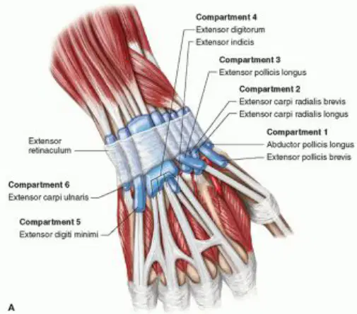
On the extensor side image, what structure holds the extensor tendons close to the wrist bones?
Palmar aponeurosis sheet
Extensor retinaculum fibrous band
Interosseous membrane band
Flexor retinaculum ligament
Which pair correctly identifies the tendons that act on the thumb?
Extensor Pollicis longus with Abductor Pollicus Longui
Extensor Carpi Radialis Brevis with Extensor Carpi Ulnaris
Abductor Pollicis Longus with Flexor Digitorum Profundus
Extensor digitorum with Extensor Pollicis Longus
Multiple structures pass through the carpal tunnel. Which nerve is correct?
Flexor carpi ulnaris tendon
Flexor pollicis longus tendon
Median nerve
Flexor digitorum superficialis tendons
Ulnar Nerve
Which structure forms the roof of the carpal tunnel?
Extensor retinaculum band
Flexor retinaculum thick band
Transverse carpal artery
Palmar fascia thin sheet
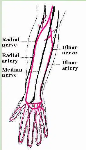
Blood supply to the hand primarily comes from which arteries?
Ulnar and brachial arteries
Median and radial arteries
Anterior and posterior interosseous
Radial and ulnar arteries
Most flexor muscles of the forearm and hand are innervated by which nerve?
Musculocutaneous nerve supply
Median nerve motor supply
Ulnar nerve motor supply
Radial nerve motor supply
Which nerve predominantly controls wrist and finger extension?
Axillary nerve motor control
Median nerve motor control
Ulnar nerve motor control
Radial nerve motor control
Match each quick motor test gesture to the nerve it assesses.
Thumbs up
Radial nerve
Power to the people (fist)
Median nerve
Peace out (V sign)
Ulnar nerve
During a sensory exam, which area tests the radial nerve?
First dorsal web space skin
Medial tip of index finger
Lateral tip of little finger
Thenar eminence skin
Identify the sacphoid bone:
Identify the lunate
Identify the 1st Metacarpal
Identify the 3rd middle phalange
Identify the metacarpals
Identify the 1st distal phalange

