
 Worksheets
Chapter 10: Ultrasonography — Worksheet Questions
Total questions: 39
Worksheet time: 20mins

Identify the primary topic of Chapter 10 shown with a veterinary professional performing a scan on a small dog using an ultrasound probe connected to a console.
Radiography
Ultrasonography
Endoscopy
Thermography
Select all items listed under the learning objectives for this chapter.
Real-time ultrasound imaging
Artifacts and limitations of ultrasonography
Transducer types and functions
Image modes
Clinical applications for body systems
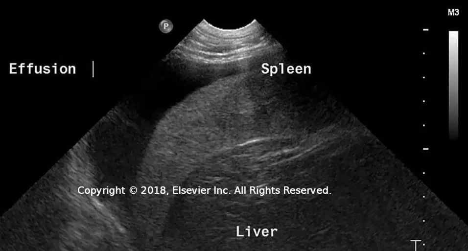
According to the applications slide with ultrasound images labeled Effusion, Spleen, and Liver, which materials or tissues are stated as visible with ultrasonography? Select all that apply.
Bone
Air
Fat
Fluid
Soft tissues
According to the applications slide, producing ultrasonographic images is relevant in which situation?
When alternative diagnostic imaging is required
Only for routine wellness examinations
Exclusively during surgical procedures
When radiography is unavailable and CT is contraindicated
Based on the principles slide, what component generates the sound waves used for imaging?
The detector array in the monitor
The ultrasound probe (transducer)
The system power supply
The thermal printer
According to the principles slide, where are the most important components of an ultrasound machine located?
Inside the display unit
Within the transducer
In the foot pedal controls
In the keyboard housing
What statement from the principles slide explains why echoes return to the probe during scanning?
Chemical reactions in tissues emit sound
Electromagnetic radiation is absorbed by organs
Sound waves are reflected back to the probe depending on various tissue densities
Heat conduction moves energy toward the transducer
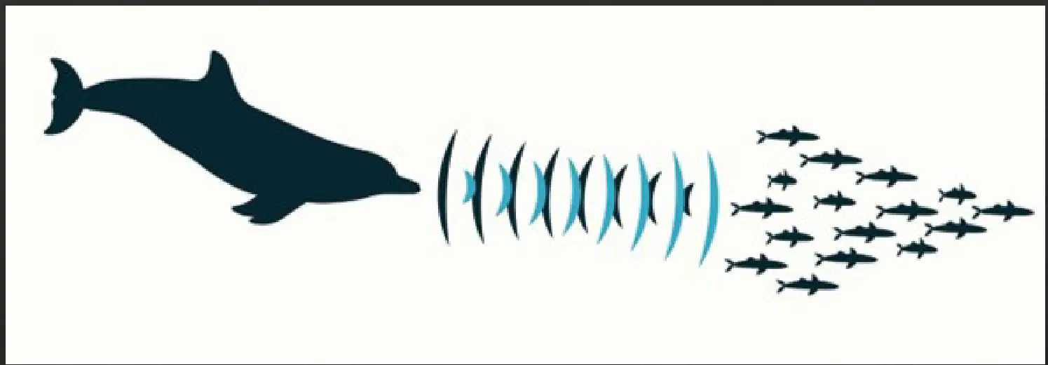
Interpreting the dolphin diagram titled Mode of Action, what fundamental concept relevant to ultrasound imaging does the illustration depict?
Continuous electrical stimulation of tissues
Emission of sound waves and reflection from targets to inform detection
Direct visualization of organs through transparent skin
Use of magnetic fields to align hydrogen nuclei

Identify the device component shown in the image: a close-up of a medical probe with a rectangular element and an orange plate, set against a mosaic background.
Ultrasound transducer (probe)
X‑ray tube head
Flexible endoscope tip
MRI surface coil
According to the slide titled "The Ultrasound Machine" under Practicality, which formats are available for ultrasound machines? Select all that apply.
Larger cart models
Laptop units
Handheld portable devices
Only desktop tower systems
From the Practicality considerations when selecting an ultrasound machine for a veterinary practice, which factors may vary by model? Select all that apply.
Available transducer types and frequencies
Computer processor speed
Quality of viewing monitors
Optional software applications
Mandatory built‑in radiography unit
Based on the Patient preparation section, which statements are required to achieve the desired imaging? Select all that apply.
The animal can be examined in any position that achieves the desired imaging
It is important that the patient remains still
The probe must have direct contact with the skin surface
The probe should hover above the skin without touching
Which benefit of ultrasound is emphasized for cardiac studies on the comparison slide?
Real‑time imaging
Superior bone detail compared with CT
Use of ionizing radiation
Fixed probe position
What is the term for computer reconstruction that allows visualization of the heart in a three‑dimensional format from multiple planes?
Multiplanar reconstruction (MPR)
Fourier transform imaging
Digital subtraction angiography
Tomographic attenuation correction
On the comparison slide, saved video recordings can be transferred into which format that allows the area of interest to be visualized over a prolonged period?
Cine loop
DICOM archive only
Static frame stack
Raster overlay
Which safety statement is correct according to the comparison slide?
Safety precautions required for ionizing radiation (x‑rays) are not required when an ultrasound is in use
Ultrasound requires the same radiation shielding as plain radiography
Ultrasound emits ionizing radiation at lower doses than x‑rays
Both ultrasound and plain radiography require dosimeter badges for probe operators
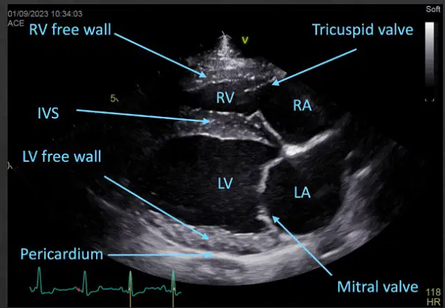
Match each cardiac abbreviation shown on the ultrasound image to its labeled structure.
RV
Right ventricle
RA
Right atrium
LV
Left ventricle
LA
Left atrium
Which term describes a structure that appears completely dark or black with no internal echoes on ultrasound?
Anechoic
Hypoechoic
Hyperechoic
Isoechoic
According to the echogenicity diagram, which items commonly appear hyperechoic? Select all that apply.
Bone
Tendon
Ligament
Nerves
Fluid in a cyst
Which progression correctly orders tissues from low density to high density as shown by the arrow on the echogenicity slide?
Skin < Fat < Soft tissue << Bone
Bone << Soft tissue < Fat < Skin
Fat < Skin < Soft tissue << Bone
Soft tissue < Fat < Skin << Bone
Match each ultrasound term to its definition as listed on the Terms slide.
Anechoic
Lacking internal echoes; appears very dark to almost black
Hyperechoic
Reflects more intense sound waves and appears brighter than surrounding tissues
Hypoechoic
Reflects fewer sound waves with less intensity; appears darker than surrounding tissues
Isoechoic
Two structures have relatively similar echogenicity toward each other
Attenuation
Loss of sound wave energy as it traverses tissue due to absorption, reflection, or scattering
Which statement best defines reverberation according to the Terms slide?
Repeated reflection of a sound wave between two highly reflective surfaces, such as air and the ultrasound probe
Loss of sound wave energy due to absorption in soft tissue
Appearance of two structures with similar echogenicity
Artifact caused by probe overheating
Which description correctly identifies the near field?
The area of a structure of interest that is closest to the probe
The area of a structure of interest that is farthest from the probe
The region beyond the focal zone only
The area outside the frame of view
Which description correctly identifies the far field?
The area of a structure of interest that is farthest from the probe
The area of a structure of interest that is closest to the probe
The probe housing immediately around the transducer
The acoustic coupling gel layer
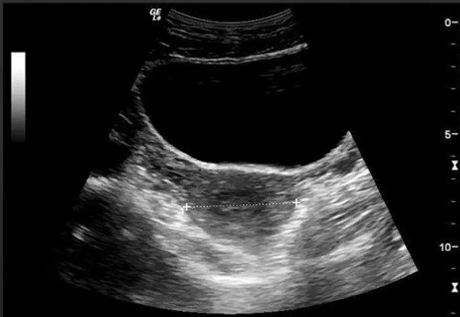
Echogenicity: Examples — What term would we use to describe this image? The ultrasound shows a fluid-filled, uniformly dark region with posterior acoustic enhancement compared to surrounding tissues.
Anechoic
Hypoechoic
Isoechoic
Hyperechoic
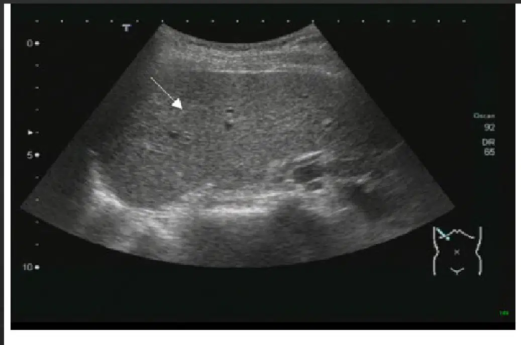
Echogenicity: Examples — What term would we use to describe this image? The ultrasound shows a focal region that appears darker than the adjacent liver parenchyma, indicated by an arrow.
Hyperechoic
Isoechoic
Hypoechoic
Anechoic
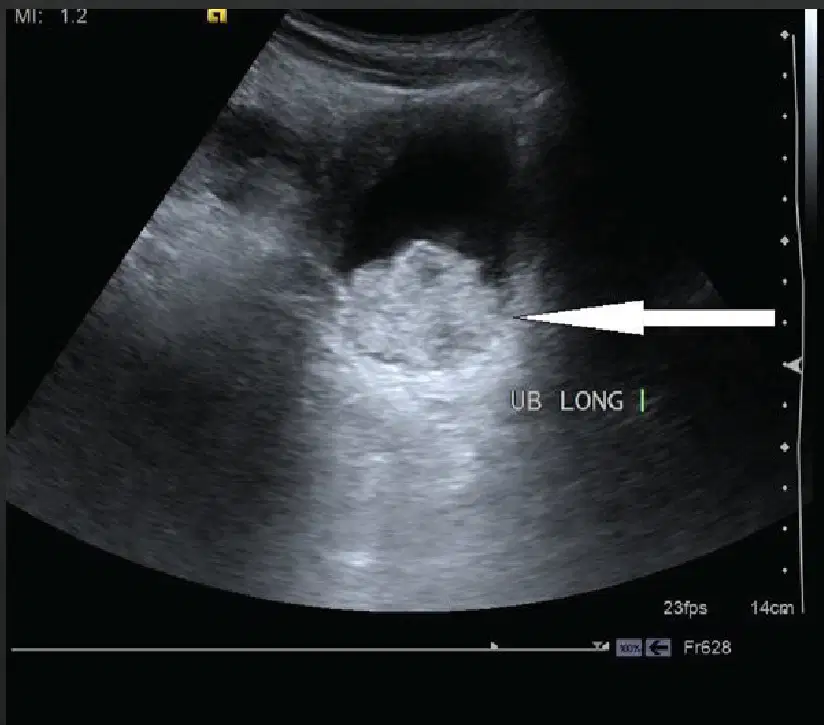
Echogenicity: Examples — What term would we use to describe this image? The ultrasound shows a solid-appearing focus that is brighter than surrounding tissues, indicated by a bold arrow.
Hypoechoic
Hyperechoic
Isoechoic
Anechoic
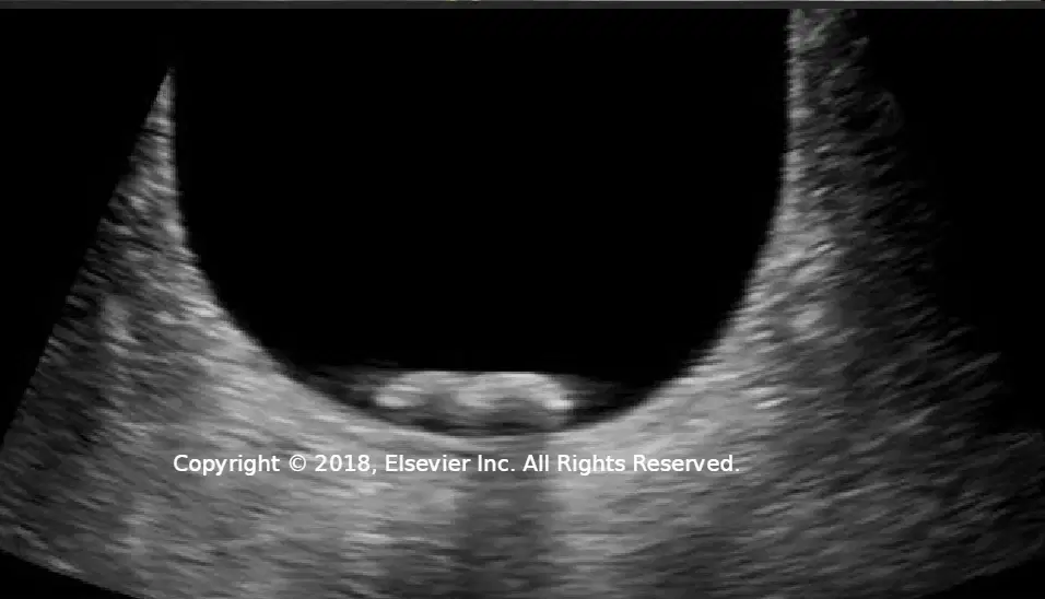
Echogenicity: Structures — Which items are classified as echogenic on ultrasound imaging?
Calcification
Gas
Fluid
Fat
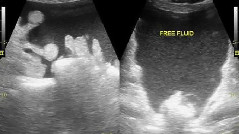
Echogenicity: Structures — Which description best characterizes fluid on ultrasound?
Anechoic to hypoechoic
Hyperechoic only
Uniformly isoechoic
Highly reflective causing shadowing
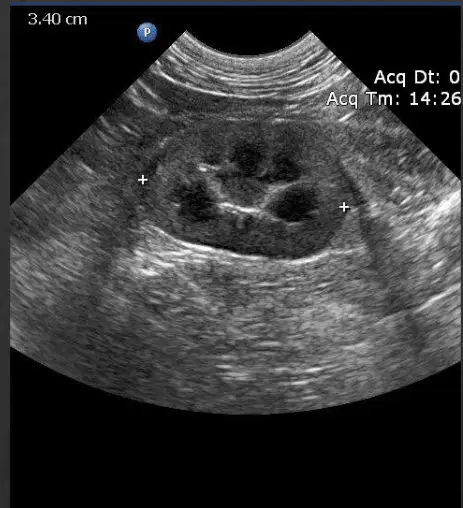
Echogenicity: Structures — In the dark regions within the displayed ultrasound images, which echogenicity category most commonly applies?
Echogenic
Variable echogenicity
Anechoic to hypoechoic
Hyperechoic

Modes in Ultrasound Imaging — What does B mode represent and primarily display?
Brightness; a 2D grayscale image classifying pixels as hyperechoic or hypoechoic
Motion; a time–depth trace of tissue movement
Flow; color-coded velocity maps
Attenuation; tissue stiffness quantification

Modes in Ultrasound Imaging — Which statements describe M mode? Select all that apply.
Represents motion
Uses a cursor line set over an area seen in B mode
Simultaneously displays a B-mode image while showing tissue motion
Focuses solely on 3D volumetric rendering
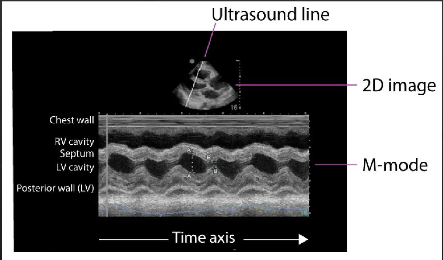
Modes in Ultrasound Imaging — In the M-mode display shown, what does the horizontal axis represent?
Time
Image depth
Ultrasound frequency
Gain settings
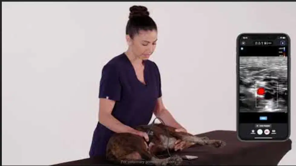
Modes in Ultrasound Imaging (Cont.) — What is the primary purpose of the Doppler modality?
To image the flow of blood and other liquids while measuring their velocity
To classify pixels as hyperechoic or hypoechoic
To create three-dimensional reconstructions
To measure tissue elasticity
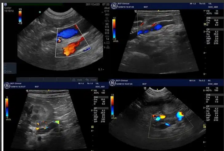
Modes in Ultrasound Imaging (Cont.) — Which are Doppler techniques? Select all that apply.
Color Doppler
Power Color Doppler
Pulse Wave Doppler
Continuous Wave Doppler
Brightness (B) mode
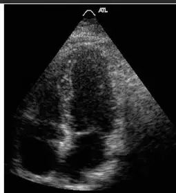
Based on the slide showing modes in ultrasound imaging, which mode helps to decrease ultrasound artifacts in very large or obese patients?
Harmonic mode
Multiplanar reconstruction
Three-dimensional/four-dimensional modes
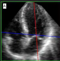
According to the description provided, what do multiplanar reconstruction (MPR) studies involve?
Reconstructing images from various planes to allow simultaneous visualization on the monitor
Reducing artifacts in very large or obese patients
Primarily imaging human fetal, obstetrical, and cardiac valves
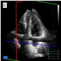
Select all statements that are accurate about three-dimensional and four-dimensional ultrasound modes as presented.
Used primarily for human fetal, obstetrical, and cardiac valve imaging
Not commonly used in veterinary medicine
Designed to decrease artifacts in obese patients
Primarily reconstruct images from various planes for simultaneous visualization

Match each ultrasound control with its function as shown on the Knobology slide.
Gain
Affects the brightness of the image
Depth
Each transducer type has set maximum and minimum depth at which it can send and receive sound waves
Time gain compensation
Allows the operator to selectively adjust the gain at various depths while they are already there
Presets
Preinstalled settings programmed by the manufacturer

