
Font size
 Worksheets
Page 1
Total questions: 40
Worksheet time: 20mins
Which sequence correctly lists structures included in airway anatomy?
Nose, pharynx, larynx, trachea, bronchi, bronchioles, alveoli
Mouth, esophagus, stomach, intestines
Sinus cavities, middle ear, eardrum, cochlea
Skull, vertebrae, ribs, sternum
What are alveoli in the context of the respiratory system?
Tiny air sacs where gas exchange occurs
Valves that prevent airflow back into the trachea
Muscles that enable inhalation
Blood vessels that carry oxygen
Which muscles help the lungs expand and contract during breathing?
Diaphragm and intercostal muscles
Biceps and triceps
Smooth muscles of the stomach
Cardiac muscle
Where is the center that regulates lung function located?
Brainstem respiratory center
Visual cortex
Cerebellar motor cortex
Limbic system
what characterizes obstructive lung diseases?
Difficulty in exhaling air fully
Difficulty in inhaling air
Reduced oxygen binding in blood
Infection localized to the alveoli
Which of the following is listed on the slide as an example of an obstructive lung disease?
Asthma
Pneumonia
Tuberculosis
Pulmonary fibrosis
Based on the bullet list shown, select all symptoms these diseases may present with.
Shortness of breath (dyspnea)
Chronic cough
Wheezing
Fatigue
Chest tightness
Refer to the slide titled “What are Pulmonary Function Tests (PFTs)?” According to the slide, PFTs are a group of tests that measure how well the lungs are working and are best described as which type of tests?
Non-invasive tests assessing lung function
Invasive surgical procedures on the lungs
Imaging scans of the chest using radiation
Blood-based biomarker assays for lung disease
Refer to the slide titled “What are Pulmonary Function Tests (PFTs)?” In pulmonary diseases, PFTs help with which clinical purposes? Select all that apply.
Diagnosing disease
Assessing severity of disease
Monitoring treatment response
Curing the disease
Screening for cardiac arrhythmias
According to the diagram titled "Types of Pulmonary Function Tests," which test is described as the basic and most commonly used?
Spirometry
Lung volume measurement
Diffusing capacity of lungs (DLCO)
Arterial blood gas (ABG) analysis
Peak Expiratory Flow Rate (PEFR)
In the list of pulmonary function tests, which measurement is stated to assess residual and total lung volumes?
Lung volume measurement
Spirometry
Diffusing capacity of lungs (DLCO)
Arterial blood gas (ABG) analysis
What does the diffusing capacity of the lungs (DLCO) assess, as indicated in the diagram?
Gas transfer
Maximum airflow during expiration
Basic pulmonary function
Oxygenation and acid–base status
Which test measures maximum airflow during expiration, as described in the diagram?
Peak Expiratory Flow Rate (PEFR)
Spirometry
Lung volume measurement
Diffusing capacity of lungs (DLCO)
Select all tests that are explicitly listed under "Types of Pulmonary Function Tests" in the diagram.
Spirometry
Lung volume measurement
Diffusing capacity of lungs (DLCO)
Arterial blood gas (ABG) analysis
Chest radiography
What is FEV1 (Forced Expiratory Volume in 1 sec) as defined on the slide?
Volume exhaled in the first second of forced expiration
Maximum volume of air exhaled after deep inhalation
Typical tidal volume during quiet breathing
Peak inspiratory flow achieved during a forced breath
What is the stated use of the FEV1/FVC ratio on the slide?
To differentiate obstructive versus restrictive patterns
To calculate total lung capacity only
To assess diffusion capacity of the lungs
To measure oxygen transport in blood
According to the Interpretation of Spirometry slide, what is the typical spirometric change in restrictive disease (e.g., pulmonary fibrosis)?
↓ FVC with normal or ↑ FEV1/FVC ratio
↓ FEV1 with ↓ FEV1/FVC ratio
Normal spirometry without any changes
↓ FEV1 and ↓ FVC with ↓ FEV1/FVC ratio
How is a mixed pattern described on the Interpretation of Spirometry slide?
Combination of both obstructive and restrictive features
Exclusively obstructive features
Exclusively restrictive features
Normal spirometry with no changes
According to the Interpretation of Spirometry slide, what is the typical behavior of the FEV1/FVC ratio in restrictive disease?
Normal or ↑ FEV1/FVC ratio
↓ FEV1/FVC ratio below 70%
Fixed at exactly 70%
Markedly ↓ FEV1/FVC ratio with normal FVC
According to the Lung Volume Measurement list, what does the abbreviation FRC stand for?
Functional Residual Capacity
Forced Respiratory Capacity
Fractional Respiratory Compliance
Functional Reserve Capacity
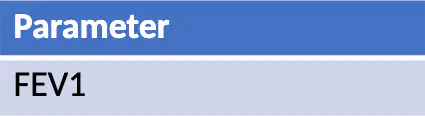
Refer to the table comparing obstructive vs restrictive pulmonary disorders. Which parameter is decreased in both obstructive and restrictive patterns?
FEV1
FVC
FEV1/FVC
TLC

Refer to the table comparing obstructive vs restrictive pulmonary disorders. What happens to the FEV1/FVC ratio in restrictive disease?
Decreased
Normal or increased
Normal only
Increased only
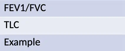
Refer to the table comparing obstructive vs restrictive pulmonary disorders. Which statement about total lung capacity (TLC) is correct?
TLC is decreased in obstructive and normal in restrictive
TLC is normal or increased in obstructive and decreased in restrictive
TLC is increased in both obstructive and restrictive
TLC is normal in both obstructive and restrictive
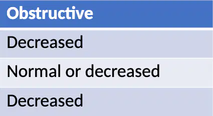
Refer to the table comparing obstructive vs restrictive pulmonary disorders. Which combination correctly describes forced vital capacity (FVC) in obstructive vs restrictive patterns?
Obstructive: decreased; Restrictive: normal
Obstructive: normal or decreased; Restrictive: decreased
Obstructive: increased; Restrictive: decreased
Obstructive: normal; Restrictive: normal

Refer to the table comparing obstructive vs restrictive pulmonary disorders. Which set of example conditions corresponds to restrictive disease?
Asthma, COPD
Fibrosis, Sarcoidosis
Asthma, Sarcoidosis
COPD, Fibrosis
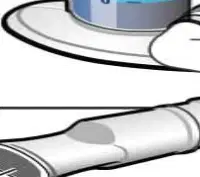
According to the spirometry diagram, which labeled part is placed in the mouth during the test?
Mouthpiece
Nose clip
Tube
Spirometer

What parameter is indicated as being recorded in the spirometry diagram?
Airflow measurement
Blood pressure
Blood glucose
Heart rate
Based on the slide titled "What are PFTs Beyond Spirometry?", which aspects do pulmonary function tests beyond spirometry help evaluate?
Lung volumes
Gas exchange across alveolar membrane
Small airway function
Bone mineral density
According to the slide titled "What are PFTs Beyond Spirometry?", these tests are used for diagnosis and monitoring of which conditions?
Restrictive lung disease
ILD (Interstitial Lung Disease)
Pulmonary vascular diseases
Peptic ulcer disease
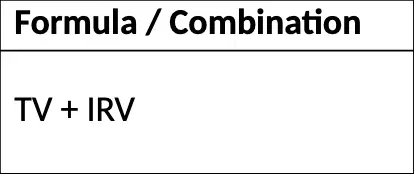
Refer to the lung capacity table that lists each capacity, its formula/combination, definition, and normal value. Which formula correctly represents Inspiratory Capacity (IC)?
TV + IRV
ERV + RV
TV + IRV + ERV
TV + IRV + ERV + RV

According to the lung capacity table, what is the definition of Functional Residual Capacity (FRC)?
Max air that can be inhaled after normal exhalation
Air remaining in lungs after normal expiration
Max air that can be exhaled after maximum inhalation
Total volume of air in lungs after maximal inspiration

From the lung capacity table, which formula correctly expresses Total Lung Capacity (TLC)?
TV + IRV
ERV + RV
TV + IRV + ERV
TV + IRV + ERV + RV (also equal to VC + RV)

According to the lung capacity table, what is the typical normal value range for Total Lung Capacity (TLC)?
~3500 mL
~2300–2500 mL
~4600–4800 mL
~5800–6000 mL
Refer to the table of pulmonary function tests and their purposes. Which test primarily measures lung volumes such as TLC, RV, and FRC?
Plethysmography
DLCO (Diffusion Capacity)
PEFR (Peak Flow)
MVV (Max Voluntary Ventilation)
According to the table, which test assesses gas exchange capacity?
DLCO (Diffusion Capacity)
PEFR (Peak Flow)
MVV (Max Voluntary Ventilation)
Nitrogen Washout / He Dilution
Which test is indicated for asthma monitoring according to the table?
PEFR (Peak Flow)
MVV (Max Voluntary Ventilation)
DLCO (Diffusion Capacity)
Plethysmography
Which test evaluates ventilatory muscle capacity in the table?
MVV (Max Voluntary Ventilation)
PEFR (Peak Flow)
DLCO (Diffusion Capacity)
Nitrogen Washout / He Dilution
According to the slide labeled Body Plethysmography, which change allows calculation of lung volumes in the body box setup?
Changes in pressure inside the box
Changes in ambient room temperature
Variations in heart rate
Fluctuations in oxygen saturation
In body plethysmography, where does the patient sit during the test?
Inside an airtight chamber called a body box
In an open room with a spirometer
On a treadmill inside a lab
In a hyperbaric oxygen chamber
Which lung volume is specifically described as air that cannot be exhaled in the Body Plethysmography slide?
Residual Volume (RV)
Total Lung Capacity (TLC)
Functional Residual Capacity (FRC)
Tidal Volume (TV)

