
 Worksheets
Exam 1 Study Guide
Total questions: 160
Worksheet time: 1hrs 20mins
Anencephaly is best described as which of the following?
Failure of spinal cord closure with intact cranial vault
Absence of cerebral hemispheres with missing skull and scalp
Partial development of the cerebellum with normal cortex
Herniation of meninges through the vertebral column
Which statement most accurately identifies anencephaly among congenital disorders?
It is the most common skeletal malformation
It represents the most frequent congenital abnormality of the CNS
It occurs less often than spina bifida worldwide
It primarily affects the spinal canal rather than the cranium
Anencephaly is most commonly found in areas where which condition is also most prevalent?
Hydrocephalus
Spina bifida
Microcephaly
Encephalocele
Placenta previa is best defined as which of the following?
Implantation of the placenta within the myometrium
Placental tissue located near the uterine fundus
Placenta partially or completely covering the internal cervical os
Separation of the placenta from the uterine wall
Placenta previa is the most common cause of which clinical finding in the third trimester?
Painful uterine contractions
Painless vaginal bleeding
Decreased fetal movement
Hypertensive crisis
Which option correctly groups the recognized types of placenta previa?
Total, partial (marginal), and low-lying
Central, lateral, and posterior
Complete, abrupt, and previa
Marginal, velamentous, and fundal
Hydranencephaly is best described as which of the following?
Partial absence of the cerebellum with intact cortex
Total or near-total absence of the cerebral hemispheres
Failure of neural tube closure in the cranial vault
Herniation of brain tissue through the skull
In hydranencephaly, which intracranial structures are typically preserved?
Cerebral cortex and lateral ventricles
Midbrain, basal ganglia, thalamus, and cerebellum
Brainstem only
Cerebellum and frontal lobes
Hydranencephaly is most commonly believed to result from which underlying mechanism?
Failure of mesodermal migration
Bilateral obstruction of the internal carotid arteries
Incomplete separation of the hemispheres
Abnormal neural crest cell development
During the first trimester, the normal growth rate of the fetal pole is approximately:
0.5 mm per day
1 mm per day
2 mm per day
4 mm per day
Organogenesis primarily occurs during which time period of pregnancy?
First 5 weeks of gestation
First 10 weeks of pregnancy
Second trimester only
After 12 weeks of gestation
By approximately 9–10 weeks of gestation, which fetal structures can typically be visualized?
Heart chambers and kidneys only
Spine, head, and developing limbs
Facial features and external genitalia
Brain ventricles and diaphragm
The umbilical cord is usually visualized at which stage of pregnancy?
Early first trimester
Mid second trimester
Latter portion of the first trimester
Third trimester only
Inevitable abortion is best characterized by which of the following?
Mild spotting with a closed cervix
Profuse bleeding with the cervix beginning to open
Absence of fetal cardiac activity without bleeding
Passage of all fetal tissue with no cervical dilation
Which placental grade shows no calcifications?
Grade 0
Grade 1
Grade 2
Grade 3
Scattered calcifications in the placenta correspond to which grade?
Grade 0
Grade 1
Grade 2
Grade 3
Basal calcifications in the placenta are characteristic of which grade?
Grade 0
Grade 1
Grade 2
Grade 3
Which placental grade may show basal and interlobar septal calcifications outlining maternal cotyledons?
Grade 0
Grade 1
Grade 2
Grade 3
Holoprosencephaly is caused by which of the following?
Failure of the prosencephalon to divide into cerebral hemispheres
Abnormal neural crest migration
Agenesis of the cerebellum
Spinal cord malformation
The thalami in holoprosencephaly are typically:
Separated
Fused
Absent
Hypoplastic
Holoprosencephaly is associated with which type of ventricular formation?
Two separate lateral ventricles
Single common ventricle (horseshoe shaped)
Normal ventricular anatomy
Multiple small cystic spaces
Facial deformities associated with holoprosencephaly commonly involve:
Eyes, nose, and upper lip
Mandible only
Ears and neck
Lower limbs
Which chromosomal abnormality is often associated with holoprosencephaly?
Trisomy 13
Trisomy 21
Turner syndrome
Klinefelter syndrome
Alobar holoprosencephaly is characterized by all of the following EXCEPT:
Single horseshoe-shaped ventricle
Fused thalami
Crescent-shaped frontal cortex
Complete separation of cerebral hemispheres
Craniofacial abnormalities associated with alobar holoprosencephaly include:
Cyclopia, proboscis, median cleft lip, hypotelorism
Micrognathia only
Low-set ears
Polydactyly
Semilobar holoprosencephaly differs from alobar type in that it has:
More cerebral tissue
Fused thalami
Crescent-shaped frontal cortex
Cyclopia
Semilobar holoprosencephaly is associated with which craniofacial anomalies?
Cleft palate and lip
Cyclopia
Proboscis
Median cleft lip
Lobar holoprosencephaly is characterized by:
Nearly complete separation of cerebral hemispheres
Single horseshoe-shaped ventricle
Fused thalami
Crescent-shaped frontal cortex
Lobar holoprosencephaly may be associated with all of the following EXCEPT:
Dandy-Walker malformation
Enlarged cisterna magna
Arnold-Chiari malformation
Cyclopia
Which abnormal structure may be absent in lobar holoprosencephaly?
Septum pellucidum
Thalami
Cerebellum
Brainstem
Ethmocephaly in holoprosencephaly refers to:
A facial abnormality with hypotelorism
Fused thalami
Horseshoe-shaped ventricle
Crescent-shaped frontal cortex
Microcephaly can be associated with which types of holoprosencephaly?
Alobar and semilobar
Lobar only
Ethmocephaly only
None of the above
Which type of holoprosencephaly shows incomplete fusion of the thalami and rudimentary occipital horns?
Semilobar
Alobar
Lobar
Ethmocephaly
Velamentous placental cord insertion (PCI) is best described as:
Umbilical cord inserting centrally into the placenta
Umbilical cord inserting at the edge of the placenta, extending into the membranes
Cord wrapped around the fetal neck
Complete absence of the umbilical cord
A major complication of velamentous PCI is:
Polyhydramnios
Vasa previa
Placental abruption
Fetal growth acceleration
Why are the umbilical vessels at risk in velamentous PCI?
They are unusually thick and prone to rupture
They are not protected by Wharton's jelly, increasing risk of rupture or thrombosis
They are always compressed by the placenta
They have additional layers of smooth muscle
IVC compression syndrome occurs due to:
Compression of the inferior vena cava by the gravid uterus when supine
Obstruction of the superior vena cava by the fetus
Umbilical cord compression during labor
Placental abruption
Common symptoms of IVC compression syndrome include:
Headache, blurred vision, and seizures
Lightheadedness, fainting, vertigo, and nausea
Abdominal pain and vaginal bleeding
Hypertension and proteinuria
The recommended treatment for IVC compression syndrome is:
Continue examination and monitor vitals
Administer IV fluids while patient remains supine
Stop the examination and turn the patient onto her left side
Immediate cesarean delivery
The fetal heart is the first organ to function at approximately:
15 days (3 weeks)
23 days (5–5½ weeks)
30 days (6 weeks)
35 days (7 weeks)
At 6 weeks of gestation, the fetal heart rate is typically:
80 bpm
110 bpm
140 bpm
175 bpm
The fetal heart rate peaks at approximately 9 weeks of gestation at:
110 bpm
140 bpm
175 bpm
200 bpm
During the second trimester, the normal fetal heart rate ranges between:
90–110 bpm
110–120 bpm
160–170 bpm
180–200 bpm
In the third trimester, the typical fetal heart rate is:
80–100 bpm
110–160 bpm
160–180 bpm
175–200 bpm
Correct femur length measurement involves which part of the bone?
The ossified femoral diaphysis (shaft)
The femoral head
The epiphyseal cartilage
The condyles
When measuring femur length, which parts should be excluded?
Ossified shaft
Epiphyseal cartilage, femoral head, and condyles
Midshaft only
None, the entire bone is measured
Measuring femur length is most accurate for which purpose?
Dating early first trimester pregnancies
Dating late pregnancy
Detecting femoral fractures
Measuring fetal weight only
Which femur should be measured during ultrasound for the most accurate length?
The femur farthest from the transducer
Both femurs averaged
The femur closest to the transducer
Either femur, position does not matter
Placenta accreta is best defined as:
Chorionic villi invade beyond the uterine serosa
Chorionic villi are in direct contact with the myometrium
Chorionic villi invade the myometrium and extend into the serosa
Placenta completely separates from the uterus
Placenta increta is characterized by:
Villi in direct contact with the myometrium
Villi invading the myometrium and extending into the serosa
Villi extending beyond the uterine serosa into adjacent organs
Placenta previa only
Placenta percreta occurs when:
Villi are in contact with the myometrium
Villi invade only the endometrium
Villi extend beyond the uterine serosa, possibly involving bladder or bowel
Placenta detaches prematurely
The amnion is best described as:
A small cavity developing from the embryonic disc
A membrane surrounding the embryo that secretes fluid to form the amniotic cavity
A projection of the hindgut
The fourth membrane that encloses all other membranes
The yolk sac is:
A membrane forming the fetal portion of the placenta
A small cavity developing from the embryonic disc
A projection of the hindgut
The fluid-filled cavity surrounding the embryo
The allantois is:
The membrane that surrounds the amniotic cavity
A projection of the hindgut
A small cavity developing from the embryonic disc
The fourth membrane enclosing all others
The chorion is:
The membrane surrounding the embryo and secreting fluid
A small cavity developing from the embryonic disc
The fourth membrane enclosing the other three, developing from the trophoblast and forming the fetal portion of the placenta
A projection of the hindgut
Using transvaginal ultrasound (TV), the gestational sac is typically visible at:
3 weeks LMP (500 mIU/ml)
4½ weeks LMP (≈1025 mIU/ml)
5 weeks LMP (1500 mIU/ml)
6 weeks LMP (1800 mIU/ml)
Using transabdominal ultrasound (TA), the gestational sac is typically visible at:
4 weeks LMP (1000 mIU/ml)
5 weeks LMP (1500 mIU/ml)
6 weeks LMP (≈1800 mIU/ml)
7 weeks LMP (2500 mIU/ml)
Organogenesis primarily occurs during which period of pregnancy?
First 4 weeks
First 10 weeks
Second trimester
Third trimester
During week 4 of fetal life, the foregut gives rise to which structures?
Jejunum, ileum, cecum, ascending colon
Esophagus, stomach, duodenum, liver, and pancreas
Colon, sigmoid, and rectum
Heart and lungs
The midgut develops into:
Esophagus, stomach, duodenum
Jejunum, ileum, cecum, and ascending colon
Colon, sigmoid, and rectum
Liver and pancreas
The hindgut forms which of the following structures?
Esophagus, stomach, and duodenum
Jejunum, ileum, cecum, and ascending colon
Colon, sigmoid, and rectum
Liver and pancreas
The decidua basalis is best described as:
The decidual lining of the uterus
The portion of decidua at the point of attachment of the blastocyst
The decidua projecting into the uterine lumen
The outer layer of the chorion
The decidua capsularis refers to:
The portion superficial to the blastocyst projecting into the uterine lumen
The lining of the uterus not involved in implantation
The decidua at the point of blastocyst attachment
The fetal portion of the placenta
The decidua parietalis (vera) is:
The decidual lining of the uterus away from the implantation site
The portion projecting into the uterine lumen
The point of attachment of the blastocyst
The layer forming the fetal placenta
The mean sac diameter (MSD) of the gestational sac is calculated by:
Adding the length, width, and height, then dividing by 2
Adding the length, width, and height, then dividing by 3
Multiplying length × width × height
Measuring only the longest diameter of the sac
The biparietal diameter (BPD) is measured in which plane of the fetal head?
Sagittal
Coronal
Axial
Oblique
BPD measurement is most accurate during which gestational period?
8–12 weeks
14–20 weeks
22–28 weeks
30–36 weeks
The correct location for obtaining a BPD measurement is:
15 degrees inferior to cathomedial and perpendicular to skull base
15 degrees superior to cathomedial and parallel to skull base
Midline only, without angulation
At the posterior fontanelle
Which of the following are the landmarks used for BPD measurement?
Cerebellum and cisterna magna
Paired hypoechoic thalami, cavum septum pellucidum, third ventricle, and midline falx cerebri
Spine and diaphragm
Fetal stomach and umbilical vein
When measuring BPD, the calipers should be placed:
From leading edge to leading edge (outer to inner) along the longest horizontal part
From inner edge to inner edge along a vertical line
From anterior to posterior fontanelle only
Along the midline falx only
The cephalic index is calculated by:
OFD + BPD
BPD ÷ OFD
(BPD + OFD) ÷ 2
BPD × OFD
Normal cephalic index values range between:
50–65%
65–75%
75–85%
85–95%
A cephalic index greater than 85–90% indicates:
Dolichocephaly (head too long)
Brachycephaly (head too wide)
Microcephaly
Normal head shape
A cephalic index less than 75% indicates:
Normal head shape
Dolichocephaly (head too long)
Brachycephaly (head too wide)
Microcephaly
A missed abortion is best defined as:
A living embryo that is spontaneously expelled
A dead embryo that has not been expelled, with no fetal heartbeat detected
Vaginal bleeding with cervical dilation
Partial separation of the placenta
The umbilical cord normally contains how many vessels?
1 artery and 2 veins
2 arteries and 1 vein
2 veins and 2 arteries
1 artery and 1 vein
Fetal weight is most heavily based on which measurement?
BPD (Biparietal Diameter)
AC (Abdominal Circumference)
FL (Femur Length)
OFD (Occipitofrontal Diameter)
The AC is considered a standard measurement for dating the pregnancy at approximately:
8–10 weeks
12–15 weeks
18–20 weeks
22–24 weeks
When evaluating the umbilical vein in an AC image, which finding indicates a persistent right umbilical vein?
Umbilical vein arches away from the stomach
Umbilical vein points toward the stomach
Umbilical vein runs along the spine
Umbilical vein is not visualized
The principal cause of first trimester fetal demise is:
Maternal hypertension
Chromosomal abnormalities
Cord accidents
Infection
Common clinical signs of first trimester fetal demise include all EXCEPT:
Uterus small for dates
Clinician cannot hear fetal heart tones
Patient reports feeling “less pregnant”
Increased fetal movement
Second trimester fetal demise is often associated with which of the following factors?
Chromosomal abnormalities only
Uterine anomalies, incompetent cervix, placental abnormalities, and autoimmune diseases (e.g., lupus)
Maternal hypotension
Excessive fetal movement
Ultrasound-specific signs of third trimester fetal demise include:
Normal fetal movement
Gas in fetal vasculature, overlapping cranial sutures (Spalding’s sign), fetal maceration
Polyhydramnios
Enlarged fetal heart
Management of fetal demise in the second trimester (<14 weeks) typically involves:
Expectant management only
Dilation and curettage (D&C)
Cesarean delivery
Oxytocin infusion only
Management considerations for third trimester fetal demise include:
Maternal hemorrhage risk (hypofibrinogenemia), D&E, oxytocin, amnioinfusion with hypertonic solution, prostaglandins
No intervention is needed
Only cesarean section is appropriate
Chromosomal testing only
In early fetal life, the lungs appear on ultrasound as:
Hyperechoic compared to the liver
Isoechoic or slightly hypoechoic compared to the liver
Anechoic
Hyperechoic to bone
Later in fetal life, the fetal lungs typically appear:
Hypoechoic to the liver
Isoechoic to the liver
Hyperechoic to the liver
Anechoic
Which biochemical measurement is commonly used to assess fetal lung maturity?
Bilirubin levels
Lecithin–Sphingomyelin (L/S) ratio
Alpha-fetoprotein
Amniotic fluid glucose
Wharton’s jelly is best described as:
The amniotic membrane surrounding the fetus
Mucoid substance that surrounds umbilical cord vessels, enclosed in the amnion
The fetal portion of the placenta
The chorionic villi connecting to the maternal decidua
A cervical cerclage is:
A loop placed in the cervix to hold it closed and help maintain pregnancy
Surgical removal of the cervix
A procedure to induce labor
A diagnostic test for cervical length
A threatened abortion is characterized by:
Vaginal bleeding with an open cervix after 20 weeks
Vaginal bleeding with a closed cervix prior to 20 weeks
Complete expulsion of the fetus
Absence of vaginal bleeding with a dilated cervix
A zygote is:
A multicellular embryo at day 5
A single cell formed during fertilization containing all genetic material from mother and father
The fetal portion of the placenta
A fluid-filled cavity within the blastocyst
The fetal stomach can usually be visualized on ultrasound by:
12 weeks
16 weeks
20 weeks
24 weeks
The fetal stomach serves as an important landmark for:
Biparietal diameter (BPD) measurement
Femur length measurement
Abdominal circumference (AC) measurement
Placental grading
Visualization of the fetal bladder on ultrasound indicates:
Normal heart function
The presence of at least one functioning kidney
Normal lung development
Correct fetal positioning
A 2-vessel cord (single umbilical artery) is seen in approximately:
0.1–0.2% of pregnancies
0.5–1% of pregnancies
2–3% of pregnancies
5% of pregnancies
A 2-vessel cord (SUA) consists of:
2 umbilical veins and 1 umbilical artery
1 umbilical vein and 1 umbilical artery
1 umbilical vein only
2 umbilical arteries only
The fetal abdominal circumference (AC) primarily reflects the development of:
Brain and cerebellum
Liver, spleen, and subcutaneous fat
Lungs and diaphragm
Kidneys and bladder
Landmarks for AC measurement include all of the following EXCEPT:
Stomach
Three ossification centers of the spine
Umbilical vein (hockey stick sign)
Cerebellum
AC can also be calculated using TAD and APD by:
Adding them together and multiplying by 1.57 (1/2 π)
Adding them together and multiplying by 2
Multiplying them together only
Averaging them without multiplication
One of the primary indications for a first trimester ultrasound is:
Evaluating placental grading
Predicting fetal viability (normal vs abnormal, including fetal heart motion)
Measuring femur length in the third trimester
Assessing fetal lung maturity
First trimester ultrasound is used to accurately determine:
Fetal weight in the third trimester
Gestational age
Cephalic index
Placental insertion
Conditions associated with hydrocephalus that can sometimes be detected in the first trimester include:
Spina bifida (most common) and cephalocele (present in 80% of cases)
Micrognathia and cleft lip
Congenital diaphragmatic hernia only
Limb-length discrepancies
Arnold-Chiari malformation is a common type of hydrocephalus associated with:
Microcephaly
Spina bifida
Holoprosencephaly
Dandy-Walker malformation
In Arnold-Chiari malformation, the spinal cord is:
Normal and ends at L4
Tethered and ends before L2
Absent
Fused with the brainstem
A characteristic feature of Arnold-Chiari malformation is:
Cerebellum pulled through the cisterna magna
Cerebellum enlarged and in normal position
Complete absence of the cerebellum
Lateral ventricles completely collapsed
The “banana sign” on ultrasound refers to:
Shape of the cerebellum appearing curved like a banana instead of a dumbbell
Shape of the lateral ventricles
Appearance of the fetal bladder
Shape of the stomach
The “lemon sign” refers to:
Narrowing of the posterior skull
Narrowing of the anterior portion of the head
Enlargement of the lateral ventricles
Flattening of the cerebellum
Dilation of the lateral ventricles in Arnold-Chiari malformation is typically:
Unilateral and mild
Bilateral and severe, called the “bat wing sign”
Absent
Only in the third trimester
Dandy-Walker syndrome is best described as:
A congenital brain malformation characterized by incomplete formation of the cerebellar vermis, dilation of the fourth ventricle, and enlargement of the posterior fossa
A spinal cord malformation associated with tethered cord
A form of holoprosencephaly
A type of microcephaly
The cause of Dandy-Walker syndrome is thought to be:
Chromosomal trisomy 13
Obstruction of the foramina of Luschka and Magendie
Maternal infection only
Umbilical cord malformation
Key features of Dandy-Walker syndrome include all EXCEPT:
Incomplete formation of the cerebellar vermis
Dilation of the fourth ventricle
Enlargement of the posterior fossa
Pulled cerebellum through cisterna magna
The “dangling choroid” sign on ultrasound refers to:
Enlarged ventricles moving toward the dependent portion of the brain, typically seen in hydrocephalus
Normal lateral ventricle anatomy
Absence of the choroid plexus
Pulled cerebellum through the cisterna magna
Spina bifida is defined as:
Overgrowth of vertebral arches
A defect in the spine due to lack of fusion of the two halves of the vertebral arch
Absence of the brainstem
A type of hydrocephalus
Which type of spina bifida involves only the meninges protruding?
Meningocele
Meningomyelocele
Occult
Arnold-Chiari
Which type of spina bifida involves both meninges and spinal cord protrusion?
Meningocele
Meningomyelocele
Occult
Hydrocephalic
Occult spina bifida is characterized by:
Large protrusion of meninges
Small defect with no protrusion of meninges or spinal cord
Protrusion of spinal cord only
Hydrocephalus
The most common location for spina bifida is:
Cervical
Thoracic
Lumbar-sacral
Sacral only
The second most common location for spina bifida is:
Lumbar
Cervical
Thoracic
Occipital
Spina bifida has a high association with which malformation?
Dandy-Walker
Arnold-Chiari malformation
Holoprosencephaly
Microcephaly
Turner’s Syndrome is caused by:
Trisomy 21
Total or partial loss of the second sex chromosome (46,X)
Trisomy 18
Mutation of the X and Y chromosomes
Turner’s Syndrome is more common in:
Males
Females
Equal in males and females
Only in twins
Turner’s Syndrome is commonly associated with:
Cystic hygroma or increased AFP
Microcephaly
Spina bifida only
Holoprosencephaly
The “Batwing sign” is associated with which condition?
Dandy-Walker syndrome
Arnold-Chiari malformation
Hydrocephalus due to aqueductal stenosis
Turner’s syndrome
The Batwing sign represents:
Absence of cerebellar vermis
Bilateral dilation of the frontal horns of the lateral ventricles
Narrowing of the anterior portion of the head
Overlapping cranial sutures
The “Lemon sign” is associated with which condition?
Dandy-Walker syndrome
Arnold-Chiari malformation
Hydrocephalus due to aqueductal stenosis
Turner’s syndrome
The Lemon sign refers to:
Narrowing of the anterior portion of the head
Bilateral dilation of the frontal horns of the lateral ventricles
Absence of the cerebellar vermis
Flattened occiput
The cavum septi pellucidi (CSP) is an important landmark for measuring:
Abdominal circumference (AC)
Biparietal diameter (BPD)
Femur length (FL)
Nuchal translucency
The CSP appears on ultrasound as:
A thick, horizontal membrane between the thalami
A thin, triangular, vertical membrane separating the anterior horns of the left and right lateral ventricles (thin parallel lines)
A round cyst in the posterior fossa
A fluid-filled cavity within the cerebellum
Cystic hygroma is best described as:
Malignant tumor of the brain
Benign lesion with lymphatic origin
Fluid collection in the fetal lungs
Cyst in the fetal abdomen
Approximately 80% of cystic hygromas originate from:
Anterior portion of the neck
Posterolateral portion of the neck
Thoracic cavity
Abdominal wall
Cystic hygroma may be associated with which condition?
Turner’s syndrome
Dandy-Walker malformation
Arnold-Chiari malformation
Spina bifida
A cephalocele is best described as:
Fluid-filled mass in the posterolateral neck
Complex paracranial mass with no bone coverage
Fluid collection in the fetal abdomen
Thickened nuchal fold
Cephalocele may be associated with which of the following findings?
Microcephaly and hydrocephalus
Turner’s syndrome only
Posterolateral neck cyst with septa
Spina bifida
Cystic hygroma is characterized by:
Solid cranial mass
Cystic mass with septa, usually located at the posterolateral neck
Posterior fossa enlargement only
Microcephaly without fluid
In cystic hygroma, what forms the midline segment?
Cavum septi pellucidi
Nuchal ligament
Cerebellar vermis
Occipital bone
The most common cause of first trimester fetal death is:
Maternal infection
Chromosomal abnormalities
Placental abruption
Turner’s syndrome
The most common cause of painless vaginal bleeding in the third trimester is:
Placental abruption
Placenta previa
Uterine rupture
Preterm labor
The most common cause of painful vaginal bleeding in the third trimester is:
Placenta previa
Placental abruption
Cervical incompetence
Uterine fibroids
The most common benign tumor of the placenta is:
Chorioangioma
Teratoma
Fibroma
Lipoma
The most common causes of oligohydramnios are:
Maternal diabetes and hypertension
PROM, IUGR, and fetal genitourinary abnormalities
Polyhydramnios and maternal anemia
Placenta previa and abruption
The most common congenital abnormalities involve which system?
Cardiovascular system
Central nervous system (CNS)
Musculoskeletal system
Renal system
The most common congenital anomaly of the CNS is:
Hydrocephalus
Spina bifida
Anencephaly (neural tube defect)
Dandy-Walker malformation
The most common congenital anomaly of the face is:
Micrognathia
Facial clefts (associated with Trisomy 13)
Cystic hygroma
Cleft palate only
The most common location for spina bifida is:
Cervical
Thoracic
Lumbar-sacral (cervical 2nd most common)
Sacral only
The menses phase of the uterine cycle occurs during which days?
1-5
6-14
15-28
14-21
During the menses phase, which of the following occurs?
Endometrium regenerates
Endometrium degenerates, sloughs off the myometrium, and is expelled as menses
Corpus luteum secretes progesterone
Ovulation occurs
The proliferative phase of the uterine cycle occurs during which days?
1-5
6-14
15-28
14 only
What hormone primarily drives the proliferative phase of the uterine cycle?
Progesterone
estrogen
LH
FSH
The secretory phase of the uterine cycle is characterized by:
Endometrial degeneration and shedding
Glandular and vascular changes in the endometrium under the influence of progesterone
Follicular growth in the ovary
LH surge and ovulation
6. During the ovarian cycle, FSH is responsible for:
A. Triggering ovulation
B. Making ovarian follicles grow
C. Degeneration of corpus luteum
D. Secretory phase of uterus
Correct answer: B
Triggering ovualtion
making ovarian follicles grow
degeneration of corpus luteum
secretory phase of uterus
Ovulation occurs approximately on which day of the menstrual cycle?
1
7
14
28
The luteal phase (days 14–28) is characterized by:
Growth of ovarian follicles
Corpus luteum formation and progesterone production
sloughing of the endometrium
follicular release of estrogen
What triggers ovulation in the ovarian cycle?
FSH surge
Estrogen surge only
LH surge
Progesterone decline
If fertilization does not occur, the corpus luteum:
Continues to grow indefinitely
Secretes increasing estrogen
Declines, causing a drop in progesterone and menstruation
Converts into a new follicle
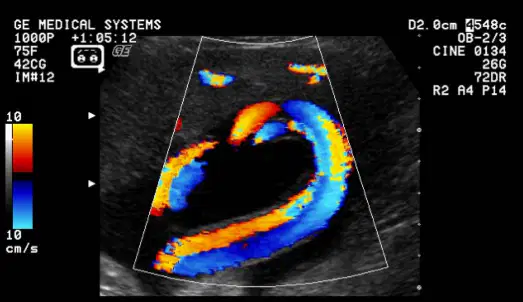
What is seen here?
1 Vessel Cord
2 Vessel SUA
Normal 3 vessel cord
Snake sign
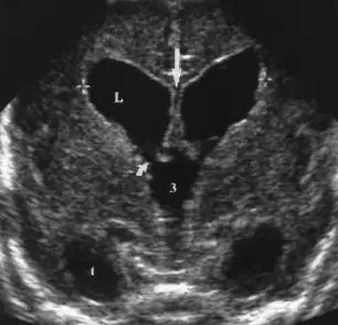
What is seen here?
Batwing sign seen with Arnold Chiari
Lemon Sign seen with Arnold Chiari
Banana Sign seen with Arnold Chiari
Dandy Walker Syndrome
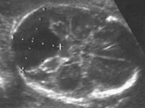
What anomaly is seen here
Dandy Walker
Hydrocephalus
Dangling Choriod
Spina Bifida
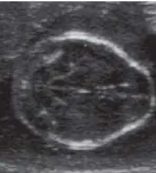
What sign is seen here?
Batwing Sign
Banana Sign
Lemon Sign
Iceberg Sign
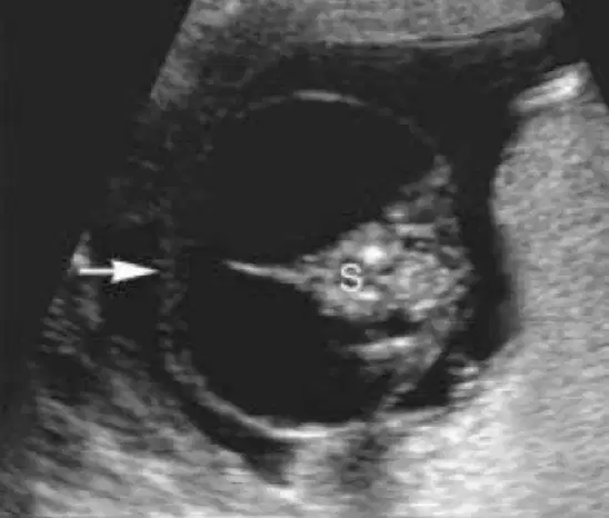
What is seen here?
Cephalocele
Cystic Hygroma
Turners syndrome
Arnold Chiari

