
 Worksheets
Seminar:Image Receptors
Total questions: 83
Worksheet time: 42mins
Which statement best distinguishes Computed Radiography from Digital Radiography in clinical workflow?
CR requires cassette scanning later in a reader
CR shows the image instantly on the monitor
DR stores exposure on a cassette for delayed reading
DR uses storage phosphor plates inside cassettes
Match each detector system to its key feature.
Computed Radiography (CR)
Uses storage phosphor plate cassette
Digital Radiography (DR)
Uses flat‑panel detector hardware
Indirect or Direct DR
Forms image on monitor without cassette
Which components are characteristic of Digital Radiography systems? Select all that apply.
Mandatory external reader scanning
Instant image display on monitor
Flat‑panel detector hardware
Storage phosphor plate cassette
Which operational step is unique to Computed Radiography and not required in Digital Radiography?
Scanning the cassette in a separate reader
Immediate image appearance on the monitor
Bypassing cassettes during acquisition
Using a flat‑panel detector for capture
Which component of a computed radiography (CR) system stores the latent image after x‑ray exposure?
Cassette housing
Laser light source
PSP imaging plate
Plate transport motor
Which materials commonly make up the photostimulable phosphor in a PSP plate?
Rare‑earth gadolinium oxysulfide
Calcium tungstate crystals
Barium fluorohalide iodides
Barium fluorohalide bromides
What is the role of the plate reader in a CR system?
Erases cassette static charges
Amplifies x‑ray beam intensity
Scans PSP to release stored light
Generates visible light for exposure
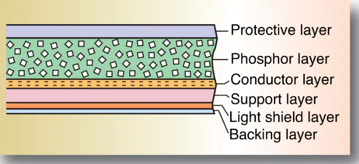
Which PSP layer protects the active phosphor from physical damage?
Support layer
Backing layer
Protective layer
Conductive layer

Which PSP layer contains the photostimulable barium fluorohalide crystals that trap energy?
Phosphor layer
Reflective layer
Color layer
Light shield layer
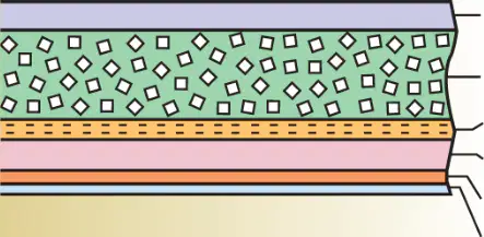
Which PSP layer prevents light from the reader from reaching the support and reflecting back, reducing noise?
Light shield layer
Backing layer
Protective layer
Conductive layer
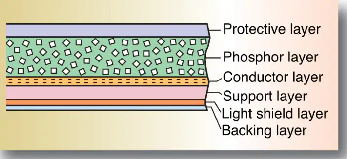
Which PSP layer dissipates static electricity to reduce artifacts during handling?
Color layer
Conductive layer
Support layer
Protective layer
Which layer of a CR imaging plate contains the material that traps energized electrons after X-ray exposure?
Support base layer
Conductive backing layer
Phosphor active layer
Protective overcoat layer
What term describes the invisible stored pattern formed by trapped electrons in the PSP plate?
Digital image
Processed image
Analog image
Latent image
In the reader, what releases the stored energy from trapped electrons to create a light signal?
High-voltage amplifier
Scanning laser beam
X-ray backscatter
Cooling fan system
Place the steps inside the CR reader in correct order.
Laser scans phosphor
First step
Light detected and amplified
Second step
ADC converts to digital
Third step
Computer displays image
Fourth step
Which statement best explains why the latent image pattern matches the X-ray exposure?
Electrons are trapped randomly everywhere
More traps fill where exposure is lower
Electron trapping density reflects dose
Traps form only after laser scanning
Which material in the active layer of the imaging plate absorbs X-ray energy through the photoelectric effect?
Cesium iodide scintillators
Calcium tungstate phosphor
Silver bromide crystals
Barium fluorohalide crystals
What role does europium (Eu) play in the CR imaging plate?
Acts as protective binder
Serves as crystal activator
Functions as optical filter
Provides mechanical support
Which process frees electrons from their normal positions within the crystal after absorption of X-ray energy?
Photoelectric effect
Thermal diffusion
Compton scattering
Pair production
Why can a single X-ray interaction release multiple photoelectrons in the phosphor crystal?
It causes lattice melting
It splits photons into pairs
It shares energy among electrons
It creates electron avalanches
During electron multiplication, what event increases the number of free electrons in regions of higher exposure?
Electrons recombine with holes
Photoelectrons collide and liberate others
Electrons are trapped immediately
Laser stimulates emission early
Where are the electron traps associated that capture energized electrons to form the latent image?
At phosphor grain boundaries
Inside the protective overcoat
At silver ion vacancies
At europium activator sites
Select all steps that occur before the ADC converts signals to digital data.
Laser stimulates trapped electrons
Visible light is emitted
Computer displays the image
Photodetector amplifies light
Match each component to its function in CR imaging.
Phosphor layer
Stores latent image
Laser scanner
Releases trapped energy
Photodetector
Converts light to signal
ADC
Digitizes electrical signal
In computed radiography, what determines how much light the imaging plate releases during reading?
The number of pixels in the scanner
The amount of X-ray exposure received
The plate’s physical thickness only
The duration of laser dwell time
A region of a CR plate that received more X-rays will produce which outcome during readout?
More emitted light from that region
Less emitted light from that region
No emitted light from that region
Equal light regardless of exposure
Which statement best describes how image data are created in CR after light is emitted from the plate?
Magnets translate light into physical density
A chemical reaction darkens the plate directly
The technologist estimates brightness manually
A computer converts collected light into digital values
Optical density (OD) on film compared with CR digital values is best characterized as
Opposite scales with inverse meaning
Identical and interchangeable concepts
Completely unrelated measurements
Similar but not exactly equivalent measures
Match each CR reader light color to its primary role.
Red laser light
Stimulates trapped electrons on PSP
Blue‑green PSL light
Carries image information to detector
White erase light
Clears remaining trapped electrons
During scanning, the red laser light mainly
Erases residual signal after readout
Displays the image on the monitor
Collects PSL at the photodetector
Excites trapped electrons to release energy
The blue‑green light observed in CR is produced when
A filter converts red light to shorter wavelengths
Electrons relax and emit photostimulable luminescence
The erase lamp floods the plate uniformly
The laser heats phosphors to high temperature
Which option contains the correct sequence from plate insertion to image appearance?
Laser scans → PSL emitted → Light collected → Signal digitized
Erase light floods → Laser scans → Image appears → PSL emitted
PSL emitted → Signal digitized → Laser scans → Light collected
Signal digitized → Laser scans → Erase light → PSL emitted
Select all actions performed by the white erase light after scanning.
Carries image information to detector
Floods the PSP plate uniformly
Clears any remaining trapped electrons
Prepares the plate to be reused
Which component directly turns the collected light into pixels for display?
The cassette housing
The red stimulation laser
The white erase lamp
The computer’s digitizer system
If a plate is not properly erased after use, the most likely artifact is
Overconstrained histogram with perfect contrast
Uniform increase in detector quantum noise
Complete loss of new exposure data
Residual ghost image from prior exam
In CR, PSL intensity from the plate is proportional to which factor during readout?
Erase lamp brightness before scan
Mechanical speed of plate removal
Stored energy from prior X-ray dose
Ambient room light level only
Which statement best describes a key advantage of digital radiography (DR) image receptors compared with computed radiography (CR)?
Require no separate processing step like CR
Use cassettes that must be read later
Send data to PACS only after scanning
Need chemical development of the image
DR systems use what primary hardware to capture the X-ray exposure?
A flat-panel detector array
A rotating anode tube
A fiberoptic image intensifier
A film-screen cassette
Faster DR workflow most directly leads to which clinical outcome?
Higher scatter and more artifacts
Greater need for cassette handling
Slower throughput with longer exams
Fewer repeats and lower patient dose
Active Matrix Flat-Panel Imagers (AMFPIs) are best defined as what?
Film holders with chemical processing
Mechanical grids that block scatter
Digital receptors using a pixel matrix array
Analog screens with fluorescent coating
In each AMFPI pixel, what component temporarily holds the signal until read out by the thin-film transistor (TFT)?
A rotating storage drum
A tungsten anti-scatter grid
A scintillator light guide
A photodiode within the pixel
Match each material type to its role in X-ray detection within flat-panel detectors.
Photoconductor
Absorbs X-rays to create charge
Scintillator
Converts X-rays into visible light
Thin-film transistor (TFT)
Opens to release stored signal
Which statements describe photoconductors used in flat-panel detectors? Select all that apply.
Emit visible light then use photodiodes
Provide charge that is collected for imaging
Create an electrical charge upon exposure
Absorb X-rays directly as energy
Which factor determines whether a detector operates via direct or indirect conversion?
The patient body habitus used
The selected exposure time setting
The type of X-ray absorption material
The orientation of detector columns
Direct versus indirect conversion in digital imaging refers to what process distinction?
Rotation of anode versus cathode
Charge created directly versus via light
Use of cassettes versus tethered panels
Chemical development versus dry processing
Which statement best describes a flat-panel detector in modern radiography?
A digital image receptor used in systems
A handheld ultrasound probe for scanning
A magnetic coil for MRI signal reception
A film screen cassette for analog systems
What benefit do flat-panel detectors provide across the entire image?
Higher dose near the periphery
Selective sharpening only at the center
Stronger magnetic sensitivity overall
Uniform quality from center to edges
Detective Quantum Efficiency (DQE) primarily indicates what characteristic of a detector?
Thickness of the scintillator crystal layer
Ability to resist mechanical vibration forces
Efficiency converting X-ray energy to signal
Speed of data processing inside ADC units
Which layer first reacts when X-rays strike in indirect capture systems?
Analog-to-digital converter stage
TFT storage capacitor array
Amorphous silicon photodiode
Scintillator layer such as CsI
In indirect capture, what is the role of the scintillator layer?
Convert X-rays into visible light
Store charge in tiny capacitors
Digitize signals into pixel data
Transmit trapped data to computer
Which material forms the photodetector layer beneath the scintillator in indirect capture?
Amorphous silicon (a-Si)
Cesium iodide (CsI)
Gadolinium oxysulfide
Lead-based phosphor
What function does amorphous silicon perform in the indirect capture process?
Act as an external magnetic shield
Perform window and level processing
Emit light from X-ray absorption
Convert light into electrical signal
Match each component in the indirect capture system to its role.
Cesium iodide scintillator
Converts X-ray energy into light
Amorphous silicon layer
Converts light into electrical charge
TFT array components
Stores and releases organized signal
Analog-to-digital converter
Turns electrical signal into pixels
Which statement about flat-panel detectors and magnetic fields is accurate?
Need shielding to avoid magnetic blur
Sensitive to ambient magnetic interference
Require strong magnets for operation
Not affected by external magnetic fields
Which material converts X-rays directly into electrical charge in direct capture detectors?
Liquid crystal panel
Gallium arsenide wafer
Amorphous selenium layer
Cesium iodide scintillator
Why do direct capture systems produce sharper images compared to indirect systems?
Higher kVp is used routinely
Images are averaged over time
No light conversion step occurs
Pixels are physically larger
In direct capture, where is the collected charge stored before digitization?
TFT array storage
Photographic film base
Cloud server memory
Lead glass plate
Select the statements that correctly describe detector elements (DELs) in a TFT array.
They emit visible light to form images
They are tiny pixels in the detector
Each contains a photodiode component
They collect energy to create charge
What is the typical width range of pixels in a TFT array used for radiography?
1–5 micrometers wide
10–50 micrometers wide
500–800 micrometers wide
100–200 micrometers wide
Which component within each pixel collects electrons from X-ray interaction to create charge?
Scintillator crystal rod
Fiber-optic plate layer
Analog integrator chip
Photodiode within the pixel
Match each component to its primary role in direct digital detectors.
Amorphous selenium (a-Se)
Converts X-rays to charge
Photodiode in pixel
Collects electrons to make charge
TFT array
Switches and routes pixel charge
Computer processor
Builds the digital image
Choose all accurate statements about reading a digital image from a TFT detector.
More than a million pixels can be read quickly
Charge routing occurs pixel by pixel
The process typically takes under one second
Light spread determines final sharpness
Direct capture avoids image blurring primarily because
No grid is used with the detector
The detector is cooled strongly
No light spreads within the layer
Exposure time is very long
Arrange the steps of image formation in a direct capture TFT system.
X-rays interact with a-Se
Initial conversion to electrical charge
Charge collected in photodiodes
Electrons gathered within pixels
TFT switches send charge
Signal routed to processor
Computer reconstructs image
Digital image formed from signals
In a flat-panel detector, what component directly holds the electrical charge for each pixel before readout?
External amplifier on the edge
CPU inside the workstation
Photodiode without capacitor
Storage capacitor connected to TFT
What happens when the thin-film transistor (TFT) switches close during image readout?
Amplifiers shut down
Pixels become isolated
Charge begins accumulating
Stored charge is released
Which statement best describes the role of data columns in the detector readout sequence?
Store binary bytes
Generate X-rays for imaging
Shield rows from noise
Collect pixel charges
Specialized silicon integrated circuits along detector edges perform which paired functions?
Control row scanning
Decide read sequence timing
Amplify weak signals
Boost with low-noise gain
Convert analog to digital
ADC into digital form
Process high-speed output
Enable fast capture/display
Why are low-noise, high-sensitivity amplifiers necessary before analog-to-digital conversion in detectors?
Prevent rows from being scanned
Store charges longer in capacitors
Shorten exposure by increasing X-rays
Boost weak signals without distortion
Binary machine language used in digital imaging represents information with which two symbols?
A and B
0 and 1
+ and −
X and Y
How many distinct values can be represented by one byte in digital imaging?
1024 different values
256 different values
16 different values
2 different values
Select all statements that correctly relate bits, bytes, and image shades of gray.
ONs and OFFs form shades of gray
Eight bits make one byte
Each 0 or 1 is a bit
One bit represents 256 values
Match each binary concept to its description.
Bit
Single 0 or 1
Byte
Group of eight bits
ON state
Electrical signal present
OFF state
Little or no electrical signal
In medical imaging, what does the field of view (FOV) describe?
Number of slices reconstructed
Pixel intensity range displayed
Total area of anatomy captured
Bit depth used for grayscale
Which statement about matrix size and spatial resolution is correct?
Larger matrix lowers resolution
Smaller matrix increases detail
Larger matrix provides better detail
Matrix size does not affect detail
A voxel is best described as which type of element?
2D pixel element
3D pixel element
Display monitor dot
Compressed file block
If FOV stays constant but matrix size increases, what happens to pixel size?
Pixel depth is reduced
Pixel size is unchanged
Pixel size decreases
Pixel size increases
Which factors directly determine spatial resolution in a digital image?
Matrix size
Field of view
Bit depth
Patient dose
Match each term to its best description.
Pixel
Single image element in 2D
Voxel
Volume element in 3D
Matrix
Grid dimension of the image
Grayscale bit depth
Number of intensity levels
Which statement about grayscale bit depth is accurate?
It determines slice thickness
It changes the anatomical coverage
It sets the physical pixel size
It defines intensity levels available
When converting images for storage and transfer, which standard ensures interoperability in healthcare?
DICOM standard
JPEG Medical
HL7 Imaging
PACS Network
Which scenario most likely yields less detail in the image?
Large matrix with fixed FOV
Small matrix with fixed FOV
High bit depth grayscale
Standard DICOM encoding
In a 4×4 matrix image, increasing the matrix to 8×8 while keeping FOV unchanged primarily affects which property?
Increases voxel thickness
Decreases grayscale levels
Reduces pixel dimensions
Expands anatomical coverage

