
TEAS Test Study Guide - Circulatory System
Presentation
•
Biology
•
University
•
Easy
+6
Standards-aligned

SN Goebel
Used 3+ times
FREE Resource
20 Slides • 124 Questions
1
2
Multiple Choice

What is the purpose of the circulatory system?
3
4
Multiple Choice

What are the main components of the circulatory system?
5
Multiple Choice

What is the main purpose of the heart?
6
Multiple Choice

Which of the following is the correct route of blood through the heart?
7
8
Match

Match the following components of the heart with their correct descriptions:
9
Match

Match the chamber of the heart to its function:
10
Match

Match the valve of the heart with its location.
11
Match

Match the layers of the heart wall to their description.
12
Multiple Choice

What is the pericardium?
13
Multiple Choice

Which of the following is NOT a function of the pericardium?
14
Match

Match the following types of blood vessels with their descriptions.
15
Multiple Choice

Which of the following is NOT a function of arteries?
16
Match

Match the following
17
Multiple Choice

Which of the following is a characteristic of arteries?
18
Multiple Choice

Which layer of arteries is responsible for vasoconstriction and vasodilation?
19
Multiple Choice

What is the purpose of the tunica externa in arteries?
20
Multiple Choice

What happens to arteries as they move further away from the heart?
21
Multiple Choice

What is the major artery that carries oxygenated blood from the heart to the rest of the body?
22
Multiple Choice

What is the main function of veins in the human body?
23
Multiple Choice

Which of the following is NOT true about veins?
24
Multiple Choice

Which statement best describes the structure of veins?
25
Match

Match the veins with their functions:
26
Multiple Choice

What is the main difference between systemic circulation and pulmonary circulation?
27
Multiple Choice

What is the pathway of blood supply to the heart muscle?
28
Multiple Choice

What does an EKG measure?
29
Multiple Choice

What is the correct sequence of events in the electrical conduction pathway of the heart?
30
Match

Match the following components of the electrical conduction system to their respective functions:
31
Match

Match the following cardiac electrical events with their descriptions.
32
Multiple Choice

What is the cardiac cycle?
33

34
35
Multiple Choice

What happens during the 'lub' part of a heart beat?
36
Multiple Choice

What happens during the dub part of a heart beat?
37
38
39
Multiple Choice

What is the difference between pacemaker potential and cardiac action potential?
40
41

42
43
44
45
46
47
Multiple Choice

What are the steps of pacemaker potential?
48
Multiple Choice

Which step of pacemaker potential allows the pacemaker cell to reach the threshold and initiate an action potential?
49
Multiple Choice

When do the fast calcium channels open during the pacemaker potential?
50
Multiple Choice

What happens during repolarization in the pacemaker potential?
51
52

53
54
55
56
57
Multiple Choice

What does the cardiac action potential ultimately lead to?
58
Multiple Choice

What are the steps of the cardiac action potential?
59
Multiple Choice

Which of the following describes the depolarization phase of the cardiac action potential?
60
Multiple Choice

Which ion channels are responsible for the plateau phase of the cardiac action potential?
61
Multiple Choice
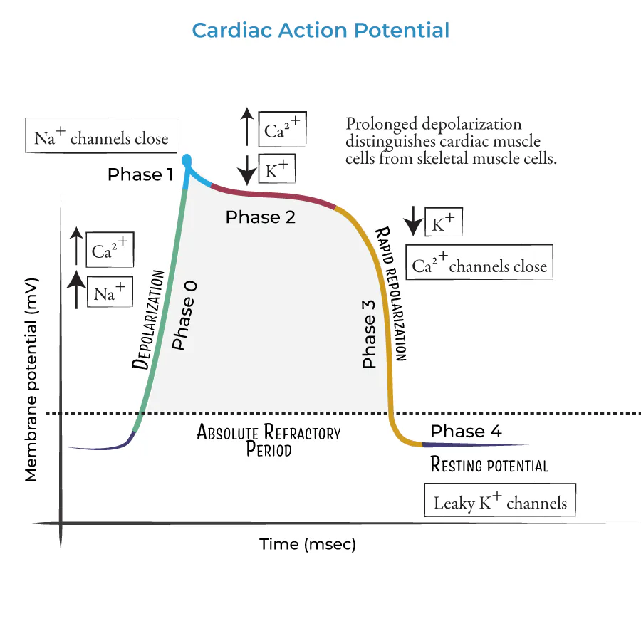
Why is the plateau phase important in the cardiac action potential?
62
Multiple Choice

What happens during the repolarization phase of the cardiac action potential?
63
Match
64
Multiple Choice

What does blood pressure measure?
65
Multiple Choice

What instrument is used to measure blood pressure?
66
Multiple Choice

What is the difference between systolic and diastolic blood pressure?
67
Multiple Choice

What units are used to measure blood pressure?
68
Multiple Choice

What is the normal range for blood pressure in adults?
69
Multiple Choice

What is hypertension?
70
Multiple Choice

What is hypotension?
71
Multiple Choice

Which of the following is a risk factor for high blood pressure?
72
Multiple Choice

What happens when blood vessels lose elasticity?
73
Multiple Choice

What type of tissue is blood?
74
Match

Match the following blood cells with their correct descriptions:
75
Multiple Choice

How much blood does the average adult human have?
76
Multiple Choice

What are the percentages of the components of blood?
77
Multiple Choice

What is hematocrit?
78
Multiple Choice

Which type of blood cell is the most abundant in the human body?
79
Multiple Choice

What are the main characteristics of the anatomy of red blood cells?
80
Multiple Choice

What is the main component of hemoglobin?
81
Multiple Choice

What is the iron-containing molecule in hemoglobin called?
82
Multiple Choice

What type of molecule is globin in hemoglobin?
83
Multiple Choice

What is the function of the heme part of hemoglobin?
84
Multiple Choice

How many globulin molecules are in each hemoglobin?
85
Multiple Select
86
Multiple Select
87
Multiple Select
88
Multiple Choice

What is the average lifespan of a red blood cell (RBC)?
89
Multiple Choice

Which organ plays a major role in the destruction of old red blood cells and acts like a recycling center of the components of red blood cells?
90
Multiple Choice

What is the primary role of white blood cells (leukocytes)?
91
Multiple Choice

Which of the following is NOT a characteristic of white blood cells?
92
Multiple Choice

What is the purpose of white blood cells having a nucleus?
93
Match

Match the following white blood cells:
94
Multiple Choice

What are the four blood types?
95
Match

Match the blood type with the antigens and the antibodies they produce.
96
Multiple Choice

What is the Rh factor?
97
Multiple Select

Rh factor can be _____________ or ______________. Pick two answers!
98
Multiple Choice

What is the blood type for this test?
99
Multiple Choice

What is the blood type for this test?
100
Multiple Choice

What is the blood type for this test?
101
Multiple Choice

What does CBC stand for in the context of human anatomy and physiology?
102
Multiple Choice
103
Multiple Choice
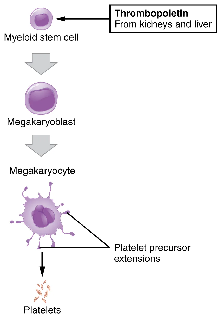
How are platelets formed?
104
Multiple Choice

Platelets are primarily involved in?
105
Multiple Choice
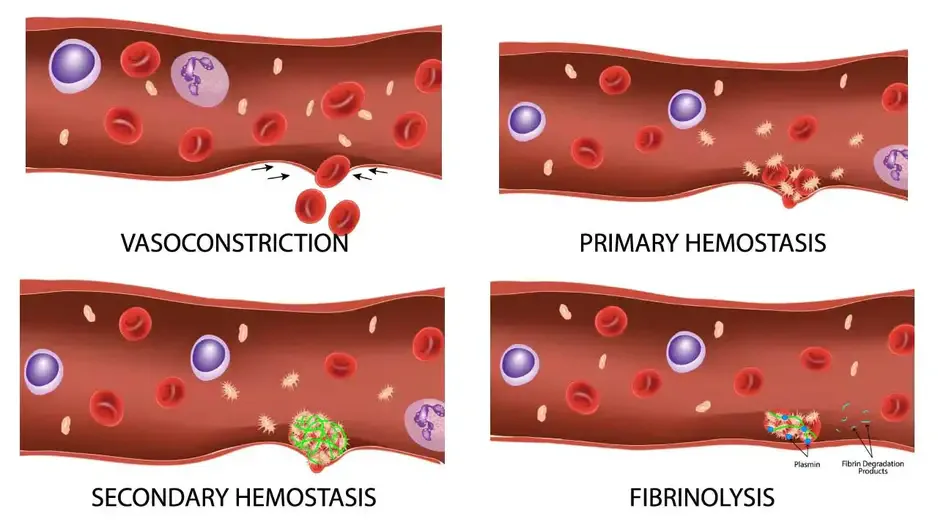
What is hemostasis?
106
Multiple Choice

Which type of blood cells are involved in clotting during hemostasis?
107
Multiple Choice

In which order do the steps of hemostasis typically occur?
108
Multiple Choice

What happens during vascular spasm?
109
Multiple Choice

What triggers the formation of a platelet plug in hemostasis?
110
Multiple Choice

During platelet plug formation, what is the first step?
111
Multiple Choice

What happens during the process of coagulation in hemostasis?
112
Multiple Choice

What is the scientific term for the process of red blood cell production?
113
Multiple Choice

What is the main difference between a thrombus and an embolus?
114
Multiple Choice

Which of the following best defines anemia?
115
Multiple Choice

What is sickle cell anemia?
116
Multiple Choice

What are some complications associated with sickle cell anemia?
117
Multiple Choice

What is Atherosclerosis?
118
Multiple Choice

What is ischemia?
119
Multiple Choice

Which of the following is a common cause of ischemia?
120
Multiple Choice

What is congestive heart failure?
121
Multiple Choice

What is angina?
122
Multiple Choice

What is cardiac arrest?
123
Multiple Choice

What is coronary heart disease?
124
Multiple Choice

What is myocardial infarction?
125
Multiple Choice

What is phlebitis?
126
Multiple Choice

What are varicose veins?
127
Multiple Choice
128
Multiple Choice

Which of the following is the correct unit used to measure pulse rate?
129
Multiple Choice

What is epistaxis?
130
Multiple Choice

What is a bruise?
131
Multiple Choice

What is another name for a bruise?
132
Multiple Choice

What is an incision?
133
Multiple Choice

What is a laceration?
134
Multiple Choice

What is a scrape injury?
135
Multiple Choice

What is the definition of throbbing?
136
Multiple Choice

What is a blood blister?
137
Multiple Choice

How does a blood blister form?
138
Multiple Choice

What is a crush injury?
139
Multiple Choice

Which of the following is NOT a symptom of a crush injury?
140
Multiple Choice

What is hemangioma (red/purple birthmark) ?
141
Multiple Choice

What is occult blood?
142
Multiple Choice

What are heart palpitations?
143
Multiple Choice

What are tarry stools?
144
Multiple Choice
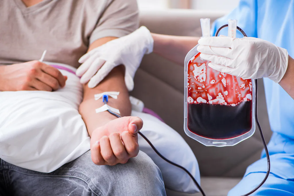
What is a transfusion?
Show answer
Auto Play
Slide 1 / 144
SLIDE
