

Foot and Ankle fractures

Presentation
•
Biology
•
University
•
Practice Problem
•
Medium

Kieran Kitchener
Used 2+ times
FREE Resource
45 Slides • 5 Questions
1

FOOT & ANKLE FRACTURES
Dr José PM Leal & Dr Kieran Kitchener
Clinical Education Fellows
George Eliot Hospital
2

LEARNING OUTCOMES
• Identification of common ankle and foot fractures
• Anatomy of ankle and foot
• Radiology interpretation
• Management of fractures
• Paediatric fractures
3

ANKLE FRACTURES
4
What four bones make up the ankle joint?
5
What four bones make up the ankle joint?
Fibula
Tibia
Talus
Calcaneus
6

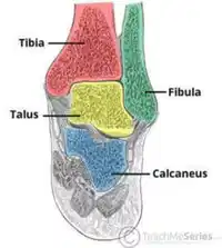
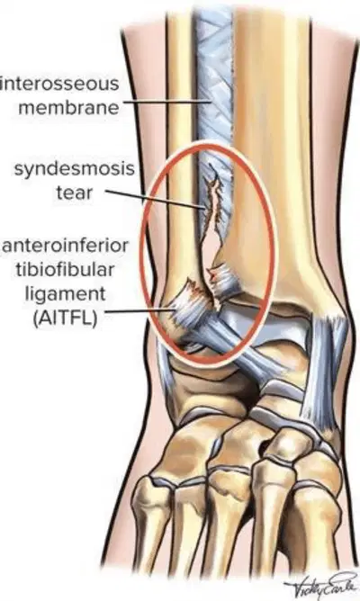
• Mortise joint: tibial plafond, medial malleolus
(tibia), lateral malleolus (fibula)
• Tibia and fibula joined at syndesmosis
(AITFL + PITFL + intra-osseous membrane)
• Stability of ankle:
• Deltoid ligament
• Syndesmosis
• Lateral ligament complex
• ATFL
• PTFL
• CFL
ANKLE ANATOMY
7

8


• Ankle fracture = fracture of any malleolus ±
disruption to syndesmosis
• Typically twisting mechanism
• Demographics:
• young, active (15-24, M>F)
• elderly (75-84, F>M)
• What are the two common methods of classifying fractures of the ankle?
ANKLE FRACTURES
9
• Ankle fracture = fracture of any malleolus ±
disruption to syndesmosis
• Typically twisting mechanism
• Demographics:
• young, active (15-24, M>F)
• elderly (75-84, F>M)
• Weber classification
• Lateral malleolus fractures
• Lauge-Hansen classification
• Based on combination of foot position and direction
of force applied at the time of injury
ANKLE FRACTURES
10

WEBER CLASSIFICATION:
CLASSIFICATION OF LATERAL MALLEOLAR FRACTURES

11


• Inferior to
syndesmosis
• Syndesmosis intact
• Usually stable
• Reduction and back
slab or CAM boot
• ORIF occasionally
needed
• At level of
syndesmosis
• Syndesmosis intact
or partially torn
• Possible medial #
or deltoid injury
• Stability variable
• ORIF may be
needed
• Superior to
syndesmosis
• Syndesmosis
damaged =
widening of joint
• Usually medial # or
deltoid injury
• Unstable
• ORIF required
WEBER CLASSIFICATION
12

WEBER A
• Below the level of the syndesmosis (infrasyndesmotic)
• Usually transverse
• Tibiofibular syndesmosis intact
• Deltoid ligament intact
• Medial malleolus occasionally fractured
• Usually stable if medial malleolus intact; treat with CAM
walker or moon boot with crutches and weight bear as
tolerated with them for 6 weeks
13

WEBER B
•Distal extent at the level of the syndesmosis (trans-syndesmotic); may
extend some distance proximally
•Usually spiral
•Tibiofibular syndesmosis usually intact, but widening of the distal tibiofibular
joint (especially on stressed views) indicates syndesmotic injury
•Medial malleolus may be fractured
•Deltoid ligament may be torn, indicated by widening of the space between
the medial malleolus and talar dome
•Variable stability, dependent on the status of medial structures
(malleolus/deltoid ligament) and syndesmosis; may require open reduction
and internal fixation (ORIF)
•Weber B fractures could be further subclassified as
•B1: isolated
•B2: associated with a medial lesion (malleolus or ligament)
•B3: associated with a medial lesion and fracture of posterolateral tibia
14

WEBER C
•Above the level of the syndesmosis (suprasyndesmotic)
•Tibiofibular syndesmosis disruption with widening of the distal tibiofibular
articulation
•Medial malleolus fracture or deltoid ligament injury often present
•Fracture may arise as proximally as the level of fibular neck and not visualised on
ankle films, requiring knee or full-length tibia-fibula radiographs (maisonneuve
fracture)
•Unstable: usually requires ORIF
•Weber C fractures can be further subclassified as
•C1: diaphyseal fracture of the fibula, simple
•C2: diaphyseal fracture of the fibula, complex
•C3: proximal fracture of the fibula
•A fracture above the syndesmosis results from external rotation or abduction
forces that also disrupt the joint
•Usually associated with an injury to the medial side
15

LAUGE-HANSEN
1. Supination-Adduction (SA) injuries: These occur when the foot is in a supinated
(turned inward) position and a force is applied in an adduction (inward) direction.
This typically results in a fracture of the lateral malleolus (fibula) and possible
rupture of the deltoid ligament on the medial side of the ankle.
2. Supination-External Rotation (SER) injuries: These occur when the foot is in a
supinated position and a force is applied in an external rotation (outward) direction.
This can result in a fracture of the fibula, followed by a rupture of the anterior
talofibular ligament and the calcaneofibular ligament.
3. Pronation-Abduction (PA) injuries: These occur when the foot is in a pronated
(turned outward) position and a force is applied in an abduction (outward) direction.
This can lead to a fracture of the medial malleolus (tibia) and possible rupture of the
lateral ligaments.
4. Pronation-External Rotation (PER) injuries: These occur when the foot is in a
pronated position and a force is applied in an external rotation direction. This can
result in a fracture of the medial malleolus, followed by rupture of the deltoid
ligament and the syndesmosis (the ligaments that hold the tibia and fibula together).
16

OTTAWA RULES
• An Ankle X-ray is only required if:
•There is any pain in the malleolar zone; and,
•Any one of the following:
•Bone tenderness along the distal 6 cm of the posterior edge of the tibia or tip of the medial malleolus
(ZONE B), OR
•Bone tenderness along the distal 6 cm of the posterior edge of the fibula or tip of the lateral
malleolus (ZONE A), OR
•An inability to bear weight both immediately and in the emergency department for four steps.
• A foot X-ray series is indicated if:
•There is any pain in the midfoot zone; and,
•Any one of the following:
•Bone tenderness at the navicular bone (C), OR
•Bone tenderness at the base of the fifth metatarsal (D), OR
•An inability to bear weight both immediately and in the emergency department for four steps.
17


OTTAWA RULES
18


19


20


21


WHAT IS SEEN HERE?
22


TRIMALLEOLAR FRACTURES
• Three-part ankle fracture
• Medial malleolus
• Posterior malleolus
• Lateral malleolus
• Unstable fracture
• Ligamentous injury highly likely
• CT scan required
• Operative planning
• Posterior malleolus assessment
23

PAEDIATRIC FRACTURES –
SALTER HARRIS

24


TRIPLANE FRACTURES
• Traumatic paediatric ankle fractures
• Complex Salter-Harris IV fracture pattern in multiple planes
• Diagnosed on plain XR
• CT scan may be required
• Management:
• Closed reduction & casting
• <2mm displacement
• ORIF
• >2mm displacement
• Complications
• Growth arrest
• Ankle pain & degeneration
25


26


27

FOOT FRACTURES
28

29
30

FOOT FRACTURES
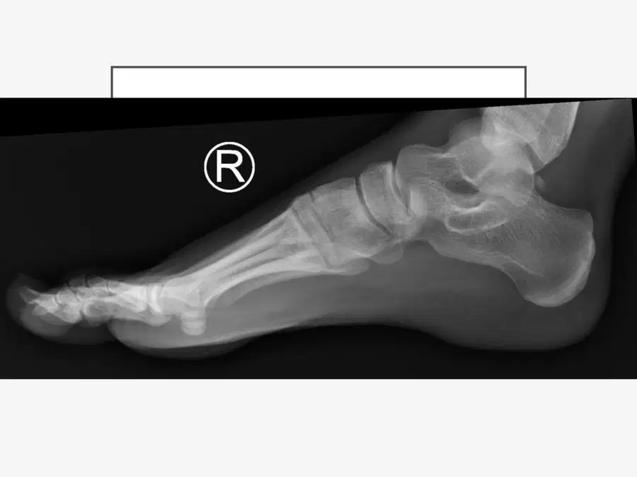
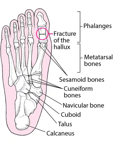
• Toe fractures
• Proximal phalanx most likely (longest)
• Conservative management, WB as able
• +/- haematoma block and manipulation
• Metatarsal fracture
• Direct blow, twisting injury
• Stress fractures
• Conservative management, WB as able
31


32


5TH METATARSAL FRACTURE
• Most common fracture of foot
• Predisposed to poor healing due to the limited blood
supply to 5th metatarsal base
• Jones fracture
33

34


LISFRANC FRACTURE
• Midfoot fracture-dislocation
• Twist and fall, direct trauma
• Weight bearing XR required
• Conservative Mx: NWB cast/boot
• Operative Mx: displaced/unstable #,
ORIF/fusion
35


LISFRANC FRACTURE
36


LISFRANC FRACTURE
37


LISFRANC FRACTURE
38


LISFRANC FRACTURE
39

CHARCOT ARTHROPATHY
• Progressive degenerative/destructive joint disorder in patients
with abnormal pain sensation and proprioception.
• Present either insidiously or as incidental findings
• Charcot joints although swollen are of normal temperature
without elevated inflammatory markers. Importantly, they are
painless.
• Management:
•Conservative – cast or boot, weight off of foot.
•Surgical – May need removal of areas of bone or fixation. Surgery if
severe and affecting mobility, or if fractures are unstable.
40
CHARCOT ARTHROPATHY
• Progressive degenerative/destructive joint disorder in patients
with abnormal pain sensation and proprioception.
• Present either insidiously or as incidental findings
• Charcot joints although swollen are of normal temperature
without elevated inflammatory markers. Importantly, they are
painless.
• Management:
•Conservative – cast or boot, weight off of foot.
•Surgical – May need removal of areas of bone or fixation. Surgery if
severe and affecting mobility, or if fractures are unstable.
41


42

ANY QUESTIONS?
43
Multiple Choice

Which ligament is torn here?
Deltoid
Spring
Anterior Talofibular
Posterior Talofibular
44
Open Ended
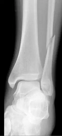
What type of fracture is seen on this x-ray?
45
Open Ended

A patient presents with an ankle injury. Given this x-ray, what is the usual treatment modality?
46


• What is seen on this xray?
47
• What is seen on this xray?
Os trigonum – accessory bone within the flexor hallicus longus bone. Can be normal variant or develop from trauma
48
Open Ended

How would you classify this fracture?
49
Multiple Choice
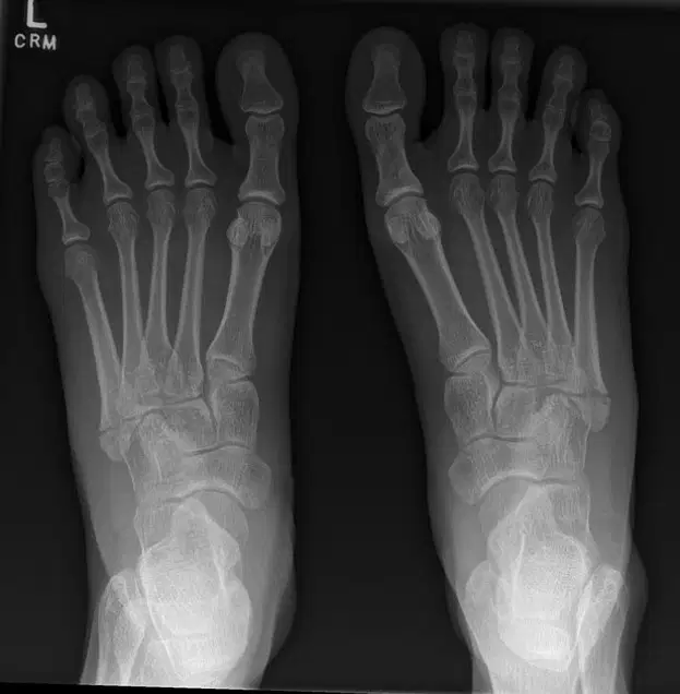
What is the fracture seen here?
No fracture seen
Charcot's foot
5th metatarsal fracture
1st metatarsal fracture
Lis Franc Fracture
50
• https://teachmesurgery.com/orthopaedi
c/ankle-and-foot/ankle-
fracture/#:~:text=The%20most%20com
mon%20classification%20used,the%20le
vel%20of%20the%20syndesmosis
• https://radiopaedia.org/articles/weber-
classification-of-ankle-fractures?lang=gb
• https://gpnotebook.com/en-
GB/pages/musculoskeletal-
medicine/ottawa-rules-regarding-
requirement-for-ankle-x-ray
• https://www.123rf.com/photo_7826177
3_four-major-pulse-points-of-the-foot-
the-pedal-pulses.html
• https://www.orthobullets.com/trauma/1
047/ankle-fractures
• https://orthoinfo.aaos.org/en/diseases--
conditions/toe-and-forefoot-fractures/
• https://www.orthobullets.com/foot-
and-ankle/7031/5th-metatarsal-base-
fracture
• https://orthoinfo.aaos.org/en/diseases--
conditions/lisfranc-midfoot-injury
• https://www.msdmanuals.com/en-
gb/home/injuries-and-
poisoning/fractures/metatarsal-
fractures#Treatment_v13967406
REFERENCES
FOOT & ANKLE FRACTURES
Dr José PM Leal & Dr Kieran Kitchener
Clinical Education Fellows
George Eliot Hospital
Show answer
Auto Play
Slide 1 / 50
SLIDE
Similar Resources on Wayground

40 questions
เรื่องที่ 8 การป้องกันการบาดเจ็บจากกีฬาเทนนิส
Presentation
•
University
39 questions
PAIN ASSESMENT ENG III
Presentation
•
University
38 questions
ORTESIS Y PROTESIS
Presentation
•
KG
43 questions
td gastro
Presentation
•
University
43 questions
Introduction to Minerals
Presentation
•
11th - 12th Grade
43 questions
Introduction to Minerals
Presentation
•
11th - 12th Grade
44 questions
untitled
Presentation
•
KG - University
51 questions
Final Cirugía Plástica
Presentation
•
University
Popular Resources on Wayground
11 questions
Hallway & Bathroom Expectations

Quiz
•
6th - 8th Grade
10 questions
HCS SCI 03 Summer School Assessment 2
Quiz
•
3rd Grade
11 questions
Home Scope
Quiz
•
7th - 8th Grade
12 questions
2026 TAP Technology in the Classroom
Presentation
•
Professional Development
15 questions
HCS SCI 05 Summer School Assessment 2 Review
Quiz
•
5th Grade
15 questions
HCS SCI 04 Summer School Review 2
Quiz
•
4th Grade
59 questions
Geometry Unit 3 Review
Quiz
•
9th - 12th Grade
14 questions
FAST ELA READING SMAPLE TEST MATERIALS

Passage
•
3rd Grade