Grade 7 eye anatomy worksheets and printables help students learn about the structure and function of the human eye through engaging practice problems, free PDF downloads, and comprehensive answer keys.
Explore printable Eye Anatomy worksheets for Grade 7
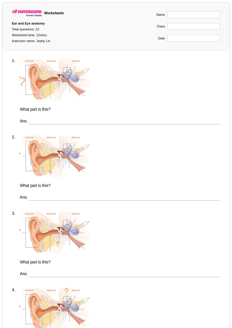
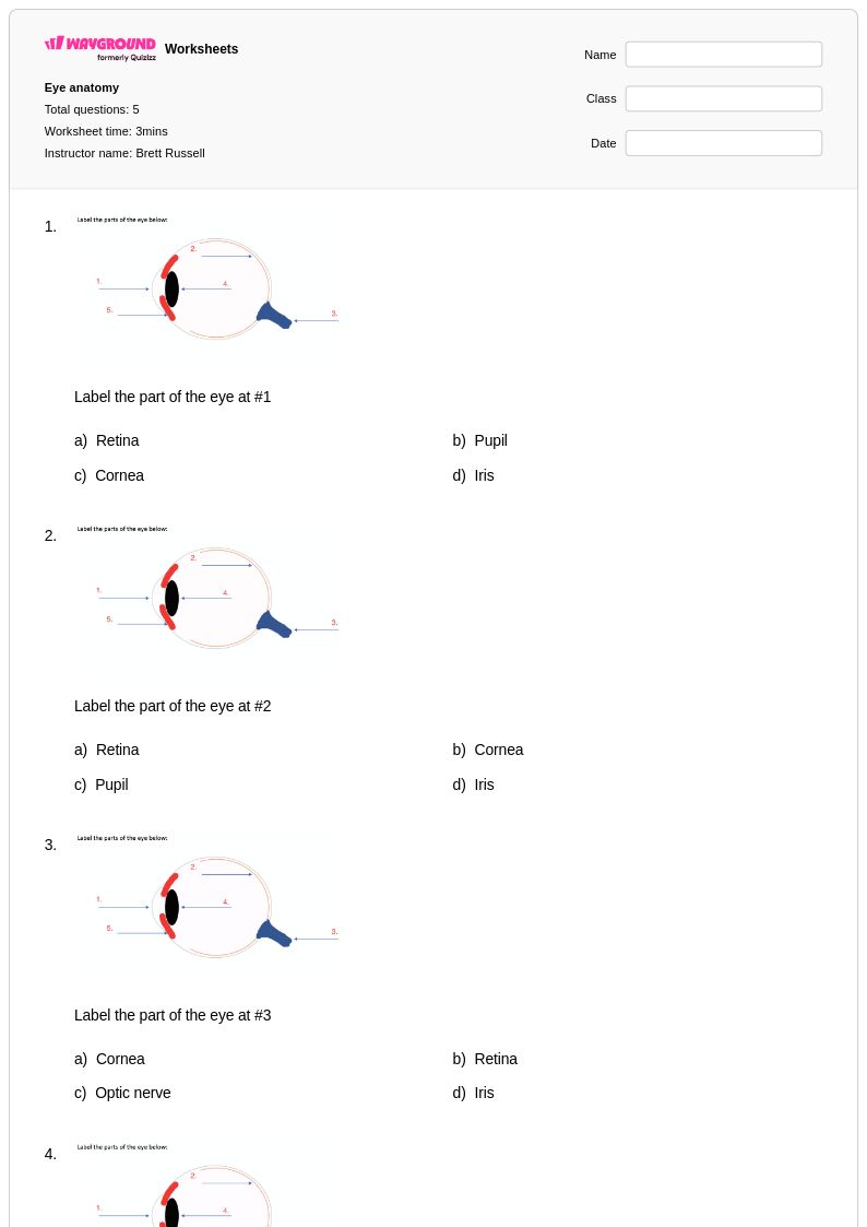
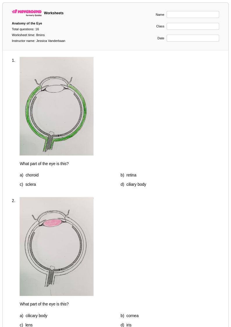
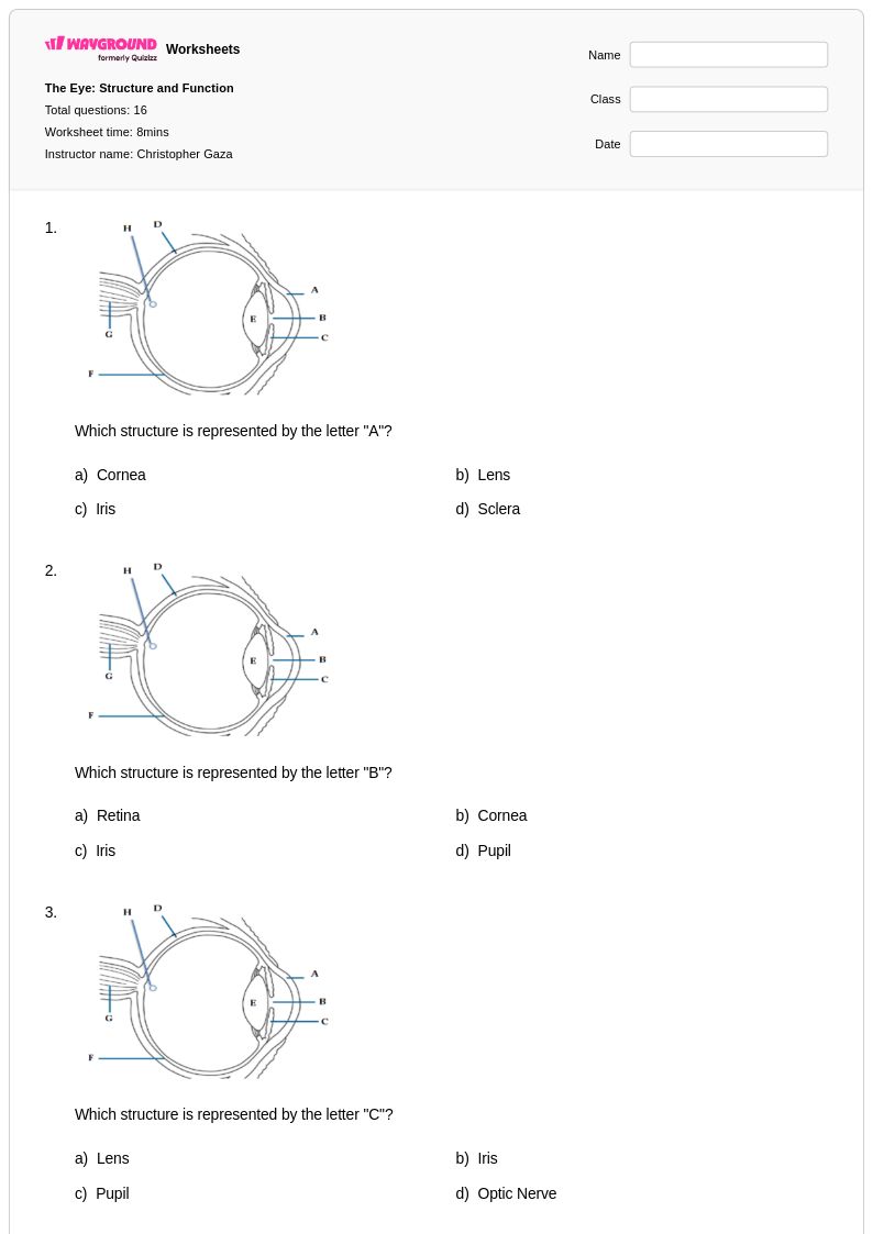
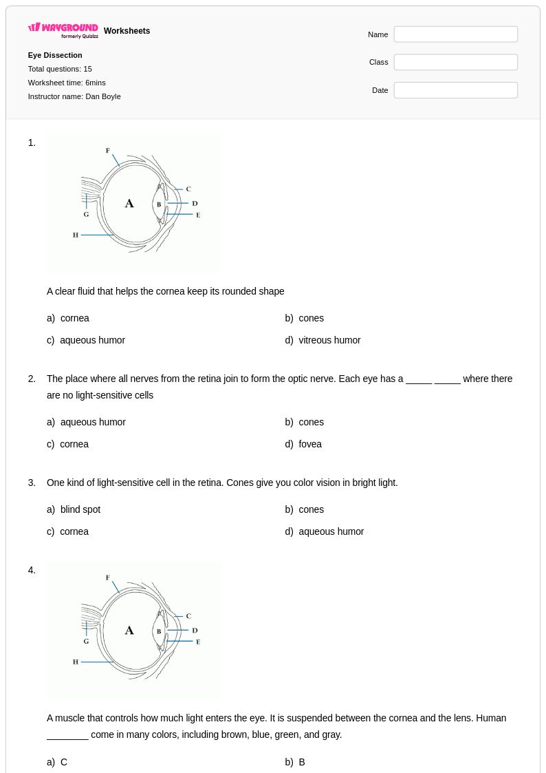
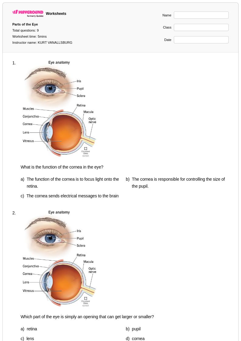
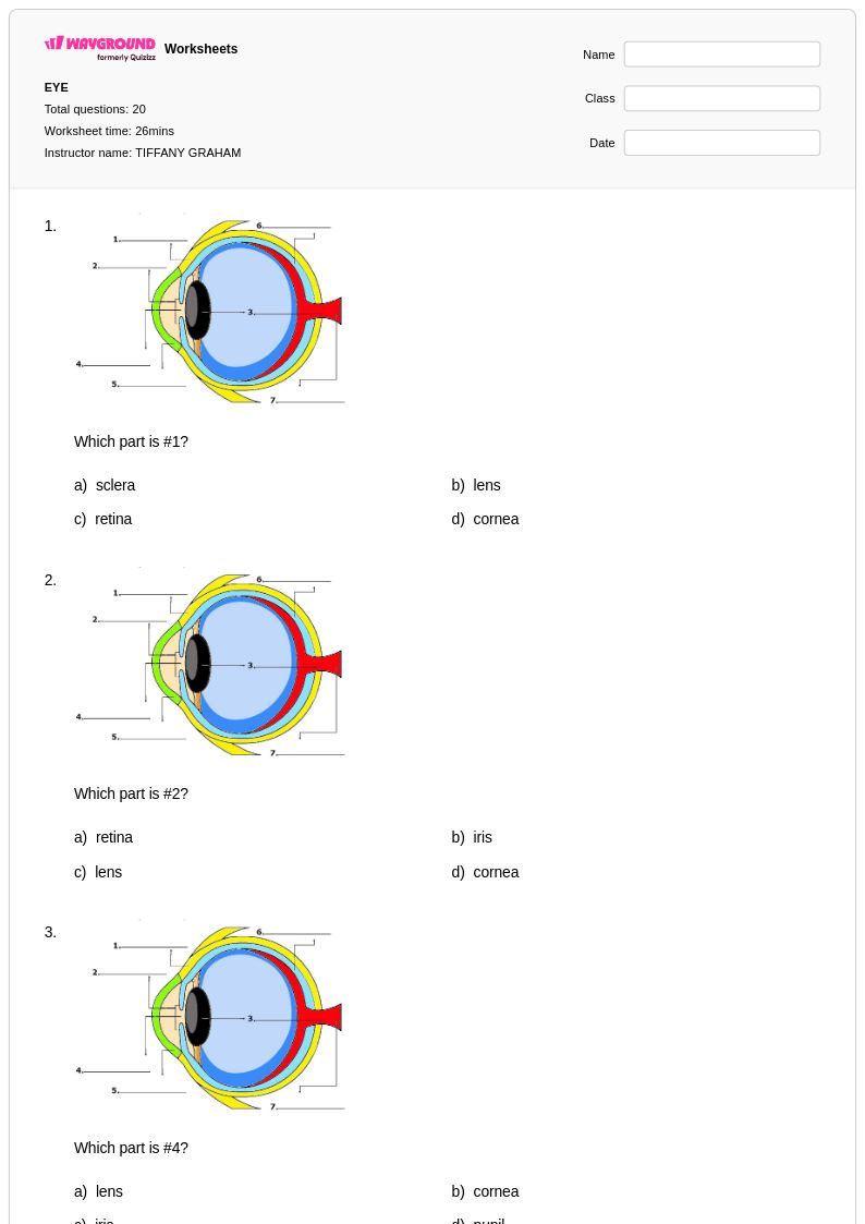
Eye anatomy worksheets for Grade 7 students available through Wayground (formerly Quizizz) provide comprehensive exploration of the human visual system's complex structures and functions. These educational resources guide seventh-grade learners through detailed examination of eye components including the cornea, iris, pupil, lens, retina, and optic nerve, helping students understand how light travels through the eye to create vision. The worksheets strengthen critical scientific skills such as anatomical identification, structure-function relationships, and biological process analysis through carefully designed practice problems that reinforce key concepts. Students benefit from varied question formats including labeling diagrams, matching exercises, and explanatory responses, with answer keys provided to support independent learning and self-assessment. These free printables serve as valuable study tools that complement classroom instruction and prepare students for assessments while building foundational knowledge essential for advanced biology studies.
Wayground (formerly Quizizz) empowers educators with millions of teacher-created eye anatomy resources specifically designed for Grade 7 science instruction, offering robust search and filtering capabilities that enable quick identification of materials aligned with specific curriculum standards and learning objectives. The platform's differentiation tools allow teachers to customize worksheets based on individual student needs, providing options for remediation support and enrichment challenges that accommodate diverse learning styles and academic levels. Teachers can access these resources in both printable pdf format for traditional classroom use and digital formats for technology-integrated lessons, streamlining lesson planning while ensuring consistent quality across all materials. The extensive collection supports various instructional approaches including direct instruction reinforcement, independent practice sessions, homework assignments, and formative assessment opportunities, helping educators effectively address the diverse learning needs within their Grade 7 biology classrooms while maintaining alignment with established science education standards.
FAQs
How do I teach eye anatomy to students effectively?
Teaching eye anatomy is most effective when students first build a mental map of the eye's layers before connecting structure to function. Start with the outer structures — cornea, sclera, and iris — then move inward to the lens, retina, and optic nerve, consistently pairing each part with its role in vision. Anatomical labeling diagrams, light pathway tracing exercises, and structure-function matching activities reinforce retention at each stage. Returning to a whole-eye diagram at the end of instruction helps students consolidate the parts into a coherent system.
What are common mistakes students make when learning eye anatomy?
One of the most frequent errors is confusing the roles of the cornea and the lens — students often attribute focusing entirely to the lens while overlooking the cornea's refractive contribution. Students also commonly misplace the retina and choroid, conflating two distinct layers with very different functions. Another persistent misconception is that the optic nerve carries light rather than electrical signals, which reflects a broader gap in understanding how sensory transduction works. Explicitly addressing these points during instruction and using answer-key-guided correction on labeling worksheets can help students self-identify and fix these errors.
What practice exercises help students learn the structures of the human eye?
Anatomical labeling diagrams are the foundational practice type for eye anatomy, requiring students to identify and place key structures such as the cornea, lens, retina, sclera, choroid, and optic nerve on a cross-sectional diagram. Light pathway tracing exercises add a functional layer by asking students to sequence how light travels from entry to signal transmission. Structure-function matching tasks and terminology reinforcement problems build the academic vocabulary students need for assessments. Varied practice across these formats ensures students can both recall and apply their knowledge.
How can I use eye anatomy worksheets in my classroom?
Eye anatomy worksheets on Wayground are available as printable PDFs for traditional classroom distribution and in digital formats for technology-integrated environments, making them flexible for in-person, hybrid, or remote instruction. Teachers can also host worksheets as an interactive quiz directly on Wayground, giving students immediate feedback and allowing teachers to track performance in real time. Each worksheet includes a complete answer key, supporting both guided classroom review and independent self-checking. This range of formats means the same resource can serve as a direct instruction tool, a homework assignment, or a formative assessment depending on how it is deployed.
How do I support students with different learning needs during an eye anatomy unit?
Wayground includes built-in accommodation tools that teachers can apply at the individual student level without disrupting the rest of the class. For students who need additional support, teachers can enable Read Aloud so questions and content are read to them, reduce the number of answer choices to lower cognitive load, or grant extended time per question. Font size and display theme adjustments through Reading Mode can also improve accessibility for students with visual processing needs. These settings are saved per student and carry over to future sessions, so teachers only need to configure them once.
How do I assess whether students understand eye anatomy versus just memorizing terms?
Surface memorization of eye anatomy terms is easy to detect — students who only memorize will typically label a diagram correctly but struggle to explain why a structure is positioned where it is or what happens when it is damaged. Assessment tasks that require students to trace the path of light through the eye in sequence, explain the consequence of a detached retina, or match structures to specific visual impairments reveal deeper conceptual understanding. Including both identification and application questions in the same assessment gives teachers a clearer picture of where each student's understanding breaks down.