
 Worksheets
Atrial Rhythms
Total questions: 58
Worksheet time: 1hrs 8mins
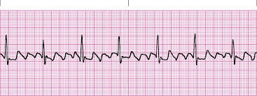
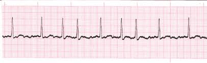
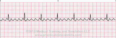
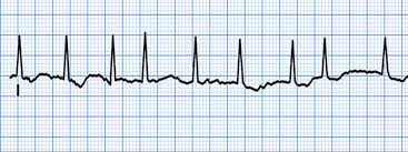
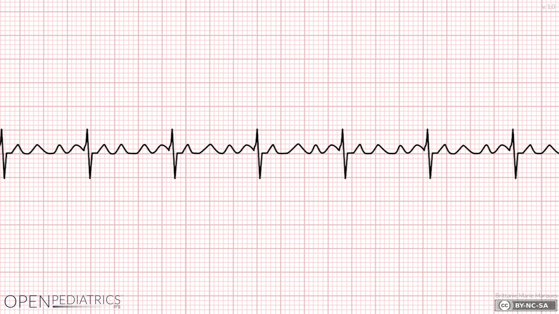
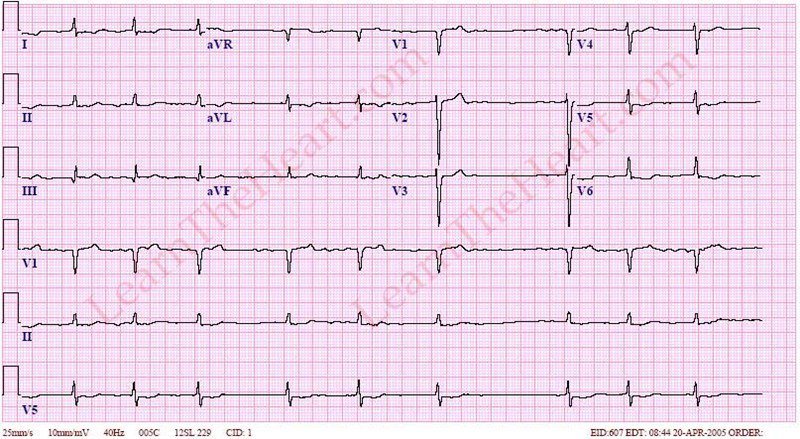
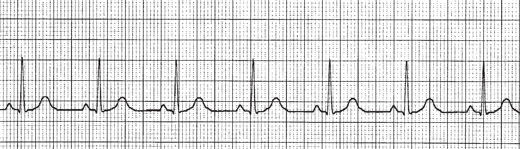
What is the heart rate of this ECG rhythm?
40
70
100
130
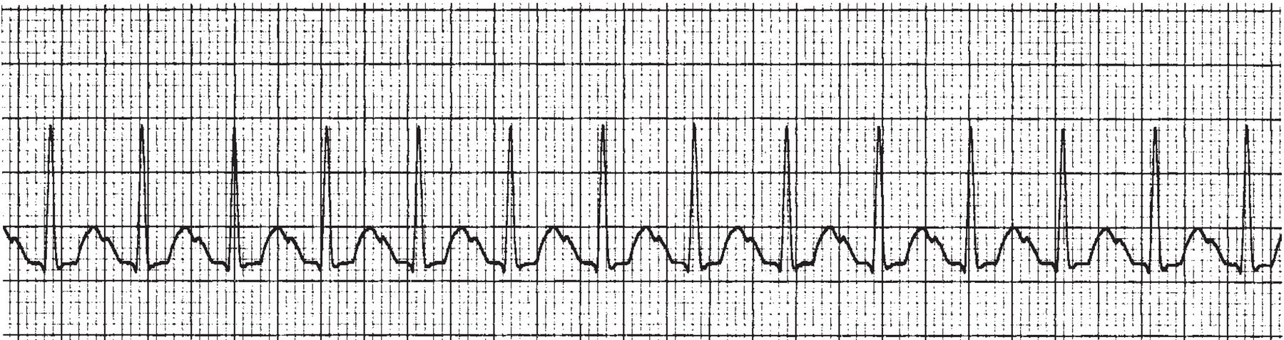
This ECG strip illustrates ____________.
Premature Atrial Contraction
Premature Ventricular Contraction
Atrial Fibrulation
Tachycardia
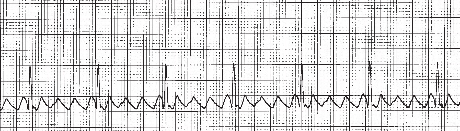
This ECG illustrates _______________.
Premature Atrial Contraction
Atrial Flutter
Atrial Fibrillation
Ventricular Fibrillation
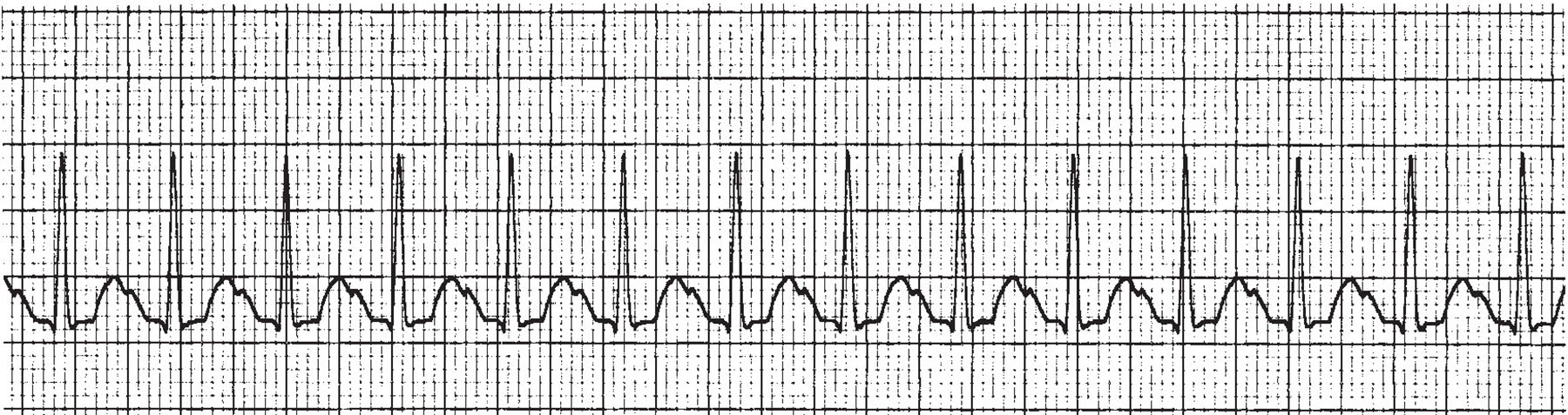
What is the HR of this ECG?
105pbm
135bpm
75bpm
165bpm
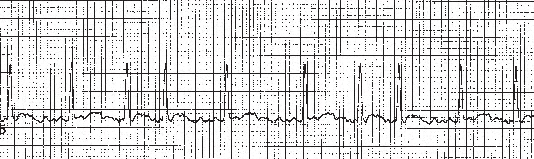
This ECG illustrates ____________________.
Atrial Flutter
Bradycardia
Atrial Fibrillation
Myocardial Infarct
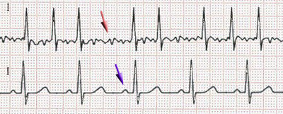
What is the ECG showing (Red Arrow)
Atrial Fibrillation
Ventricular Fibrillation
Asystole
Premature Ventricular Contraction
Heart Block
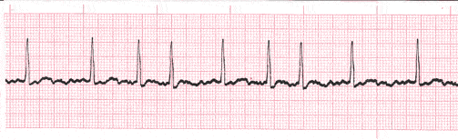
which atrial abnormality results in an atrial rate of 130-250 bpm with a normal ventricular rate
atrial tachycardia
atrial flutter
atrial fibrillation
atrial bigeminy
on an ECG when the ventricular heart rate is regular, the rate can be determined by counting the number of large squares between two consecutive QRS complexes then
dividing 600 by that number of large squares
dividing 300 by that number of large squares
dividing 400 by that number of large squares
dividing 200 by that number of large squares
which abnormal atrial mechanism is often one of the first signs of CHF ?
atrial fibrillation
atrial bigeminy
atrial tachycardia
atrial flutter
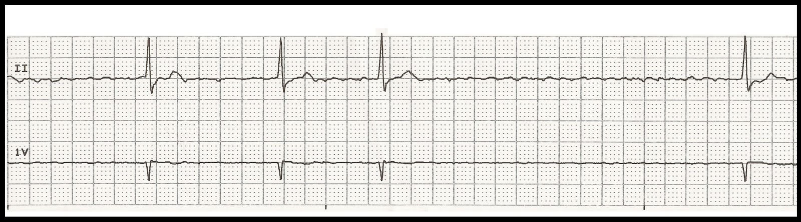
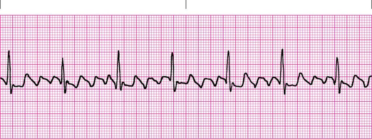
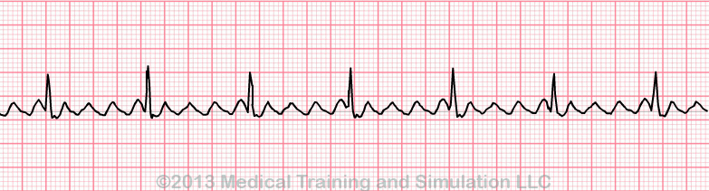
What is this rhythm?
Atrial Tachycardia
Atrial Fibrilation
Wandering Atrial Pacemaker.
Atrial Flutter
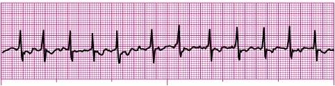
Identify the following rhythm.
Atrial flutter
Atrial fibrillation
Supraventricular tachycardia
Sinus tachycardia
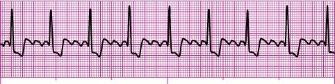
Identify the following rhythm.
Atrial flutter
Atrial fibrillation
Supraventricular tachycardia
Junctional tachycardia
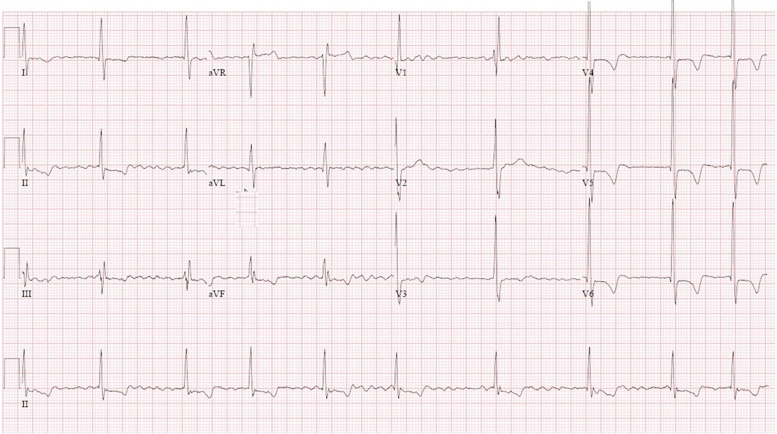
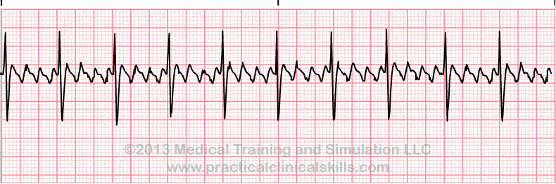
This strip is interpreted as:
Atrial fibrillation
Normal Sinus Rhythm
Atrial Flutter
Ventricular Fibrillation
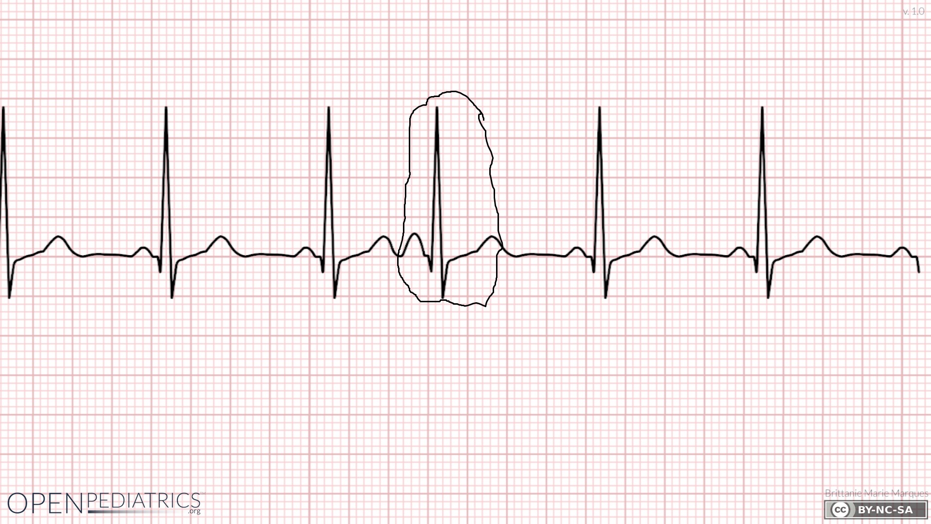
Identify what the waveform that is circled?
Premature atrial contraction
Premature ventricular contraction
Sinus bradycardia
Sinus tachycardia
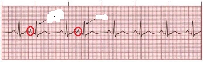
Identify what is occurring at the red circled areas?
Asystole
Normal Sinus Rhythm
Premature Ventricular Contraction
Premature Atrial Contraction
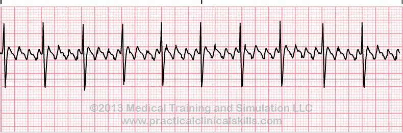
Identify the cardiac rhythm strip seen above:
Ventricular Fibrillation
Atrial Flutter
Ventricular Tachycardia
Normal Sinus Rhythm
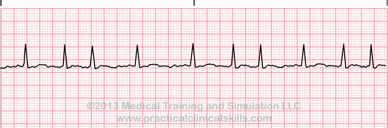
Identify the cardiac rhythm strip pictured above
Ventricular Tachycardia
Atrial Fibrillation
Normal Sinus Rhythm
Ventricular Fibrillation
This ECG illustrates _______________.
Premature Atrial Contraction
Atrial Flutter
Atrial Fibrillation
Ventricular Fibrillation
What is the Heart Rate of this ECG?
105pbm
135bpm
75bpm
165bpm
This ECG illustrates ____________________.
Atrial Flutter
Bradycardia
Atrial Fibrillation
Myocardial Infarct
Select all of the options which are true of atrial systole.
Atria contract
Blood is pumped from ventricles into atria
AV valve is open
SL valve is open
A common cardiac dysrhythmia characterized by premature heartbeats originating in the atria.
A-Fib
A-Flutter
PAC's
SA Block
Medical condition where SA node ceases to generate the electrical impulses that normally stimulate the tissues for heart conduction.
Sinus Arrhythmia
1st Degree AV Block
Sinus Arrest
Sino-Atrial Block
Lead II is the standard lead in viewing the EKG.
True
False
Common in elderly and is associated with exacerbations of chronic obstructive pulmonary disease (COPD).
V-Tach
PAC's
MAT
PJC's
Causes: alcohol, medications/drugs, caffeine, tobacco, exercise, anxiety, or any chemical imbalances
PAC's
PJC's
PVC's
MAT
Occurs when the natural pacemaker site shifts between SA node, the atria, and/or the AV node.
1st Degree AV Block
SA Block
Sinus Pause
Wandering Atrial Pacemaker
An atrial pacemaker site that changes from location to location has
P waves that continually change in appearance
One pacemaker site from which the impulse arise
Inverted P’ waves
A chaotic baseline preceding each QRS complex
When the atria fire faster than 350 beats per minute, the P waves
are indiscernible; instead, there is a chaotic looking baseline
appear in a saw-toothed pattern
are inverted (in lead II)
are buried in the T wave of the preceding beat
__________, __________. & _________ place pressure on the atria and cause atrial dysrhythmias to occur. Select all that apply
Myocardial infarction
valvular problems
neurological influences
angina
MAT (multifocal atrial tachycardia) is sometimes confused with which dysrhythmia?
WAP (wondering atrial pacemaker)
Atrial Flutter
PAC (premature atrial complexes)
Atrial Fibrillation
What is the rate of WAP?
40-60 BPM
60-100 BPM
100-150 BPM
Greater than 150 BPM

