
 Worksheets
skeletal system test
Total questions: 165
Worksheet time: 2hrs 35mins
Example of a flat bone.
trapazoid
humerous
vertebrae
patella
sternum
Example of a sesmoid bone.
trapazoid
humerous
vertebrae
patella
sternum
Example of a irregular bone.
trapazoid
humerous
vertebrae
patella
sternum
Blood cells are formed in...
red marrow
yellow marrow
osteon systems
osteocytes
Rickets is caused by a deficiency of which vitamin?
C
A
D
E
An abnormal lateral curve of the spine is called...
greenstick
scoliosis
osteoperosis
arthritis
The spaces of the trabeculae are filled with what material?
red marrow
yellow marrow
insterstiatial fluid
synovial fluid
The process of bone forming from cartilage is called
ossification
callus zone
growth
fracture repair
A part of a bone that sticks out for muscle attachment.
process
sinus
meatus
foramen
The tough outer covering of a bone.
compact bone
medullary cavity
spongy bone
periosteum
All of these joints are synovial except...
saddle joints
gliding joints
cartilaginous joints
hinge joints
Where is spongy bone located?
diaphysis
epiphysis
haversian canals
medullary cavity
The joints of the skull called sutures are
hinge joints
fibrous joints
synovial joints
gliding joints
_________ bones include: carpals and tarsals
Irregular
Flat
Short
Long
________ bones include: ulna, tibia and femur.
Irregular
Flat
Short
Long
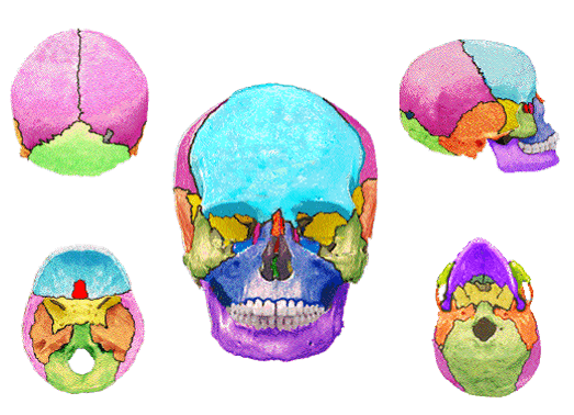
Identify the bone that is sea/light blue.
Frontal
Occipital
Temporal
Parietal
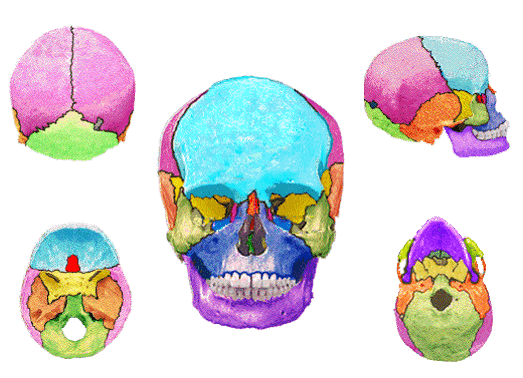
Identify the rust/orange colored bone.
Frontal
Occipital
Parietal
Temporal
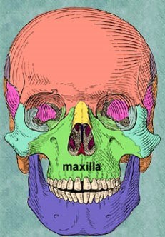
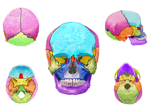
Identify the darker blue bone in the illustration.
Maxilla
Molar
Mandible
Malaphin
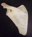
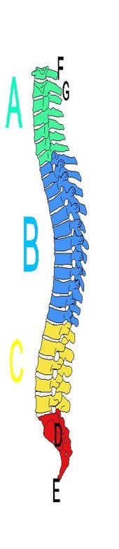
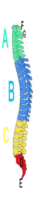
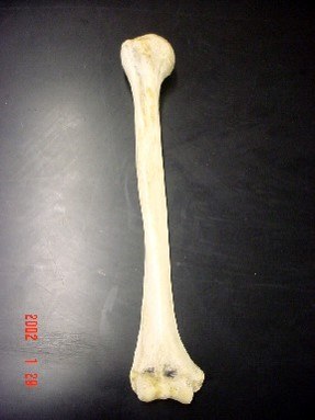
The upper arm bone.
Femur
Coccyx
Humerus
Ulna
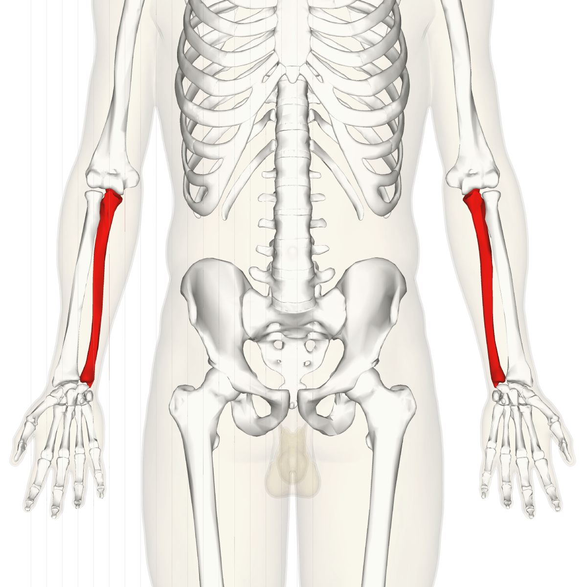
The lateral lower arm bone. White bone in illustration.
Humerus
Femur
Ulna
Radius
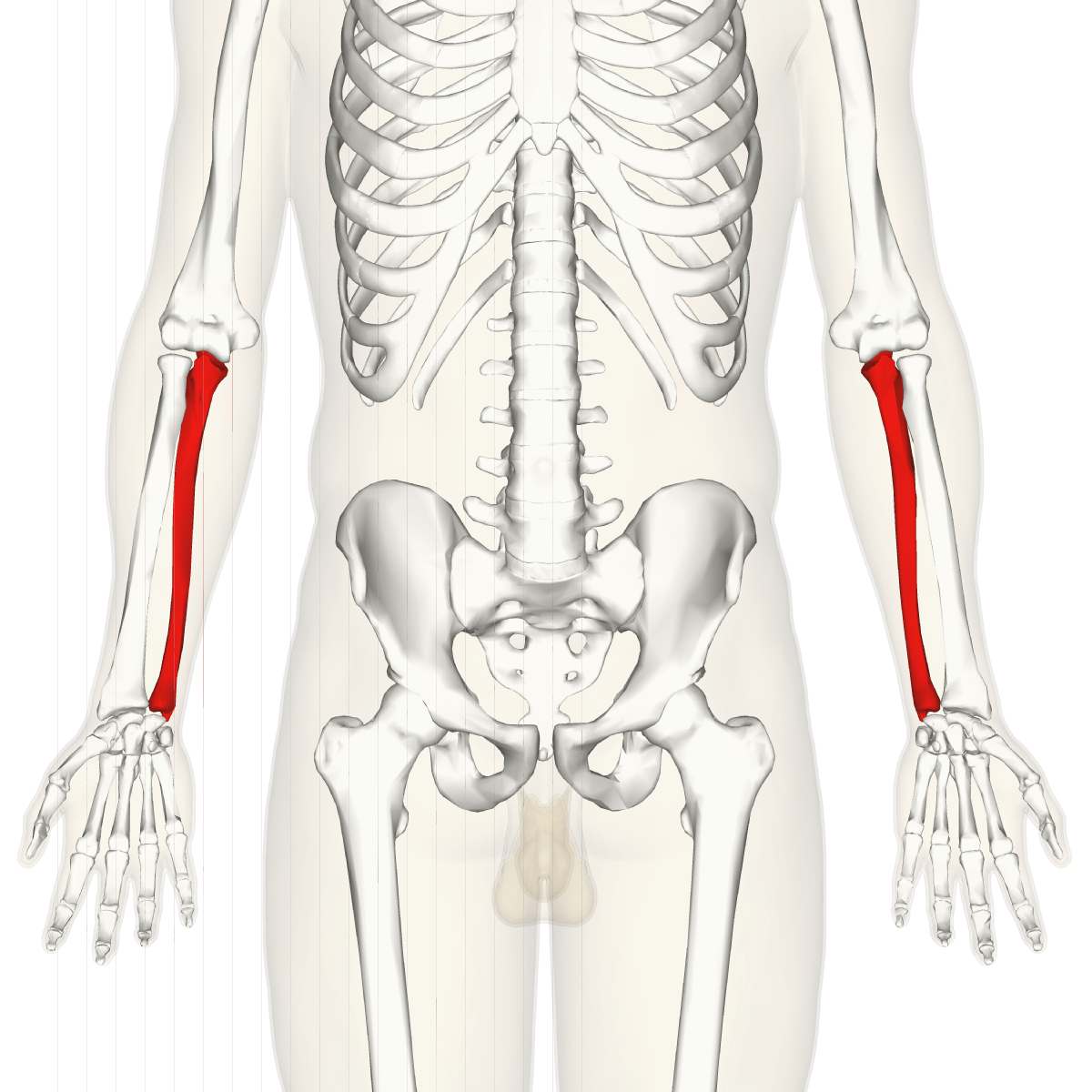
The medial bone of lower arm that also makes the elbow. appears Red in illustration.
Femur
Humerus
Ulna
Radius
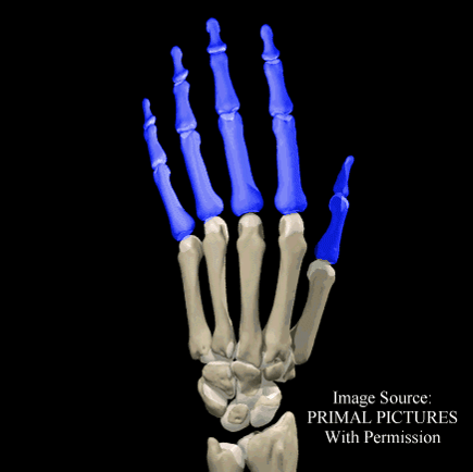
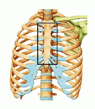
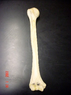
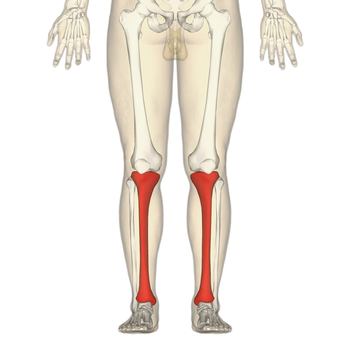
The shin bone that is weight bearing as identified in the illustration in red.
Tibia
Fibula
Femur
Radius
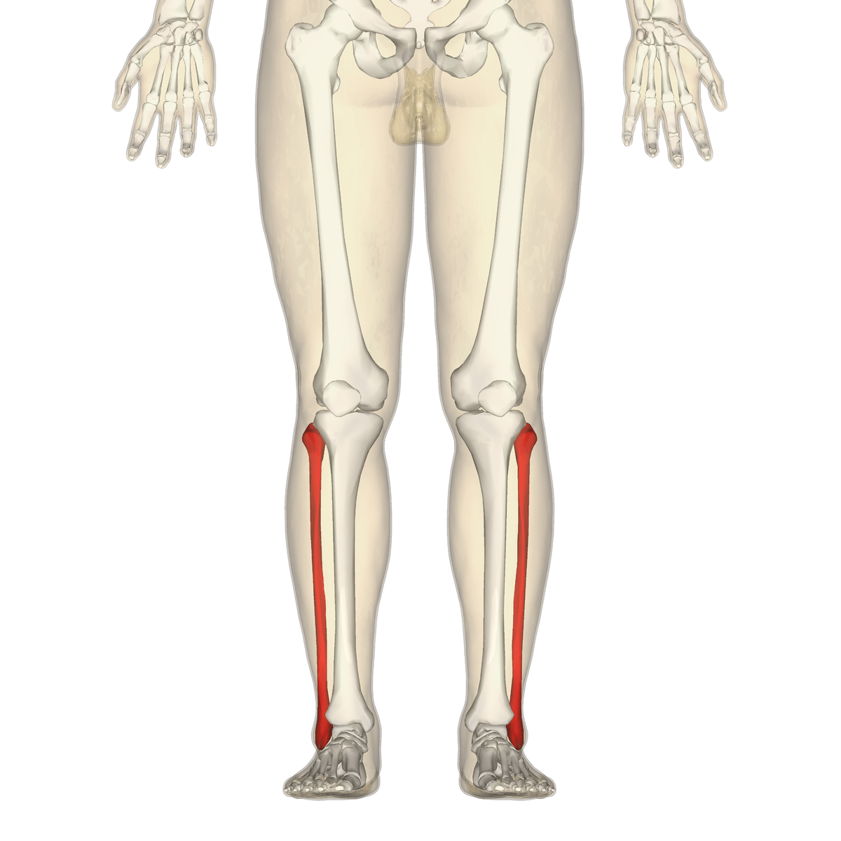
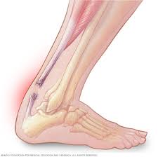
A flexible but inelastic cord of strong fibrous collagen tissue attaching a muscle to a bone.
Tendon
Ligament
Cartilage
Bone
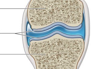
It acts as a cushion between bones at a joint and protects the bones. Structure appears light blue in illustration and has the middle lines going to it.
Tendon
Ligament
Cartilage
Bone
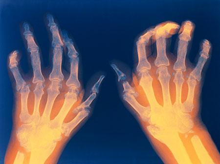
Autoimmune disorder that causes painful inflammation and stiffness of the joints which can lead to disfigurement.
Osteoporosis
Rheumatoid Arthritis
Osteoarthritis
Sprain
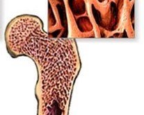

Select the letter designated to the canaliculi
A
B
C
D
Select the letter designated to the osteocyte
A
B
C
D
Select the letter designated to the osteon
A
B
C
D
Select the letter designated to the central haversian canal
A
B
C
D
Bone forming cells are called:
diaphysis
fontanels
osteoblasts
osteocytes
John fell on an outstretched arm while running outside. John's fracture caused the bone to break through the skin. What type of fracture does John have?
comminuted
compound
greenstick
simple


The cells that destroy/breakdown bone are called.
Osteoclasts
Osteoblasts
Osteocytes
Erythrocytes

Shaking your head "no" using C1 and C2 is an illustration of using what type of joint in your neck?
hinge
immoveable
ball and socket
pivot
If cartilage remains in this "specific" area, then growth is still occurring; it is located at the end of long bone. Where the secondary ossification center is.
Epipyseal plate
Medulary Cavity
Diaphysis
Endosteum

Infection in a bone...
gout
osteomyelitis
rickets
scoliosis
Bone cancer...
osteoporosis
osteomyelitis
osteosarcoma
scoliosis
area of the skull that has not yet fused in children under two; "soft spot"
fontanel
foramen
ramus
line
immovable joint of the skull after fusion
fontanel
suture
notch
tuberosity

What is the structure in the picture?
foramen magnum
occipital condyles
external auditory meatus
zygomatic process
sella turcica

What is the structure in the picture?
foramen magnum
occipital condyles
external auditory meatus
zygomatic process
sella turcica

What bone of the pelvic girdle is colored purple (#1)?
ilium
ischium
pubis
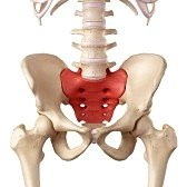
sacrum
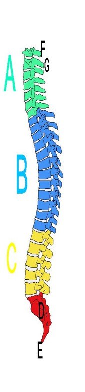
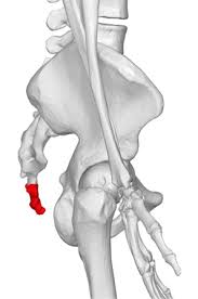
coccyx
What bone of the pelvic girdle is colored beige (#2)?
ilium
ischium
pubis
sacrum
coccyx

What is A? articulates directly with the skull
atlas
axis
What is B? acts as a pivot to move the skull.
atlas
axis

What part of the thoracic vertebrae is D?
pedicle
body
lamina
transverse process
spinous process
What part of the thoracic vertebrae is B?
pedicle
body
lamina
transverse process
spinous process
What part of the thoracic vertebrae is E?
pedicle
body
lamina
transverse process
spinous process

In the lumbar vertebra pictured, what is B?
body
lamina
pedicle
vertebral foramen
transverse process

28
Head
Neck
Lesser Trochanter
Greater Trochanter
Shaft

B
Facet
Crest
Fossa
Line

G the rounded end
Tubercle
Epicondyle
Head
Fossa
Trochlea
Bone destroying cells known as osteoclasts are activated by the hormone ______________when levels of calcium in the blood are low.
calcitonin
parathyroid hormone
insulin
growth hormone
A bone fracture where the bone is broken into many fragmetns is a __________________.
Compound fracture
Simple fracture
Comminuted fracture
Compression fracture
Greenstick fracture


The purple portion is the..
manubrium
xiphoid process
body
Cervical Vertebrae


What is the name of the three fused bones in the picture that is part of the appendicular skeleton?
Sacrum
Coccyx
Lumbar Vertebrae
Os coxae

Bone marking D
crest
medial malleolus
lateral malleolus
Bone marking E
medial malleolus
crest
lateral malleolus

Structure H articulates with the clavicle to form part of the shoulder joint
spine
acromion process
coracoid process
glenoid fossa

Which marking is this?
obturator foramen
ilium
ischium
acetabulum
The green bone marking in this picture...
Manubrium
Sacrum
Body
Xiphoid Process

What bone is represented in the picture? Only bone not attached to another bone in the body.
Manubrium
Hyoid
Sternum
Carpals

What is the marking at the top of ulna called? articulates with humerus. your elbow bone.
styloid process
olocranon process
head

What marking is 10?
greater trochanter
lesser trochanter
head
patellar surface
Bones can form in two ways, either
intramembranous and intermembranous
endochondral and exochondral
intramembranous and endochondral
endochondral and intermembranous
Bone creating cells known as osteoblasts are activated by the hormone ______________when levels of calcium in the blood are high.
calcitonin
parathyroid hormone
insulin
growth hormone
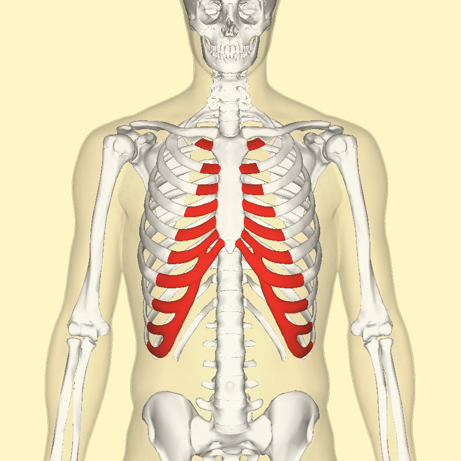
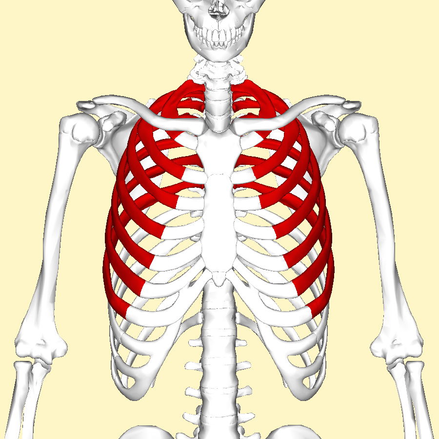
Which ribs are highlighted
true ribs
false ribs
floating ribs
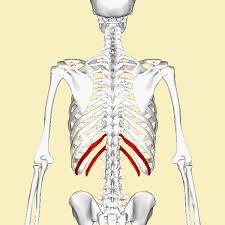
Which section of the rib cage is highlighted
true ribs
false ribs
floating ribs

Looking at the diagram how do true ribs differ from false ribs?
True ribs are attached directly to the sternum but the false ribs are not directly attached.
True ribs are not directly attached to the sternum but the false ribs are attached directly to the sternum
True ribs are not directly attached to the sternum but the false ribs are completely detached.

Zygomatic
Temporal
Parietal
Maxilla

Zygomatic
Vomer
Ethmoid
Lacrimal

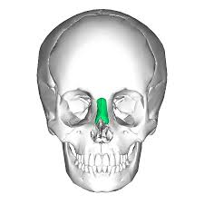
The thin, flat bone that forms the back and inferior portion of the nasal septum. It is #3 on the image.
ethmoid
lacrimal
inferior nasal conchae
vomer

The knee and elbow joints are?
Hinge
Gliding
Ball and socket
Pivot

Name this joint at the pollex.
gliding
saddle
condyloid
pivot
pivot, ball and socket and gliding are all what kind of joint.
fibrous
cartilaginous
synovial
All __________ joints are reinforced by ligaments
fibrous
cartilaginous
synovial
LIQUID that reduces friction between cartilaginous ends
Articular Cartilage
Synovial Membrane
Synovial Fluid
Articular Capsule
Ligament
Produces synovial fluid
Articular Cartilage
Synovial Membrane
Synovial Fluid
Articular Capsule
Ligament
Contains synovial fluid
Articular Cartilage
Joint Cavity
Synovial Fluid
Articular Capsule
Fibrous Layer
Select the two layers that make up the articular capsule.
Synovial Membrane
Joint Cavity
Synovial Fluid
Articular Cartilage
Fibrous Layer
A ___________ is an articulation between two or more bones
flexion
tendon
joint
tissue
two structural joints with no cavity
fibrous and cartilaginous
fibrous and synovial
synovial and cartilaginous
there is only one joint with no cavity

Label #1
Joint Cavity
Joint Capsule
Articular Cartilage
Synovial Membrane
Label #3
Joint Cavity
Fibrous Capsule/ligaments
Articular Cartilage
Synovial Membrane
Label #4
Joint Cavity
Joint Capsule
Articular Cartilage
Synovial Membrane
Label #2
Joint Cavity
Joint Capsule
Articular Cartilage
Synovial Membrane

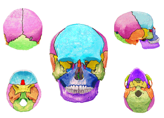
suture highlighted in red
frontal
sagittal
coronal
squamosal

suture highlighted in red
lamboidal
frontal
sagittal
squamosal
Which of the following is NOT a function of the skeletal system?
Helps your body move
Send oxygen to the body
Protect your organs
Give your body shape and structure
Make your blood
What bone of the pelvic girdle is colored pink (#3)?
ilium
ischium
pubis
sacrum
coccyx


What joint is displayed in picture?
hinge joint
plane joint
pivot joint

Which is one of the functional ways a joint is classified
Fibrous—tend to be immovable
Amphiarthroses—slightly movable joints
Synovial –tend to be freely movable

Where is the lamboidal suture located
between right and left parietal bones
next to the temporal bones
between the parietal and occipital bones

Where is the coronal suture?
between the parietal and occipital bones
between right and left parietal bones
between the frontal and parietal bones


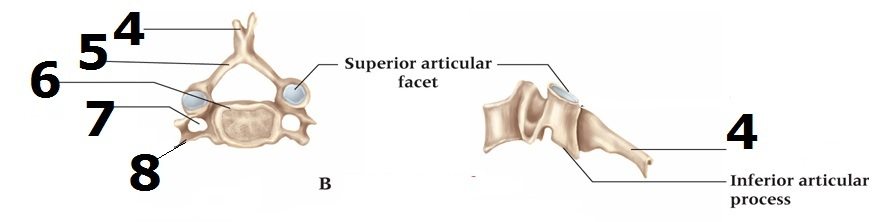
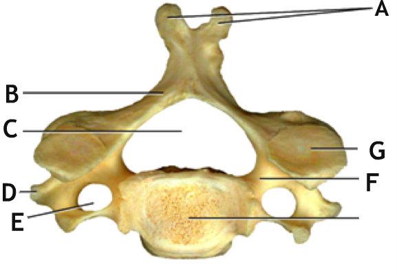
what type of vertebrae is pictured here?
cervical
thoracic
lumbar
sacral
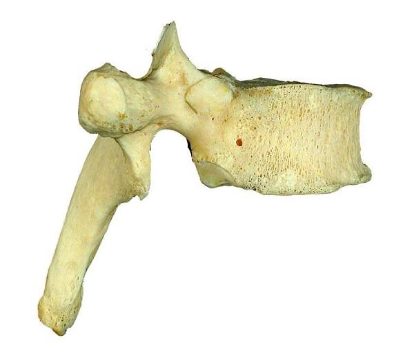
The function of the vertebra is to articulate with ribs.
thoracic
lumbar
cervical
sacral

