
 Worksheets
A&P Muscles & Sliding Filament Theory
Total questions: 50
Worksheet time: 29mins
Actin is the ...
Thick Filament within a Muscle Fibre
Thin Filament within a Muscle Fibre
Protein that covers myosin-binding sites
Molecule that causes muscle contraction
Myosin is the ...
Thin Filament of a muscle fibre
Segment of a muscle fibre
A molecule that changes shape when bound with Calcium Ions
Thick Filament of a muscle fibre
True or False: Cross-bridges is when a myosin head attaches to an actin during contraction
True
False
The Z Line provides attachment for the ...
Troponin
Myosin
Actin
ATP
Myofibril is an arrangement of ___________ within a muscle fibre.
Bands
Sacromeres
Proteins
Crossbridges
_________ carries the impulse deep into the myofibrils.
T-Tubules
Synapses
Sarcoplasm
Neurotransmitters
True or False: Calcium Ions bind to Tropomyosin to change the shape of Troponin.
True
False
What does the Myosin Heads release to relax and pull the Actin filament?
ATP
ADP
CNS
ETC
What binds to the Myosin Head to cause it to detach from the Actin?
ATP
ADP
CNS
ETC
What causes the muscle to stop contracting?
Lack of effort
Myosin-Heads fail to attach
Calcium Ions are pumped away
Muscle fibre tears
What starts a muscle contraction?
Release of Calcium Ions
Attachment of Myosin-Heads
An Action Potential
Release of ADP

Calcium:
causes ATP binding to actin
increases the action potential along the sarcolemma
binds to myosin
triggers troponin and tropomyosin to uncover the myosin binding sites on actin
What is a crossbridge?
Where myosin binds to actin
Where the calcium crosses into the cytoplasm
Where calcium binds with troponin
None of the answers are correct.

The neurotransmitter stored in the axon terminal identified as #3
Calcium Ion (Ca2+)
Protein Channel
Acetylcholine
Synaptic Cleft
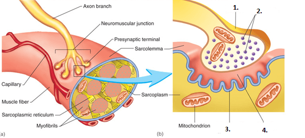

The Ion that is responsible for removing Troponin from Tropomyosin.
the neuron that transmit impulse from central nervous system to muscle effector is called
sensory neuron
motor neuron
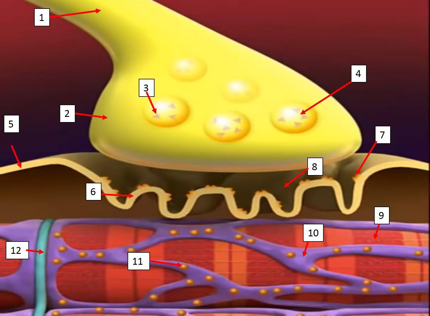
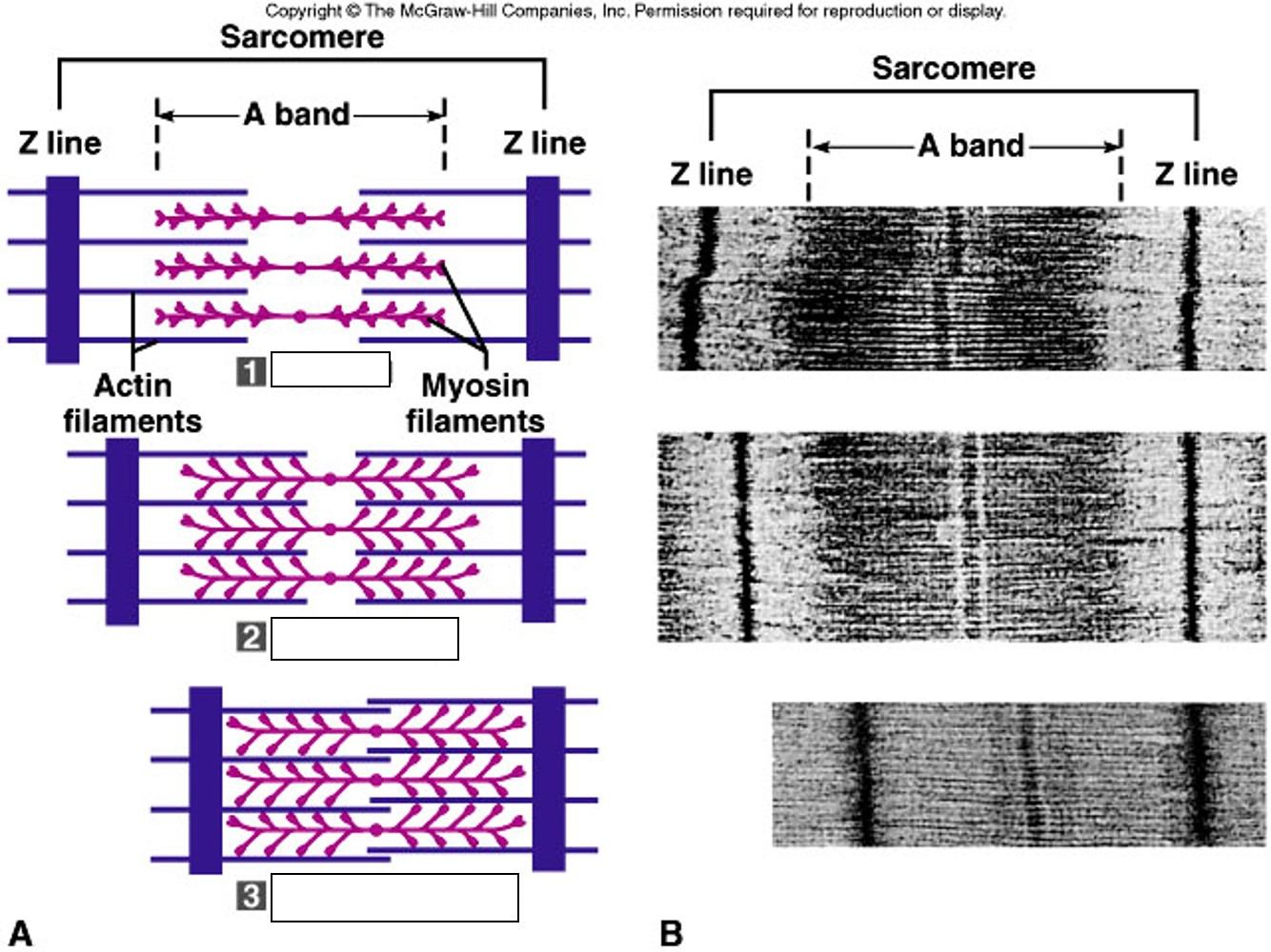

which skeletal muscle is represented in the image?
brachioradialis
biceps brachii
biceps femoris
deltoideus

which skeletal muscle is represented in the image?
external obliques
serratus anterior
rectus abdominis
pectoralis major

which skeletal muscle is represented in the image?
gastrocnemius
tibialis anterior
fibularis longus
vastus medialis

which skeletal muscle is represented in the image?
gluteus medius
gluteus maximus
semitendinosus
external obliques

which skeletal muscle is represented in the image?
latissmus dorsi
teres major
infraspinatus
deltoideus

which skeletal muscle is represented in the image?
pectoralis major
serratus anterior
rectus abdominis
deltoideus

which skeletal muscle is represented in the image?
pectoralis major
serratus anterior
rectus abdominis
deltoideus

which skeletal muscle is represented in the image?
rectus femoris
vastus lateralis
vastus medialis
sartorius

which skeletal muscle is represented in the image?
sternocleidomastoid
trapezius
zygomaticus
depressor anguli oris

which skeletal muscle is represented in the image?
sternocleidomastoid
trapezius
zygomaticus
depressor anguli oris

which skeletal muscle is represented in the image?
triceps brachii
latissimus dorsi
deltoideus
brachioradialis
Which type of muscle is voluntary and striated?
Cardiac
Smooth
Skeletal
Which type of muscle is involuntary and striated?
Cardiac
Smooth
Skeletal
Which type of muscle is involuntary and non striated?
Cardiac
Smooth
Skeletal
What is the name of the connective tissue sheath that surrounds each muscle fiber?
Endomysium
Epimysium
Perimysium
Which membrane covers several fibers to form a fascicle?
Endomysium
Epimysium
Perimysium

What's the name of this muscle?
Pectoral
Deltoid
Bicep
Abdominal

Name this muscle
Gastrocnemius
Quadricep
Gluteus Maximus
Hamstring

Name the highlighted muscle
quadraceps
hamsting
rectus abdominals
deltoid


What is the muscle that allows you to close and protrude lips
temporalis
orbicularis oris
orbicularis oculi
buccinator

Muscle that elevates the eyebrows
occipitals
frontalis
temporalis
corrugator

The all-or-none principle of muscle contraction states what?

