
 Worksheets
Anatomy & Physiology Midterm Review
Total questions: 159
Worksheet time: 2hrs 54mins
Specialized plasma membrane surrounding the muscle fiber.
Myofibril
Myofilament
Sarcolemma
Sarcomere

What is another name #2? (Hint: It is a muscle layer)
Skeletal muscle
Epimysium
Perimysium
Endomysium
What is another name #6? (Hint: It is a muscle layer)
Skeletal muscle
Epimysium
Perimysium
Endomysium
What is another name for #9 (cell membrane)?
Sarcomere
Sarcolemma
Myofibril
Myofiber
What is another name for #10?
Sarcomere
Sarcolemma
Myofibril
Myofiber
Skeletal muscles are responsible for moving
The heart
The skeleton
the pancreas
The brain

A "bundle" of muscle fibers is called:
Perimysium
Endomysium
Myofibril
Fascicle

Which of the following is found around a fascicle?
Endomysium
Perimysium
Epimysium
Myofibril

Attaches muscle to bone: _______
Endomysium
Myofibril
Tendon
Ligament
Which part of the osteon contains the osteocytes?
Lacunae
Lamellae
Canaliculi
Central Canal

Letter B
Lacunae
Lamellae
Canaliculi
Osteon
Where is red bone marrow located?
Spongy tissue
Compact tissue
Where is yellow bone marrow located?
Spongy Tissue
Compact tissue

Number 1 or 3
Diaphysis
Epiphysis
Periosteum
Articular cartilage

Number 8
Epiphyseal line
Spongy bone
Articular cartilage
Diaphysis

What can be found in the central canal of the haversian canal system?
osteocytes
lamella
blood vessels and nerves
red blood cells
Tiny, hairlike canals, that transport blood, nutrients and wastes, connecting lacunae to each other and to the Haversian canal
Concentric Lamella
Volkmann's Canal
Lacunae
Canaliculli
Osteon
Channels lying at right angles to the central canal, connecting blood and nerve supply of the periosteum to that of Haversian canals
Concentric Lamella
Volkmann's Canal
Lacunae
Canaliculli
Osteon

What is this image pointing to and circled?
Concentric Lamella
Lacunae
Canaliculli
Osteons
Volkmann's Canal




Which muscle is labeled 2
orbicularis oculi
platysma
frontalis
occipitals

Which muscle is labeled F
zygomaticus
masseter
temporalis
platysma

Which muscle is labeled I
platysma
sternocleidomastoid
trapezius
masseter
Making one's jaw move side-to-side will activate the
masseter
Pterygoids
Infrahyoids
platysma
temporalis

This muscle is named for the chewing action it produces
buccinator
masseter
lateral pterygoid
medial pterygoid

This circular muscle surrounds the mouth
orbicularis oris
orbicularis oculi
zygomaticus major
buccinator

This large, flat muscle inserts along the mandible and pulls your lip down as in pouting
buccinator
platysma
medial pterygoid
lateral pterygoid

These muscles around your eye help you bat your eyes~ closes the eyelids
orbicularis oculi
orbicularis oris
frontals
corrugator

Muscle that elevates the eyebrows and wrinkles the forehead
occipitals
frontalis
temporalis
corrugator

This muscle help you to smile!
buccinator
masseter
zygomaticus major
lateral pterigoid

This muscle aids in chewing by keeping food between the cheek and teeth, it is named based on location.
buccinator
masseter
zygomaticus minor
lateral pterigoid

Muscle that elevates the upper lip and assists in flaring of the nostrils.
depressor septi nasi
levator labii superioris
levator labii superioris alaeque nasi
aponeurosis
Which of the following muscles allow you to purse your lips (selfie pose duck lips)
nasalis and buccinator
buccinator and orbicularis oris
buccinator and orbicularis oculi
orbicularis iris and orbicularis oculi
How many muscles function in moving your mouth?
4
7
9
14


Identify the highlighted muscle.
Tricep
Quadriceps
Biceps
Pectorals

Identify the highlighted muscle.
Gastrocnemius
Gluteius Maximus
Triceps
Pectorals

Identify the highlighted muscle.
Biceps
Abdominals
Deltoids
Quadriceps

Identify the highlighted muscle.
Gluteus Maximus
Hamstrings
Quadriceps
Pectorals

Identify the highlighted muscle.
Hamstrings
Gastrocnemius
Quadriceps
Pectorals

Identify the highlighted muscle.
Abdominals
Pectorals
Triceps
Quadricpes

Identify the highlighted muscle.
Abdominals
Latissimus Dorsi
Gastrocnemius
Gluteus Maximus

Identify the highlighted muscle.
Hamstrings
Quadriceps
Pectorals
Biceps

What muscle is the arrow pointing to?
Pectoralis (Pecs)
Biceps
Triceps
Lats


What is the scientific name of the 'jaw bone'?
Maxilla
Mandible
The bones at the end of our fingers and toes are known as _______________.
Tarsals
The phalanges
The scientific name for the skull is the _____________.
Cranium
Zygomatic process
The scientific name of the collar bone is the ____________.
Scapula
Clavicle
The vertebra is a gliding joint.
True
False
The shoulder and hip are ball and socket joints.
True
False
Examples of a hinge joint are the elbow and knee.
True
False
Which of the following is NOT a bone area associated with the axial skeleton?
pelvic girdles
the skull
rib cage
vertebral column
Which of the following is NOT a function of bones?
hematopoiesis
storage
maintenance of body temperature
protection
What is the number of bones that compose the adult skeleton?
205
206
208
200
Bone forming cells:
osteocytes
osteoblasts
osteoclasts
osteomasts
The process by which bones increase in diameter:
ossification
hematopoiesis
appositional growth
longitudinal growth
Occurs when the bones of inactive people tend to lose mass:
atrophy
ossification
shingles
osteoarthritis
Bone destroying cells:
osteocytes
osteoblasts
osteomasts
osteoclasts
Must be present in the blood at all times for the nervous system to transmit messages, for muscles to contract, and for blood to clot:
calcium
adipose tissue
phosphorus
iodine
Bone tissue composed of small needle-like pieces of bone with open spaces:
lacunae
compact bone
canaliculi
spongy bone
Process of bone formation before birth:
ossification
hematopoiesis
appositional growth
atrophy
Fat stored in the internal cavities of bones:
red marrow
osseous tissue
adipose tissue
spongy bone
How many bones are in the human body?
200
206
205
204


Bone cells are known as
Lacunae
Osteocytes
Collagen
Osteon
Secondary ossification centers appears in
Epiphyses
Diaphysis
Epiphyseal plate
Endosteum

What bone is this?
scapula
humerus
radius
clavicle

What bone is this?
clavicle
scapula
sternum
humerus

What bone of the pelvic girdle is colored purple (#1)?
ilium
ischium
pubis
sacrum
coccyx
What bone of the pelvic girdle is colored beige (#2)?
ilium
ischium
pubis
sacrum
coccyx
What bone of the pelvic girdle is colored blue (#5)?
ilium
ischium
pubis
sacrum
coccyx
Two bones, the ___ and the ulna, form the skeleton of the forearm.
radius
tibia
fibula
femur
The skeleton of the hand consists of the ___, the metacarpals, and the phalanges.
tarsals
metatarsals
carpals
fibula
The tibia and ___, form the skeleton of the leg.
femur
fibula
radius
ulna
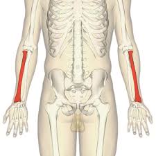
Which bone is highlighted
humerus
radius
ulna
femur
fibula
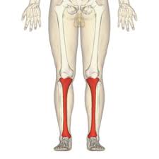
Which bone is highlighted
fibula
tibia
ulna
radius


Name Bone 2
cuneiform
cuboid
calcaneus
talus
navicular
Name Bone 5
lateral (3rd) cuneiform
medial (1st) cuneiform
intermediate (2nd) cuneiform


