
 Worksheets
Unit 2 Quizizz Review
Total questions: 93
Worksheet time: 57mins



E
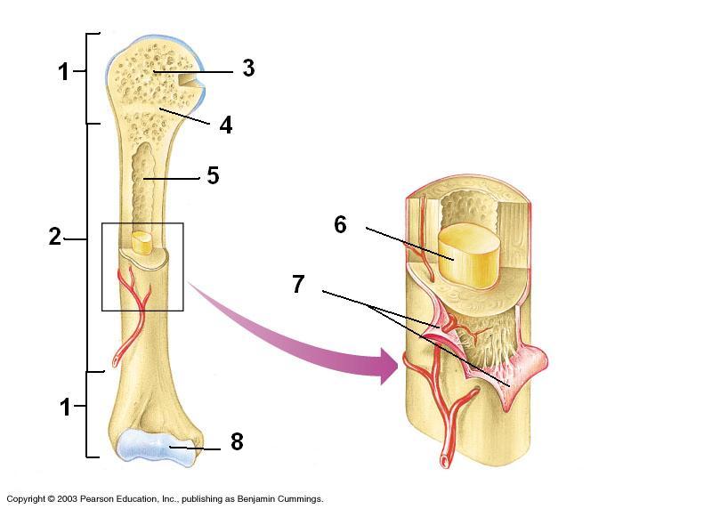
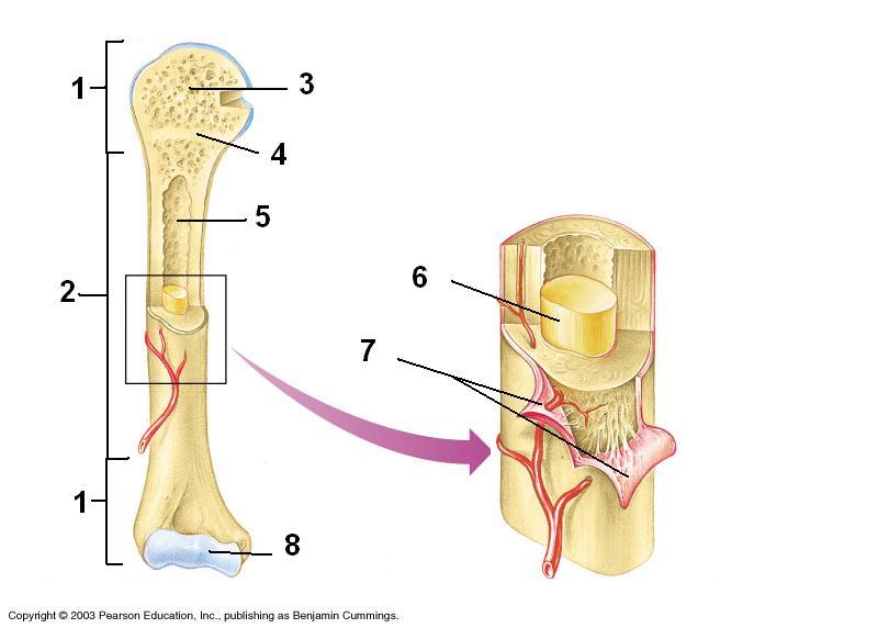
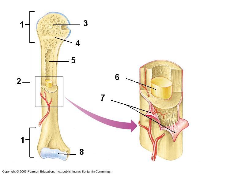
Compact Bone
Spongy Bone
Diaphysis
Articular Cartilage
Epiphyseal Line / Plate

Which bone is highlighted?
Clavicle
Sacrum
Tibia
Mandible

Which bone is highlighted?
Fibula
Ulna
Humerus
Sternum
Which bones make up the wrist and hand?
Tarsals
Metacarpals
Metatarsals
Carpals

Which bone is this?
Coccyx
Rib cage
Mandible
Femur

Which bone is this?
humerus
femur
skull
backbone/spine

Which bone is this?
humerus
ribs
tibia
radius

Which bone is this?
humerus
ulna
radius
carpals

Which bone is this?
femur
tibia
backbone/spine
fibula

Which bone is this?
ulna
radius
scapula
femur

The bone highlighted in green is the what?
Zygomatic
Patella
Cranium
Phalanges

What is the section of the hand highlighted in red?
Phalanges
Meta-Tarsals
Meta-Carpals
Cranium
The bones in the fingers are
carpals
tarsals
phalanges
clavicles

What bone is highlighted?
Skull (Cranium)
Mandible
Scapula
Clavicle
Choose the bones that are found in the leg.
Patella
Fibula
Sternum
Humerus
Femur
Choose the bones that are found in the arm.
Ribs
Humerus
Carpals
Tibia
Ulna

The blue bone(s) on the posterior end of the skull are called
maxillary bones
parietal bones
nasal bones
temporal bones

The green bone(s) inferior to the parietal bones are called
temporal bones
frontal bones
ethmoid bones
sphenoid bones

The large pink bone anterior to the parietal bones is called
the nasal bone
the temporal bone
the vomer
the frontal bone

The orange bone on the anterior side of the skull is termed the
vomer
maxilla
mandible
sphenoid

The paired blue bones are called
the zygomatic bones
the temporal bones
the occipital bones
the parietal bones

Which bone is posterior and inferior on the skull? (hint: it is not pictured in this diagram)
the ethmoid
the sphenoid
the lacrimal
the occipital

The bone indicated by #3 is the
frontal bone
nasal bone
Parietal
temporal bone
The bone indicated by #3 is the
frontal bone
nasal bone
Parietal
temporal bone

The bone indicated by #1 is the
parietal bone
temporal bone
frontal bone
occipital bone

What part is #10?
Incus/Anvil
Malleus/Hammer
Cochlea/Snail Shell
Stapes/Stirrup
What part is #12?
Incus/Anvil
Malleus/Hammer
Cochlea/Snail Shell
Stapes/Stirrup


What is does the picture show?
An osteon
A hair follicle
Hyaline cartilage
A flat bone
Large multi-nucleated cells - It is responsible of resorption
Osteogenic cells
Osteoblasts
Osteocytes
Osteoclasts

Identify the specific structure at arrow 7
osteon
perforating Volkmann's canal
concentric lamellae
central Haversian canal
medullary cavity

The following is NOT one of the 3 Joint Classifications..
Cartilaginous
Cartilage
Fibrous
Synovial
The joints that allow the most movement are called
Fixed
Fibrous
Cartilaginous
Synovial
Examples of a Ball & Socket Joint are found in the
Shoulder
Knee
Hip
Ankle
A hinge joint allows
A wide range of movements
Circumduction
Rotation
Movement in only one direction (ie. backwards & forwards)
An example of a Saddle joint is in the
Knee
Hip
Thumb
Vertebral column


Which type of joint gives you the largest range of motion?
Pivot joint
Ball-and-socket joint
Hinge joint
Gliding joint
A disease called osteoarthritis occurs when the cartilage between joints wears away. What can you infer about osteoarthritis?
It prevents joints from moving smoothly
It causes joints to dislocate easily
It causes bones to become more brittle
It reduces joints' range of motion
The main purpose of synovial fluid is:
Keeping bones moist
Lubrication
Strengthening tendons and cartilage
Protecting the spinal cord

What is the slippery surface that cover the bones?
synovial membrane
synovial fluid
ligament
cartilage

When kicking a football the knee
Flexes
Extends
Abducts
Adducts


What type of fracture is this?
Compound
Comminuted
Simple
Spiral

What type of fracture is this?
Compound
Comminuted
Simple
Spiral

What type of fracture is this?
Hairline
Greenstick
Comminuted
Simple

What type of fracture is this?
Hairline
Greenstick
Comminuted
Simple

What type of fracture is this?
Simple
Open
Greenstick
Hairline
Which type of fracture is considered the most serious?
Comminuted
Spiral
Open
Closed
3. Which of the following hormone regulates calcium when levels drop too low?
a) Parathyroid hormone
b) calcitriol
c) calcitonin
d) thyroid hormone
Which of the following hormones is not important in calcium homeostasis?
Calcitonin
Parathyroid hormone
Thyroid hormone
Calcium is a mineral that people need to help with
Strong bones & teeth
blood clotting
muscle contraction
none of the above
all of the above
Bone forming cells
Osteocytes
Osteoclasts
Osteoblasts
Bone cells that break down the bone surface
Osteoblasts
Osteoclasts
Osteocytes
When the blood calcium level is too low the (a) Gland stimulates osteoclasts to break down bone and release calcium.
When there is an increase in calcium level the (a) gland is activated and releases calcitonin that stimulates calcium deposition in bones and reduces calcium uptake in kidneys that brings calcium level back to set point.

