
 Worksheets
A&P 1st Semester Review 22-23
Total questions: 62
Worksheet time: 36mins
The study of internal and external structures is
anatomy
physiology
disease
pathology
The study of internal and external functions is
anatomy
physiology
disease
pathology
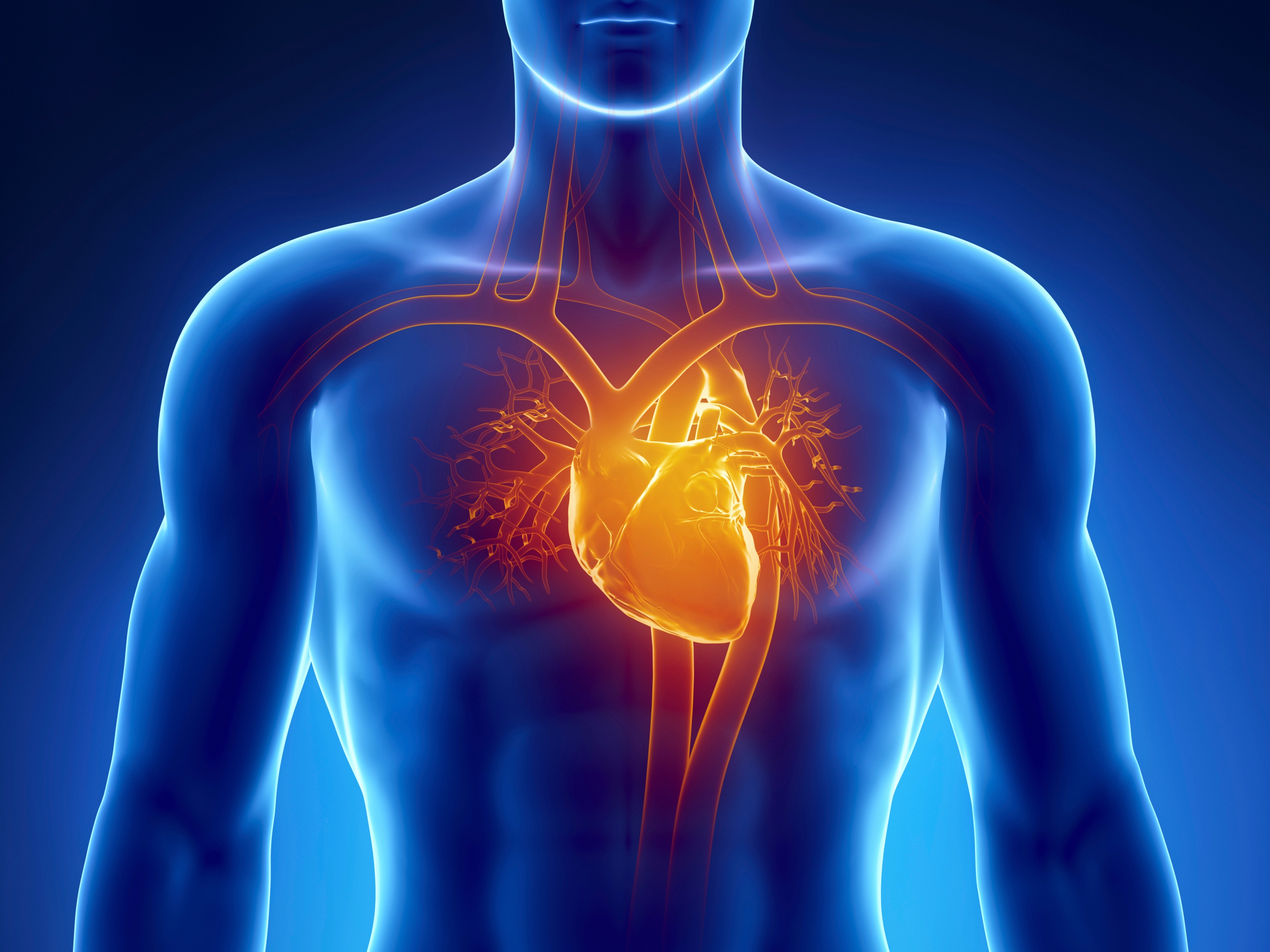
The heart is part of the _____ system.
respiratory
nervous
muscular
cardiovascular
_____ is the body's ability to maintain a relatively stable internal environment.
physiology
anatomy
homeostasis
kinesiology
The ______ plane divides the body into left and right halves.
transverse
frontal
sagittal
The fingers are _____ to the wrist.
proximal
distal
Which picture below best represents the anatomical position?


What plane is this?
midsagittal
frontal
transverse
parasagittal

What plane is this?
parasagittal
oblique
transverse
frontal
Towards the head or above.
Superior
Proximal
Inferior
Medial
Toward the middle/midline of the body:
Medial
Intermediate
Lateral
Ipilateral
AWAY from the middle/midline of the body:
Medial
Lateral
Contralateral
Ipsilateral
Closer to the surface of the body
Deep
Medial
Superior
Superficial
Back of the body:
Ventral/ Anterior
Dorsal/ Posterior
Inferior
Sagittal
Towards the feet or lower:
Cranial
Superficial
Inferior
Deep
Farther into the body, away from the surface:
Superficial
Deep
Inferior
Superior
The living human which represents the sum total of all its organ systems working together to maintain life.
Cell
Organelle
Organ
Organism
A group of organs that work together to perform a vital body function.
Organelle
Organism
Organ system
Organic
A part of the body formed of two or more tissues and adapted to carry out a specific function.
Organ
Organelle
Organ system
Organism
A group of similar cells.
Organelles
Tissues
Organs
Cell groups
Basic structural and functional unit of the body.
Organelle
Organism
Tissue
Cell
The function of the integumentary system includes all of these except what?
Homeostasis
Cooling system
Synthesis of Vitamin A
Excretion of wastes


Blood cell that protect the body against infection
Platelets
Plasma
Hemoglobin
White Blood Cells
Blood
Types of cells in the blood that causes blood clots to form
Platelets
Blood
Plasma
Hemoglobin
White blood cells
A wall of tissue that separates the 4 chambers of the heart
Pulmonary Circulation
Valves
Atria
Ventricles
Septum
The lower two chambers of the heart
Pulmonary Circulation
Valves
Atria
Ventricles
Septum
Blood vessels that return blood to the heart
Arterioles
Arteries
Venules
Veins
Capillaries
Blood vessels that carry oxygenated blood away from the heart
Arterioles
Arteries
Veins
Venules
Capillaries
The fluid in which other parts of the blood are suspended
Platelets
Blood
Plasma
Hemoglobin
White Blood Cells
What is a nerve cell?
axon
neuron
dendrite
myelin
Which 2 are parts of the central nervous system?
brain
nose
spinal cord
left big toe
What part of the nervous system is responsible for slowing down breathing and pulse?
Spinal Nerves
Sympathetic
Parasympathetic
Spinal Nerves
Dopamine and Serotonin are examples of _______________
Axon
Neurotransmitters
Somatic Nerves
Autonomic Nerves
The branch like figures that receive impulses & conducts them to the cell body
Dendrite
Axon
Cell Body
Myelin Sheath
The main function of the nervous system
send messages to and from the brain and spinal cord to and from the body
break down food to be used by the body
remove wastes from the body
all of these
Which part of the neuron transmits impulses to other neurons?
axon
cell body
dendrite
node
The part of the body that integrates the information it receives from all over the body in order to make decisions is called the __________________
peripheral nervous system
sympathetic nervous system
central nervous system
parasympathetic nervous system
What contains the nucleus of the neuron?
Terminal Bouton
Cell Body
Axon Hillock
Myelin Sheath

Compared to arteries, veins
are more elastic.
have more smooth muscle in their tunica media
hold their shape better when cut.
have thinner walls
Valve between the left atrium and the left ventricle.
tricuspid valve
bicuspid valve
pulmonary valve
aortic valve
Thickest walled chamber of the heart.
left atrium
right ventricle
left ventricle
right atrium
Blood in the left atrium:
Oxygenated
Deoxygenated
Blood in the right atrium:
Oxygenated
Deoxygenated

Identify:
Left Ventricle
Right Atrium
Right Ventricle
Left Atrium
Why is the muscle in the left ventricle thicker than that of the right ventricle ?
The left ventricle pumps oxygenated blood round the entire body.
The right ventricle pumps oxygenated blood round the entire body.
The right ventricle pumps blood to the lungs which is a long way.
They are both the same.

Blood enters the heart from the body from the inferior vena cava/superior vena cava at the ....
When blood exits the left ventricle, it must go through which valve?
Aortic
Pulmonic
Mitral
Tricuspid
What portion of the conduction system is known as the pacemaker?
AV node
Purkinje fibers
SA node
Bundle of His

