
 Worksheets
Open Stax Microbiology Ch. 3 Review
Total questions: 86
Worksheet time: 53mins
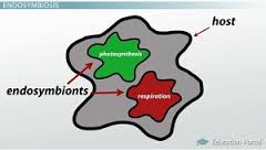
Which of the following is evidence to support the endosymbiotic theory?
Mitochondria and chloroplasts have their own DNA, which is similar to prokaryotic DNA.
Fossil evidence shows that the first forms of life were prokaryotic cells.
All eukaryotic cells have a double-layered membrane composed of phosplipids and proteins.
Prokaryotes have the same number and types of enzymes as eukaryotes.
Which type of cell? (It has a nucleus, membrane-bound organelles). Check all that apply.
prokaryotic cell
eukaryotic cell
plant cell
bacteria
Are eukaryotes multicellular or unicellular?
multicellular
unicellular
both unicellular and multicellular

Two students are reading an article about the evidence of the endosymbiotic theory. As they are reading, they have drawn images of what they believe to be occurring on index cards. The two students are engaging in a discussion on how the index cards should be ordered to accurately show the evolution of eukaryotic cells.
Student A claims the order should be B, E, D, A, C. Their reasoning is that since the small circles without lines in the index cards represent chloroplasts, photosynthesizing organisms evolved before aerobic organisms.
Student B claims the order should be B, A, C, E, D. Their reasoning is that since the small ovals with lines in the index cards represent mitochondria, respiratory organisms evolved before photosynthesizing organisms
Use the diagram to answer the question. Which statement below is accurate about the discussion above?
student A has a correct ordering of index cards but incorrect reasoning.
student B has a correct ordering of index cards and correct reasoning.
Student A has a correct ordering of index cards and correct reasoning.
Student B has a correct ordering of index cards but incorrect reasoning.
____________ & _________________ are organelles that definitely arose from endosymbiosis.
Nucleus and cell membrane
Chloroplast and mitochondria
Mitochondria and nucleus
Cell wall and chloroplast
"Endosymbiosis" means
Living together
Entering together
Living separately
Joining together
Both eukaryotic and prokaryotic cells have:
ribosomes
cell membranes
false
true
Mitochondria and chloroplasts have their own DNA.
true
false
Mitochondria , chloroplast , and bacteria are similar in size.
true
false
Mitochondria and chloroplast do not divide on their own.
false
true
Eukaryotes are thought to have evolved by a prokaryote engulfing one heterotrophic bacteria and one photosynthetic bacteria.
false
true
Which statement below correctly explains and gives the importance of the endosymbiotic theory?
The endosymbiotic theory states that the inner and outer membrane of the endoplasmic reticulum indicates these were once free living organisms which were taken into an early cell via endocytosis. The theory was further supported by the presence of prokaryotic ribosomes and replication of the endoplasmic reticulum via binary fission.
The endosymbiotic theory states that the inner and outer membrane of the mitochondria indicates these were once free living organisms which were taken into an early cell via endocytosis. The theory was further supported by the presence of prokaryotic ribosomes and replication of mitochondria via binary fission.
The endosymbiotic theory states that through endocytosis, early prokaryotic cells were able to bring in additional nutrients. These additional nutrients provided important energy that drove evolution of these prokaryotic cells into the eukaryotic cells seen today in more advanced organisms.
The endosymbiotic theory states that through endocytosis, early prokaryotic cells were able to bring in early energy rich eukaryotic cells. The energy provided drove evolution of these prokaryotic cells into the eukaryotic cells seen today in more advanced organisms.
Why type of cell does not contain membrane bound organelles?
Animal Cells
Prokaryotic Cells
Eukaryotic Cells
Plant Cells

What do you see?
a prokaryotic cell
the bottom of the ocean
a white blood cell
an eukaryotic cell

What do you see?
a white blood cell
an eukaryotic cell
DNA
a prokaryotic cell
Which cells have cytoplasm?
Prokaryotic
Eukaryotic
Both


The shape of this bacterial cell is:
bacillus
coccus
spirillum
A cell that does not contain a membrane-bound nucleus is:
prokaryotic
eukaryotic

The shape of this bacterial cell is:
coccus
bacillus
spirillum

The shape of this bacterial cell is:
bacillus
coccus
spirillum
A prokaryotic cell has a cell membrane.
False
True
Bacterial cells can have one of three shapes. Which of the following is not a common shape of bacterial cell?
Spirilla
Cocci
Bacilli
Cube
Which of the following is the largest organism type?
Viruses
Eukaryotes
Prokaryotes
Bacteria are treated with:
Antibiotics
Vaccines
Antifungals
Chemotherapy

The organelle described as a long, thin-like projection that helps with movement.
Ribosome
Nucleoid
Flagella
Cell Membrane

Organelle that creates proteins in bacteria.
Ribosome
Cell Wall
Mitochondria
Pili

Where is the DNA located in prokaryotic cells?
Nucleus
Cell Membrane
Cytoplasm
Chloroplast
In the bacterial cell, the chromosome or DNA is located floating freely in the cytoplasm in a region called the:
Ribosome
Nucleoid
Flagella
Cell Membrane
Bacteria have small, circular chromosomes separate from the nucleoid that they can easily exchange with other bacteria called:
Ribosomes
Plasmids
Capsules
Membranes

Bacteria that line up in chains have the prefix:
strepto-
staphylo-

Bacteria that arrange in clusters have the prefix:
strepto-
staphylo-
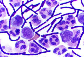
These bacteria are:
gram positive
gram negative
both gram positive and gram negative
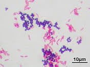
These bacteria are:
both gram positive and gram negative
gram positive
gram negative
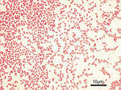
These bacteria are:
both gram positive and gram negative
gram positive
gram negative

What would be the gram stain results of a bacteria with this cell wall? (Hint: look at the peptidoglycan layer in the cell wall.)
gram positive
gram negative

Select all that apply to this bacteria:
cocci
bacilli
strepto-
staphylo-
Gram negative
These bacteria have a thin peptidoglycan cell wall and therefore have an outer membrane (capsule) to help protect them.
True
False

Which part of a bacteria cell wall "holds" the Gram stains?
plasma membrane
pili
peptidoglycan cell wall
cytoplasm
These bacteria are
gram positive
gram negative
This diagram shows a bacterium that will be _________________ when stained with the Gram staining method.
gram positive
gram negative

Select all that apply to the above bacterial species that were stained using the Gram Stain method. (There are three).
coccus
bacillus
Gram positive
Gram negative
staphylo-

Select all that apply to the above bacterial species that were stained using the Gram Stain method. (There are three)
strepto-
bacillus
Gram positive
Gram negative
staphylo-
The term that describes bacteria being found virtually "everywhere"
ubiquitous
astrobiologist
specific epithet
microbes
this term indicates that the morphology of bacteria is rod shaped
bacillus
coccus
spirilla
none of the above
this term indicates that the morphology of bacteria is spherical shaped
bacillus
coccus
spirilla
none of the above

What bacterial arrangement is represented in this picture?
Coccus
Diplo
Staph
Strep

What bacterial morphology is present in the image?
Bacillus
Coccus
Spirillum
Vibrio
True / False:
Gram negative bacteria has MORE layers of protection than Gram positive bacteria
True
False
Gram positive bacterial cell walls are so thick that when they are dyed, they are able to absorb more crystal violet and appear purple during Gram Stain.
True
False
Gram negative bacteria have an extra outer layer of ____________ which help give them more protection and antibiotic resistance.
cell membrane
peptidoglycan
murein
all of the above
This scientists finally disproved spontaneous generation once and for all.
Needham
Miller and Urey
Pasteur
Spallanzani
Pasteur proved the spontaneous generation theory wrong and also demonstrated microorganisms are everywhere.
TRUE
FALSE


This image depicts what flagellar arrangement?
monotrichous
amphitrichous
polar monotrichous
peritrichous
amphilophotrichous

This image depicts what flagellar arrangement?
monotrichous
amphitrichous
polar monotrichous
peritrichous
amphilophotrichous
What is the difference between the cilia and flagella?
Cilia are short, hair like appendages extending from the surface of a living cell. Flagella are long, threadlike appendages on the surface of a living cell.
Eukaryotic cilia and flagella are motile organelles built on a scaffold of doublet microtubules and powered by dynein ATPase motors.


Match the following:
stabilizes and maintains flexibility of cell membrane
Cholesterol
2 layers; allows small & non-polar molecules to pass through the membrane
Phospholipid bilayer
regulates what goes in and out of the cell
Plasma or Cell Membrane
allows large and polar molecules to pass through the membrane
Transport or Channel Proteins
located outside of the membrane; helps with cell communication and cell-to-cell connections
Carbohydrates

