
 Worksheets
Anatomy Chapter 6 Muscular System Review
Total questions: 87
Worksheet time: 1hrs 9mins

#11
trapezius
latissimus dorsi
external obliques
pectoralis major

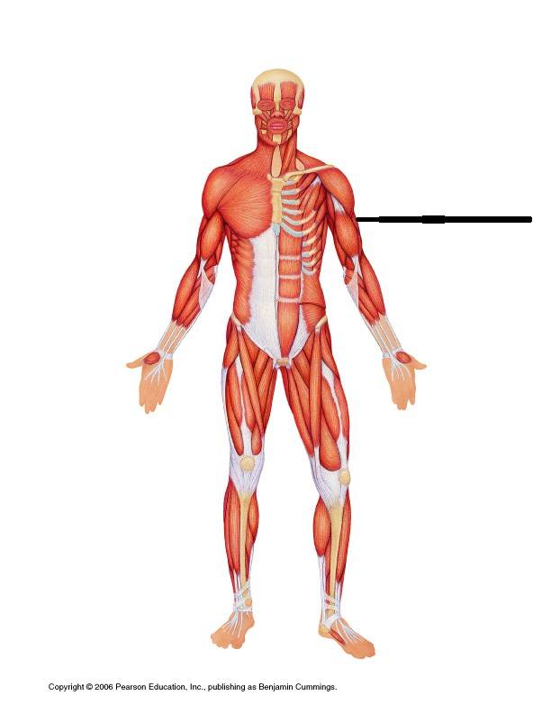
brachialis
biceps brachi
triceps brachi
brachioradialis
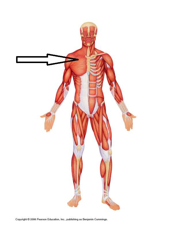
intercostal
rectus abdominus
pectoralis major
masseter

What muscle is represented by #19?
Gastrocnemius
Sartorius
Adductor Longus
Serratus Anterior

What muscle is represented by #7?
Biceps Brachii
Triceps Brachii
Brachioradialis
Latissimus Dorsi

What muscle is highlighted in red?
deltoid
tarpezius
rectus abdominus
vastus lateralis

What muscle is highlighted in red?
(a)
What muscle is highlighted in red?
triceps brachii
deltoid
biceps femoris

What muscle is highlighted in red?
erector spinae
external oblique
rectus abdominus
serratus anterior

What muscle is highlighted in red?
(a)
What muscle is highlighted in red?
(a)
What muscle is highlighted in red?
(a)
What muscle is highlighted in red?
(a)
What muscle is highlighted in red?
(a)
What muscle is highlighted in red?
(a)
What muscle is highlighted in red?
(a)

What muscle is highlighted in red?
(a)

What muscle is highlighted in red?
(a)
Contractile unit of a muscle which is also the functional unit of a muscle is ...
sarcomere
motor unit
fascicle
myofibril

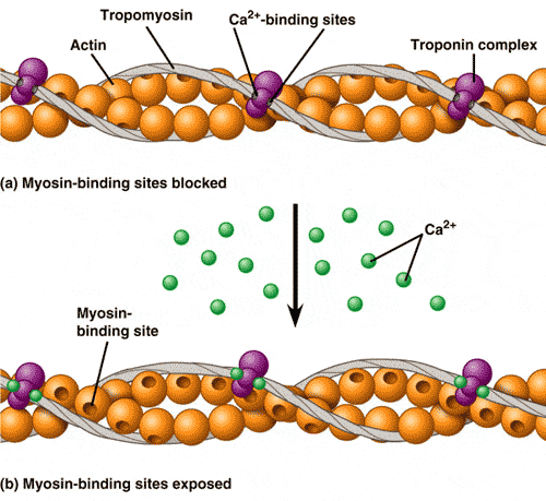
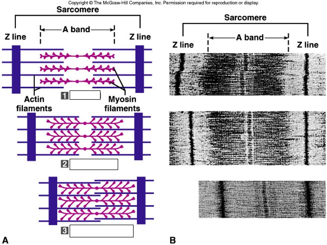
One neuron and all the skeletal muscle cells it stimulates is the ____ ___.
Neurotransmitter
Neuromuscular junction
Motor unit
Synaptic cleft
Once sodium is pumped into the sarcolemma, what happens?
Action potential is transferred to the sarcolemma to cause contraction.
The process ends and ACh breaks down.
Calcium is pumped into the axon terminal.
ACh is released into the synaptic cleft.
Which filament slides inward during muscle contraction
Actin
Myosin
During full muscle contraction, which region disappears?
I band
M line
H zone
A band

Which region consists only actin (thin filament)?
I band
M line
H zone
A band

Which part separate one sarcomere to the next?
M line
I band
H zone
Z line

Do the lengths of the thick and thin filaments change during contraction?
Yes
No
Calcium binds to ___of the troponin-tropomyosin complex.
troponin
tropomyosin
actin
myosin
A ___extends from one Z disc to another Z disc.
Sarcolemma
Neuromuscular junction
Sarcomere
Motor end plate

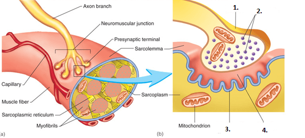
Which number represents the sarcomere?
1
2
3
4

Which number represents the actin filament?
1
2
3
4

Which number represents the myosin filament?
1
2
3
4
What are the three types of muscle tissue in the body:
Skeletal, cardiac, and smooth muscle
Tendons, Ligaments, Smooth Muscles
Endomysium, perimysium, epimysium
Myofilaments, troponin, tropomysium

What type of muscle cell is shown:
Cardiac Muscle Cell
Skeletal Muscle Cell
Smooth Muscle Cell
This is not a muscle cell, a tendon is visualized

What type of muscle cell is shown:
Cardiac Muscle Cell
Skeletal Muscle Cell
Smooth Muscle Cell
This is not a muscle cell, a tendon is visualized

What type of muscle cell is shown:
Cardiac Muscle Cell
Skeletal Muscle Cell
Smooth Muscle Cell
This is not a muscle cell, a tendon is visualized
The connective tissue coverings of a skeletal muscle, listed from superficial to deep, are:
endomysium, perimysium, and epimysium
endomysium, epimysium, and perimysium
epimysium, endomysium, and perimysium
epimysium, perimysium, and endomysium
The contraction of a muscle exerts a pull on a bone because
muscles are attached to bones by ligaments
muscles are directly attached to bone
muscles that are attached to bones by tendons
All of the above are correct
What do the epimysium and perimysium contain?
Blood Vessels and Nerves
Muscle Fibers and Blood Vessels
Muscle Fibers and Nerves
Only Muscle Fibers
True or False:
Each muscle cell/muscle fiber is composed of a singular myofibril.
True
False

Which is pictured here?
epimysium
perimysium
endomysium

Structure A is on either side of the sarcomere, it is called the
A band
I band
Z disc
fascia

The unit between A and A is called
a sarcomere
The I band
the Z disc
fascia
The impulse that triggers a contraction travels deep into a muscle fiber by means of
Sarcoplasmic reticulum
Transverse Tubules
Mitochondria
Motor end Plates

Identify X? A protein that covers the mysoin binding sites on the actin filament in a relaxed muscle, preventing contraction.
actin
myosin
tropomyosin
troponin
cross bridge

Identify the pink structures? It is a globular protein complex, occuring with tropomyosin in the thin filaments of muscle tissue. Binds to calcium ions, moving tropomyosin away from the myosin binding sites on actin filaments.
actin
myosin
tropomyosin
troponin
cross bridge

When the sarcomeres contract, what happens to the length of the I band?
Increases
Stays the same
Decreases

When the sarcomeres contract, what happens to the length of the A band?
Increases
Stays the same
Decreases
Which of the following muscle types are voluntary?
Skeletal
Cardiac
Smooth muscle

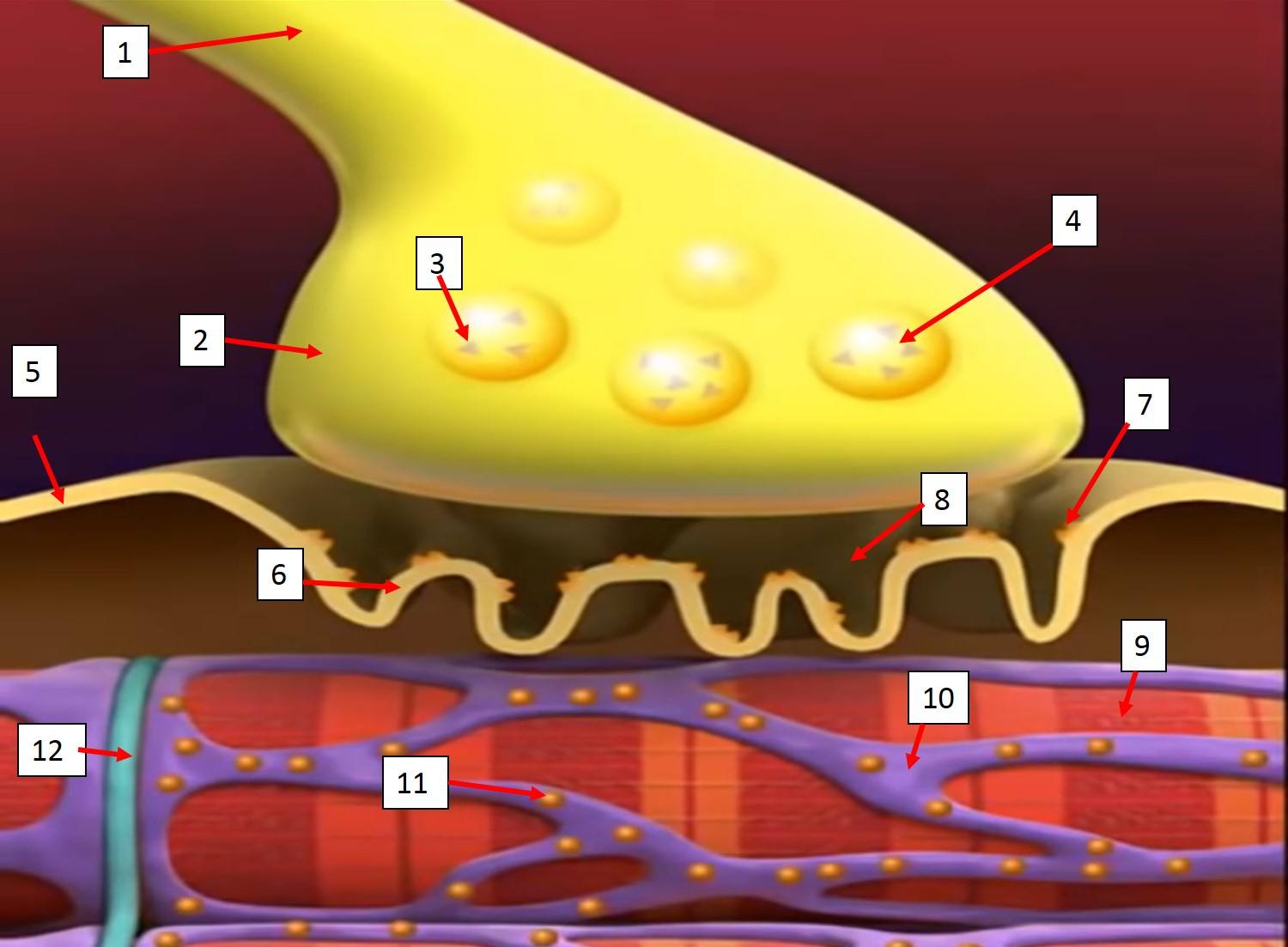
In the image, what are the small purple dots (D in the legend)?
Calcium Ions
Sodium Ions
Neurotransmitter
Acetylcholine
What happens that causes calcium channels to open?
Action Potential on motor neuron
Depolarization of muscle fiber
Influx of sodium ions
Neurotransmitter moves out of the nerve cell.
What is the space between the motor nerve and the muscle called?
Neuromuscular Junction
Synaptic Cleft
Chemical Gap
Motor End Plate
What is structure B in the image?
Neurotransmitter
Acetylcholine
Acetylcholinesterase
Sodium ions (Na+)
In the image, which structure is acetylcholine going to directly affect?
Structure C
Acetylcholine Receptors
Sodium ion channels
Sodium ions (Na+)
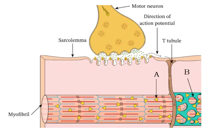
What is the purpose of T-tubules in muscle cells?
to carry sodium ions into the muscle cell
To carry electricity into the muscle cell
to carry calcium ions into the muscle cell
What is structure B in the image?
(a)
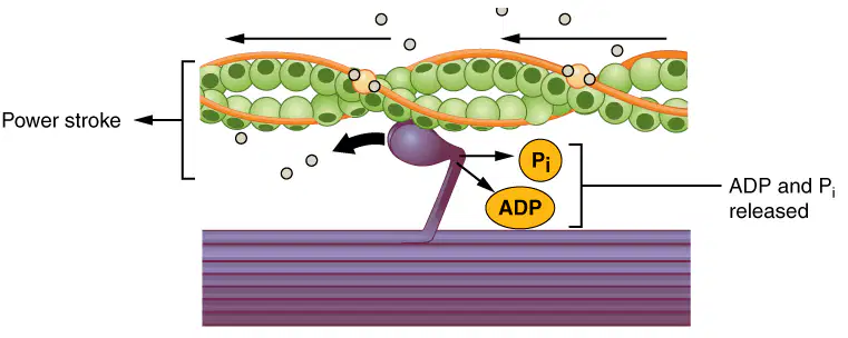
The last step in the contraction cycle is to reset the original position of the myosin head to the stretched position. What event causes this to happen?
ATP is split into ADP and Pi
ADP and Pi reattach to the myosin head
Calcium ions attach to the myosin head
sodium ions attach to the myosin head
What happens to cause myosin heads to detach from he actin?
Calcium ions attach to the myosin head
ADP and Pi reattach to the myosin head
ATP attaches to the myosin head
sodium ions attach to the myosin head
An electrical impulse within a single neuron is called a
synapse
membrane potential
action potential
cell body
An enzyme that break down neurotransmitter acethycholine?
lysosome
receptors
cholinesterase
synaptic cleft
A synaptic connection between a neuron and a muscle cell during movement is called
synaptic cleft
junctional folds
myasthenia gravis
neuromascular junction
Part of a neuron cell that carries impulses away from the cell body is called the
vesicle
receptor
dendrite
nerve axon
A model used to describe the process of how actin and myosin filaments slide over each other during muscle contraction.
synaptic cleft
neural junction
neuromascular junction
sliding filament theory
It is like the plasma membrane of muscle cells.
sarcoplasmic reticulum
sarcolemma
sarcomere
receptors
The neurotransmitter that crosses the synaptic gap during neuromascular junction. It send messages/signals to the muscles during movement.
acetylcholine
cholinesterase
motor end plate
neuromascular junction
A connective tissue that surrounds the entire muscle is called
endomysium
perimysium
epimysium
fascicle
A connective tissue that surround each bundle of muscle fibers is called
endomysium
epimysium
perimysium
sarcomere
One bundle of muscle fibers is called
myofibril
fascicle
myofilament
sarcoplasm

