
 Worksheets
A&P II Exam 2
Total questions: 107
Worksheet time: 4hrs 1mins
Pacemaker potential is characterized as
Na+ channels open and Na+ influx causes interior to
become more positive
Ca2+ channels open and Ca2+ influx causes rising phase of
action potential
K+ channels open and K+ efflux causes interior to become
negative again
Na+ channels open and Na+ efflux causes interior to become more negative
Depolarization is characterized as
Na+ channels open and Na+ influx causes interior to
become more positive
Ca2+ channels open and Ca2+ influx causes rising phase of
action potential
K+ channels open and K+ efflux causes interior to become
negative again
Na+ channels open and Na+ efflux causes interior to become more negative
Repolarization is characterized as
Na+ channels open and Na+ influx causes interior to
become more positive
Ca2+ channels open and Ca2+ influx causes rising phase of
action potential
K+ channels open and K+ efflux causes interior to become
negative again
Na+ channels open and Na+ efflux causes interior to become more negative
The Intrinsic Conduction System begins where?
the AV node
the AV bundle
the SA node
the Purkinje fibers
If the SA node is damaged, the heart can function adequately with which of the following?
the AV node
the AV bundle
the SA node
the Purkinje fibers
How is the SA node and AV node different from each other?
AV nodes are smaller in diameter with fewer gap junctions
SA node generates impulses about 75x/min
AV nodes generate impulses about 50x/min without the SA node's input
both SA and AV nodes generate the same impulse
What is the AV bundle?
the only electrical connection between the atrial and ventricles
nonconducting fibrous cardiac skeleton insulates rest of AV junction
Complete pathway through interventricular septum into apex and ventricular walls
depolarizes only about 30x/min in absence of AV node
What is the Purkinje fibers?
the only electrical connection between the atrial and ventricles
more elaborate in the left ventricle
Complete pathway through interventricular septum into apex and ventricular walls
depolarizes only about 30x/min in absence of AV node
How does the Sympathetic Nervous System affect the heart?
Cardioacceleratory center
increase force and rate
Cardioinhibitory center
Stimulates SA and AV
nodes, heart muscle, and
coronary arteries
How does the Parasympathetic Nervous System affect the heart?
Cardioacceleratory center
inhibits SA and AV nodes
via vagus nerves
Cardioinhibitory center
increase rate
Which brain region affects the heart's force and/or rate?
Cerebral cortex
Adrenal medulla
Medulla oblongata
Pons
How is the cardiac contractile cells' action potential similar to the pacemaker cells' action potential?
it follows the same ion movement (Na influx, Ca influx, K efflux)
the action potential graph is the same
both have a resting period
both are similar to skeletal muscle cells
What are the steps for the cardiac contractile cells' action potential?
plateau
depolarization
repolarization
pacemaker potential
What is the name for the pacemaker cells' action potential?
plateau
depolarization
repolarization
pacemaker potential
How are contractile cells different than skeletal muscle cells?
plateau happens in the contractile cells
contractile cells have longer refractory period
contractile cells have longer AP and contraction
contractile cells are stimulated by cardiac cells instead of neurons
The ECG/EKG is used for which of the following?
detect electrical currents generated by skeletal muscle
detect electrical currents generated by the heart
detect the conductivity of a specific body part
separates the composition of the blood
What is detected in the ECG graph?
P waves, or atrial depolarization
P waves, or atrial repolarization
QRS complex, or ventricular depolarization and atrial repolarization
QRS complex, or ventricular repolarization and depolarization
T waves, or ventricular repolarization
The intervals between the waves are:
P-R interval
Q-T interval
R-R interval
Q-S interval
The segments in the ECG are
P-R segment
S-T segment
T-P segment
R-R segment
Which of the following graphs represents an junctional rhythm?
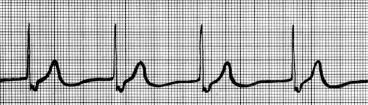

Which of the following graphs represents a normal sinus rhythm?
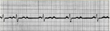
Which of the following graphs represents a second-degree heart block?
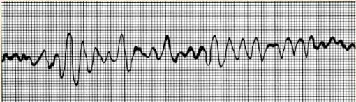
Which of the following graphs represents a ventricular fibrilation?
Atrial systole means that...
Ventricular diastole means that...
The mechanical events that happen in the heart are...
ventricular filling
isovolumetric contraction
ventricular ejection
isovolumetric relaxation
What occurs in the ventricular filling phase?
80% of the blood fills the ventricles due to gravity (passive)
atrial repolarization causes the remaining 20% to be pushed into the ventricles
80% of the blood is forcefully pushed into the ventricles
right before the ventricles contract, end diastolic volume occurs can be measured
What occurs during the isovolumetric contraction phase?
atria relaxes; ventricle begins to contract
falling pressure in atria causes closure of AV valves
at one point, both chambers are closed, so the volume will remain constant
rising pressure in ventricles causes SL valves to open
Which mechanical event has the most pressure in the aorta?
Ventricular ejection
Isovolumetric contraction
Isovolumetric relaxation
Ventricular filling
When pressure in the ventricular chambers exceed the pressure in the arteries, which of the following phases will occur afterwards?
Ventricular ejection
Isovolumetric contraction
Isovolumetric relaxation
Ventricular filling
Which event has the least pressure in the ventricle?
Isovolumetric contraction
Isovolumetric relaxation
Ventricular filling
Ventricular ejection
In a split second, all valves are open and pressure is constant. What event am I talking about?
Isovolumetric contraction
Isovolumetric relaxation
Ventricular filling
Ventricular ejection
Which event follows P Waves?
Isovolumetric contraction
Isovolumetric relaxation
Ventricular filling
Ventricular ejection
Which event follows T Waves?
Isovolumetric contraction
Isovolumetric relaxation
Ventricular filling
Ventricular ejection
Which events follows QRS Waves?
Isovolumetric contraction
Isovolumetric relaxation
Ventricular filling
Ventricular ejection
Which of the following creates the "lub-dup" sound?
S1 is the closing of AV valves
S1 is the closing of SL valves
S2 is the closing of AV valves
S2 is the closing of SL valves
S1 is associated with ventricular filling to isovolumetric contraction. Is this true; if not, what is the correct events?
True
False
Isovolumetric contraction to beginning of ventricular ejection
Ventricular contraction to isovolumetric relaxation
In an ECG test, S2 is associated with which waves?
T waves
P waves
QRS complex
R interval
If S2 is associated with isovolumetric relaxation, then what is its' ventricular volume level?
ESV
EDV
SV
mid point
The amount of blood pumped in each ventricle in 1 minute is
systole
diastole
ventricular output
cardiac output
What is the relationship between Cardiac Output, Heart Rate, and Stroke Volume?
CO = HRSV
CO = SVHR
CO = HR x SV
They are not related to each other at all
____ ____ can be used to explain why during fight or flight, a person's CO is increased, but after the fight or flight experience, their CO is lowered.
cardiac output
stroke volume
cardiac reserve
preload
Which of the following regulates stroke volume?
preload
afterload
load
contractility
The major intrinsic factor of stroke volume is which of the following terms?
An increase in preload means stroke volume will be ____?
increase
decrease
An increase in contractility means stroke volume will be ____?
increase
decrease
A hypertensive adult is experiencing afterload. What is happening to their ESV and SV?
ESV increases
ESV decreases
SV increases
SV decreases
What are the factors that regulate heart rate?
autonomic nervous system
chemicals
other factors
the force
When norepinephrine binds to the heart, what happens to the heart on a cellular level?
Pacemaker cells fire more rapidly -- increases heart rate
pacemaker cells fire more slowly --- decreases heart rate
increases contractility
decreases contractility
SV remains unchanged
How does the parasympathetic nervous system affect the heart?
slows the HR; opposes the autonomic nervous system
little affect to contractility
dominant influence on heart rate
both parasympathetic and sympathetic nervous system causes the same results in the heart
What are the hormones that affect the heart rate and how does it affect the heart?
epinephrine and norepinephrine increases HR and contractility
thyroid hormone increases heart rate
oxytocin decreases heart rate
gonadal hormones increases the heart rate
Which ions may affect the heart?
Ca ions may cause arrhythmia
K ions may stop the heart
Fe ions will make the heart more red
P ions may turn the heart bananas
What other factors may contribute to our heart rate?
gender
age
exercise
body temperature
Congestive heart failure occurs when the heart---
is an inefficient pump
weakened myocardium
CO is so low
got stolen
A patient is feeling a sharp pain in their heart. Taking a look inside shows that they have a clogged coronary artery. What is the cause of this?
peripheral atherosclerosis
coronary atherosclerosis
persistent high blood pressure
multiple myocardial infarcts
dilated cardiomyopathy
Which of these terms is best defined as: the aortic pressure exceeds 90 mm Hg. Prolonged high aortic pressure will cause the cardiac cells to weaken as it exerts more force.
peripheral atherosclerosis
coronary atherosclerosis
persistent high blood pressure
multiple myocardial infarcts
dilated cardiomyopathy
A patient unfortunately suffered from many heart attacks and has a build up of scarred tissues in their heart. What term best describes what the patient has?
peripheral atherosclerosis
coronary atherosclerosis
persistent high blood pressure
multiple myocardial infarcts
dilated cardiomyopathy
What is it called when ventricles stretch and become flabby, and the
myocardium deteriorates
peripheral atherosclerosis
coronary atherosclerosis
persistent high blood pressure
multiple myocardial infarcts
dilated cardiomyopathy
Failure on the right side of the heart results in
Peripheral congestion
Fluid pools far from the heart (like limbs)
Failure on the left side of the heart results in
Pulmonary congestion
Fluid accumulation in the lungs therefore difficulty in gas exchange
As a fetus, which two structures were created to bypass the lungs?
fossa ovalis - from right atrium to left atrium
foramen ovale - from right atrium to left atrium
ductus arteriosus - from pulmonary trunk to aortic arch
ligamentum arteriosum - from pulmonary trunk to aortic arch
What age-related changes can affect the heart?
Sclerosis
Decline in cardiac reserve
Fibrosis
Atherosclerosis
Exercise and diet
A blood vessel that contains tight junctions, an endothelium, and the innermost tunica is called?
A blood vessel does NOT contain lumen is called?
None of them
Artery
All of them
A blood vessel that carries oxygenated blood away from the heart. Except for the pulmonary and fetal circulation. Is called?
A blood vessel that carries deoxygenated blood towards from the heart. Except for the pulmonary and fetal circulation. Is called?
This blood vessel directly interacts with tissues.
Which tunic contains the endothelium and a subendothelial layer?
Does the tunica media contain smooth muscles, cardiac muscles, and/or elastin fibers?
it contains smooth muscles
it contains cardiac muscles
it contains elastin fibers
it contains skeletal muscles
This tunic has collagen fibers and its function is to protect and reinforce the wall and anchor the structure.
The veins lack these structures that are seen in the arteries.
internal elastic membrane
external elastic membrane
vasa vasorum
collagen fibers
This type of artery has the most elastin fibers, substantial amount of smooth muscles, act as pressure reservoirs for continuous flow.
Elastic Arteries aka conducting arteries
This type of artery delivers blood to the organs and has the thickest tunica media. It's also active in vasoconstriction
Muscular artery aka distributing artery
This type of artery leads to the capillaries and controls the flow into the capillaries to make sure it doesn't rupture.
Arteriole aka resistance artery
This type of capillary is abundant in the skin, muscles, and CNS. It helps form the blood-brain barrier.
Continuous capillary
This type of capillary has endothelium with pores for increased permeability. These blood vessels can be seen kidneys, intestines, or endocrine organs.
This type of capillary has few tight junctions, larger intercellular clefts, and incomplete basement membrane. They have larger lumens and can be seen in liver, bone marrow, spleen, and adrenal medulla.
What are the structures that make up capillary beds?
Terminal arterioles
Capillaries
Postcapillary venules
Arterioles
What structure do capillaries drain into?
Postcapillary venule
Venule
What are the adaptations that make up the veins?
large lumens & thin walls = blood reservoir
large diameter = little resistance
venous valves = prevents blood from pooling downwards due to gravity
no valves = even less resistance
Pick all of the correct anastomoses:
Vascular anastomoses - special interconnections of vessels
Arterial anastomoses - provide alternate pathways (collateral channels) for continuous flow
Venous anastomoses - very abundant
Shunts - can be diverted into other areas
Portal system - blood flows through two capillary beds separated by portal veins
What is the term that means the volume of blood passing through a vessel, organ or entire body. May be associated with cardiac output.
blood flow
blood pressure
resistance
What is the term that means the force per unit exerted on the walls of a blood vessel? Usually refers to systemic arterial pressure and goes down the pressure gradient.
blood flow
blood pressure
resistance
What is the term that means the amount of friction blood encounters as it passes through vessels? The most impactful influence of this is the diameter of the blood vessel.
blood flow
blood pressure
resistance
What is the correlation between blood flow, blood pressure, and resistance?
blood flow is directly proportional to blood pressure
blood flow is inversely proportional to resistance
blood pressure is directly proportional to resistance
blood pressure is inversely proportional to resistance
Identify the pulse points.
superficial temporal artery
facial artery
common carotid artery
brachial artery
femoral artery
What is the formula to calculate MAP?
diastolic pressure + 3pulse pressure
arterial pressure - 3pulse pressure
diastolic pressure - 3pulse pressure
systolic pressure + 3pulse pressure
What are the steps to take blood pressure?
use the auscultatory method via a sphygmomanometer
hear the sounds of Korotkoff
use an automated sphygmomanometer
hear the sounds of the heart
What are the changes in blood pressure in the systemic pathway?
when blood reaches to the arterioles, the pressure drops significantly
greatest pressure is in the arteries
lowest pressure is in the vena cavas
low pressure blood then returns to the right atrium
What are some adaptations that help with venous return?
Muscular Pump - contraction of skeletal muscle milks blood back into heart
Respiratory Pump - pressure changes during breathing move blood to heart
Sympathetic Vasoconstriction - tunica media constricts from sympathetic control
The 3 factors that regulate blood pressure are
Cardiac output
Resistance
Blood volume
Hormone levels
An increase in blood volume, CO, or resistance would ____ blood pressure
increase
decrease
How would a neural regulatory control regulate high blood pressure?
baroreceptor receives stimulus
baroreceptor decreases vasomotor center = vasodilation
baroreceptor increases vasomoter center = vasoconstriction
baroreceptor decreases sympathetic impulses
Chemoreceptors detect a high amount of CO2, how would chemoreceptors regulate this?
increased blood pressure speeds the return of blood to heart and lungs
activate cardioacceleratory and vasomotor centers
Which hormones increase blood pressure in a short amount of time?
Epinephrine and norepinephrine
Angiotensin II stimulates vasoconstriction
Atrial Natriuretic Peptide causes vasodilation
ADH conserves water/vasopressin
Which hormones decreases blood pressure in a long amount of time?
Epinephrine and norepinephrine
Angiotensin II stimulates release aldosterone and ADH
Atrial Natriuretic Peptide antagonizes aldosterone
ADH conserves water/vasopressin
What does tissue perfusion mean?
The extrinsic factors for blood flow are? and how do they affect blood flow
parasympathetic
sympathetic
hormones
throughout the entire body
maintain a constant MAP
Intrinsic factors for the rate of blood flow are
Metabolic
Neural
Hormonal
Myogenic
How would metabolic controls autoregulate blood flow?
some tissues may release nitric oxide (powerful vasodilator)
increase tissue chemical reactions stimulate vasodilation = increased blood flow
How would myogenic controls autoregulate blood flow?
local vascular walls stretch to keep perfusion constant
passive stretch = increased MAP stretches vessel walls more than normally
reduced stretch = decreased MAP means less stretch
the entire body controls all the blood vessels to keep everything organized in one place
Velocity is highest in where?
aorta
arteriole
veins
capillaries
The greatest cross-sectional area is?
veins
capillaries
aorta
venules
Velocity is ____ ____ to cross-sectional area
Bulk flow is
forcing blood out of capillaries and returning to the venule end
A positive NFP means?
Hydrostatic pressure is greater than Colloid Osmotic pressure
Colloid Osmotic pressure is greater than Hydrostatic pressure
more fluids is leaking out
more fluids are getting sucked back into the capillaries
As we age, what happens to our blood vessels?
varicose veins
athersclerosis
increased blood pressure
as infants: ductus venosus drains umbilical vein into inferior vena cave

