
 Worksheets
Practical 1 Quiz study guide BIO-168
Total questions: 124
Worksheet time: 31hrs 0mins
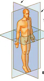
What are the three planes indicated in the picture? (Make sure to separate answers with a comma and use an uppercase letter at the beginning of each word.)
(a)

Stratified squamous epithelium
Simple cuboidal epithelium
Simple squamous epithelium
Stratified cuboidal epithelium

Simple cuboidal epithelium
Simple columnar epithelium
Stratified columnar epithelium
Pseudo stratified epithelium

Simple squamous epithelium
Stratified columnar epithelium
Simple cuboidal epithelium
Stratified squamous epithelium

neuron
white blood cell
bone
cardiac muscle tissue

Skeletal muscle tissue
cardiac muscle tissue
smooth muscle tissue
bone

skeletal muscle tissue
cardiac muscle tissue
smooth muscle tissue
hyaline cartilage

hyaline cartilage
cardiac muscle tissue
smooth muscle tissue
skeletal muscle tissue


What is A?
Dorsal/posterior
spinal
cranial
ventral/anterior
What is B?
Dorsal/posterior
spinal
cranial
ventral/anterior
What is C?
Dorsal/posterior
spinal
cranial
ventral/anterior
What is L?
Dorsal/posterior
thoracic
abdominopelvic
ventral/anterior
What is K?
abdominal
thoracic
abdominopelvic
ventral/anterior
What is J?
abdominal
thoracic
abdominopelvic
pelvic
What is i?
abdominal
thoracic
abdominopelvic
pelvic
What is H?
abdominal
thoracic
abdominopelvic
diaphragm
What is G?
abdominal
thoracic
abdominopelvic
diaphragm
What is F?
cranial
oral
nasal
orbital
What is E?
cranial
oral
nasal
orbital
What is D?
cranial
oral
nasal
orbital

Which cellular structure looks like short "eyelash" extensions and functions to help with locomotion and movement of substances?
cytoskeleton
flagella
cilia


Name the phase pictured here:
Anaphase of mitosis
Anaphase I of meiosis
Anaphase II of meiosis

What phase is pictured here?:
Anaphase of mitosis
Anaphase I of meiosis
Anaphase II of meiosis

What phase is pictured here?:
Telophase of mitosis
Telophase I of meiosis
Telophase II of meiosis

What phase is pictured here?:
Metaphase of mitosis
Metaphase I of meiosis
Metaphase II of meiosis

What phase is pictured here?:
Anaphase of mitosis
Anaphase I of meiosis
Anaphase II of meiosis

Name this structure:
Chromosome
Chromatin
Chromatid
Tetrad

Name 1 side of this structure:
Chromosome
Chromatin
Chromatid
Tetrad

Name the structure the blue arrow points to:
Chromosome
Chromatin
Chromatid
Tetrad

Name this phase
Interphase
Prophase
Metphase
Anaphase
Telophase
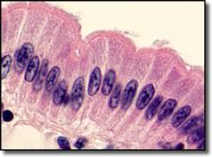
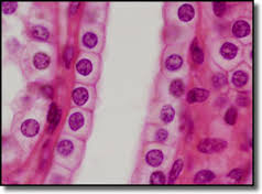


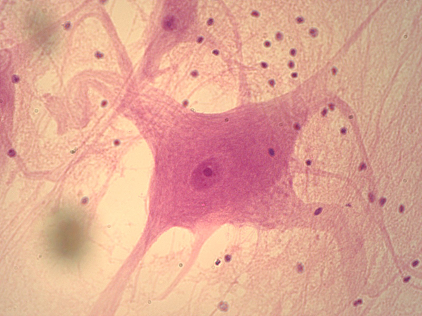


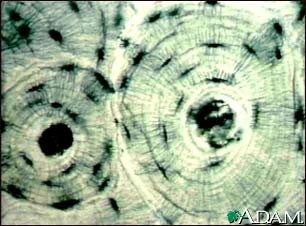
smooth muscle
bone
nerve
blood


Name the tissue
Skeletal
Cardiac
Smooth


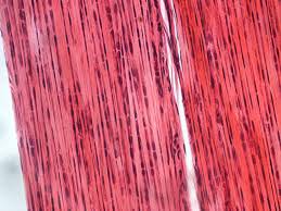
What type of tissue is this?
Dense connective tissue
Elastic connective tissue
Irregular connective tissue
Hyaline connective tissue
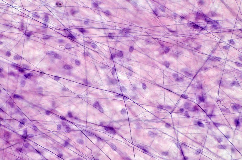
What type of tissue is this?
Loose connective tissue
Dense connective tissue
Elastic connective tissue
Hyaline connective tissue

Identify the tissue type.
Elastic cartilage
Hyaline Cartilage
Fibrocartilage
Dense regular connective tissue

Identify the tissue type.
Elastic cartilage
Hyaline cartilage
Fibrocartilage
Elastic connective tissue

Identify the tissue type.
Dense regular connective tissue
Dense irregular connective tissue
Elastic connective tissue
Adipose connective tissue
The top layer of the skin is called the ---
dermis
oil gland
subcutaneous fat
epidermis
____ is the coloring in skin and is found in the epidermis.
Melanin
Oil
Perspiration
Pores
What are the three main layers of the skin?
The epidermis, the dermis, and the subcutaneous fat
The epidermis, the dermis, and the sebaceous glands
The dermis, the sebaceous glands , and the subcutaneous fat
The follicles, the dermis, and the epidermis
What is the name of tube that hair grows from?
Follicle
Root
Shaft
Sebaceous
Which layer contains accessory structures such as sweat glands, sebaceous glands, and hair follicles?
epidermis
dermis
subcutaneous fat
hyperdermis
Oil glands that are found all over the skin are called ---
merocrine glands
sebaceous glands
apocrine glands
sweat glands
What causes acne?
clogged sebaceous glands
chocolate
peanut butter
salty foods
Which layer of the skin does NOT contain blood vessels?
epidermis
subcutaneous fat
dermis
Sweating and shivering are your body's way of regulating its ---
Water loss
Temperature
Hair growth
Vitamin D absorption

#1 on this diagram is the ---
epidermis
dermis
subcutaneous fat
follicle
#2 on this diagram is the ---
epidermis
dermis
subcutaneous fat
follicle
#3 on this diagram is the ---
epidermis
dermis
subcutaneous fat
follicle

What are the 3 layers of the skin?
Epidermis, dermis, hair
Dermis, hypo dermis, nails
Epidermis, dermis, fatty layer

What are the 4 parts of the Integumentary System?
Skin
Hair
Heart
Nails
Sweat Glands

This tough protein is found in hair and nails for structural stability.
melanin
sebum
keratin
Merkle cells

The 5 layers of the epidermis from outer to inner layers are: Stratum -
basale, spinosum, granulosum, lucidium, corneum
corneum lucidium, spinosum, granulosum, basale
corneum, lucidium, granulosum, spinosum, basale
basale, granulosum, spinosum, lucidium, corneum

These are your sweat glands with a large concentration under your arms, on your forehead, on your palms, and on the soles of your feet.
sebaceous glands
sudoriferous glands
keratinocytes
Merkel cells

This part of the dermis includes the nerve endings, glands, and hair follicles of the skin.
papillary layer
recticular layer
sebaceous glands
sebum

The white part at the base of your nail is called the
moon
lunule
lacunae
arc de moon

*The muscle that makes you hair stand on end is called the -
gluteus maximus
standupus straightus
arrector pili
nerousus scaredus

These glands are a teenager's worst nightmare because they empty an oily substance called sebum all over the face. It also empties into the hair follicles make the hair soft.
sebaceous glands
sudoriferous glands
keratinocytes
Merkel cells

Which of the following has the layers of the integumentary system in the correct order? (From outer to inner)
Hypodermis, Dermis, Epidermis
Epidermis, Dermis, Hypodermis
Dermis, Epidermis, Hypodermis
Hypodermis, Epidermis, Dermis

Which term describes #3?
lamella
central canal
osteon
periosteum
Which term describes #1?
lamella
central canal
osteon
periosteum
Which term describes #2?
lamella
central canal
osteon
periosteum


What four bones make up the knee joint?
Tibia
Fibula
Patella
Femur
All of the above

What is L?
Medial Collateral Ligament
Lateral Collateral Ligament
Anterior Cruciate Ligament
Posterior Cruciate Ligament

What is M?
Medial Condyle
Meniscus
Lateral Condyle
Patella

What is N?
Quadriceps Tendon
Lateral Collateral Ligament
Patellar Tendon
Tibia

What structure is highlighted in blue?
Neck
Shaft
Linea Aspera
Head
Lateral Supracondylar Line

What structure is highlighted in blue?
Neck
Shaft
Linea Aspera
Head
Lateral Supracondylar Line

What structure is highlighted in blue?
Neck
Shaft
Linea Aspera
Head
Lateral Supracondylar Line

What structure is highlighted in blue?
Greater Trochanter
Intercondylar Fossa
Intertrochanter Crest
Head
Lesser Trochanter

What structure is highlighted in blue?
Greater Trochanter
Intercondylar Fossa
Intertrochanter Crest
Head
Lesser Trochanter

What structure is highlighted in blue?
Greater Trochanter
Intercondylar Fossa
Intertrochanter Crest
Head
Lesser Trochanter

What structure is highlighted in blue?
Medial Epicondyle
Medial Condyle
Lateral Epicondyle
Lateral Condyle
Intercondylar Fossa

What structure is highlighted in blue?
Medial Epicondyle
Medial Condyle
Lateral Epicondyle
Lateral Condyle
Intercondylar Fossa

What structure is highlighted in blue?
Medial Epicondyle
Medial Condyle
Lateral Epicondyle
Lateral Condyle
Intercondylar Fossa

What bone is this?
Patella
Radius
Femur
Tibia
Fibula

What does both T and C represent?
Talus & Cuboid
Trapezoid & Capitate

What do the letters written represent?
Capitate, Clavicle, Navicular
Calcaneus, Navicular, Cuneiform

In numerical order what are these bones referred to as?
Tarsals, Metatarsals, Phalanges
Metatarsals, Tarsals, Phalanges
Phalanges, Tarsals, Metatarsals
Phalanges, Tarsals, Metatarsals

In numerical order what are these bones referred to as?
Carpals, Metacarpals, Phalanges
Phalanges, Metacarpals, Carpals
Metacarpals, Carpals, Phalanges
Carpals, phalanges, Metacarpals

What are the phalanges in numerical order?
Proximal, Intermediate, Distal
Distal, Intermediate, Proximal

