
 Worksheets
Anatomy and Physiology Final Review
Total questions: 57
Worksheet time: 44mins
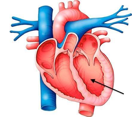
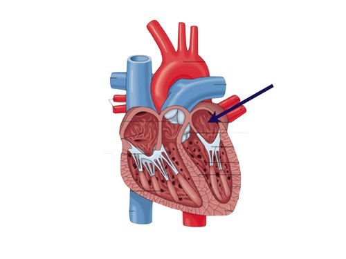

The correct label for part "A" is?
pulmonary veins
pulmonary artery
aorta
vena cava
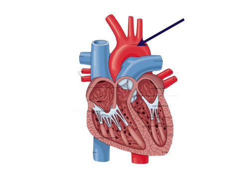

Identify the circled valve
Tricuspid
Mitral/bicuspid
Pulmonary
Aortic
The thickest and strongest blood vessel is the
vein
artery
capillary
both veins and capillaries
The blood vessel that has valves at intervals...
capillary
artery
vein
none of the above
Valves in veins
support them
protect them
prevent the backward movement inside them
provide them elesticity

Name structure A
Pupil
Iris
Cornea
Retina

Name Structure B
Lens
Pupil
Sclera
Cornea

Name structure G
Lens
Pupil
Optic Nerve
Ciliary Muscles

What structure is responsible for carrying sensory impulses from the eye to the brain?
Retina
Iris
Optic Nerve
Rods & Cones
substance that fills the posterior cavity of the eye
aqueous humor
vitreous humor
lysozyme
conjunctiva
Photoreceptors that detect the level of light but not the color
rods
cones
bipolar neurons
ganglion
Photoreceptors that detect colors and give a fine detailed image
cones
rods
ganglion neurons
bipolar neurons
The process when the lens changes shape to focus close or far away
sensory adaptation
accommodation
Transduction
globe luxation

What is the function of the auditory canal?
to block sound waves that could be harmful
to direct sound waves to the eardrum
to detect sensations of movements
to equalize air pressure between the outer and middle ear

When sound waves cause the eardrum to vibrate, these vibrations are immediately passed on to _________.
the auditory canal
the cochlea
the semi-circular canals
the ossicles

Which part of the ear allows balance when in motion and detects rotary (turning) sensations?
the vestible
the cochlea
the semi-circular canals
the ossicles
the eardrum
Sound is produce by
Vacuum
Sound wave
Hitting
Vibration
Identify the Auditory Canal (external Acoustic Meatus).
Identify the Cochlea.
Identify the Auricle (Pinna).
Sequence the following structures from the start to the finish for the sound pathway.
Pinna catches the sound waves
Sound waves travel through the auditory canal
Sound waves vibrate the ear drum
Sound waves vibrate through the ear ossicles
Sound waves vibrate on the oval window causing fluid to move inside the cochlea
Which of the following is not a taste receptor
Salty
Umami
Pungent
Sweet
The (a) in the cochlea transmit sound information via the (b) to the (c) lobe of the brain.
Label the diagram of the ear
Which of the following is an example of Sensorineural deafness?
Damage to the hair cells
Punctured tympanic mebrane
Build up of cerumen (earwax) in the auditory canal
Damaged ear ossicles
During this stage, K+ channels open and K+ Ions leave the neuron.
Match the following
Depolarization
+30 mV
Resting membrane potential
-70 mV
Threshold potential
-55 mV
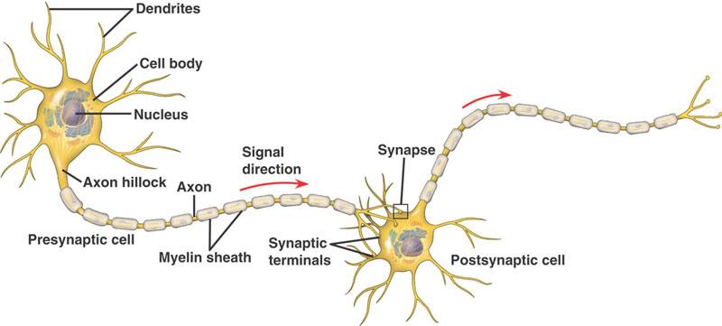
Identify the Axon of the Neuron
Identify the dendrite of the Neuron
Identify the myelin sheath of the Neuron
Identify the cell body of the Neuron
Sends impulses AWAY from CNS to muscle and glands.
Motor Neuron
Sensory Neuron
Interneuron
What direction does an electrical signal move?
Dendrite, Cell body, Axon
Cell body, Axon, Dendrite
Cell body, Dendrite, Axon
Axon, Cell Body, Dendrite
Match the following structure and function
axon
carry nerve impulse away from cell body
dendrite
receives incoming stimuli
cell body (soma)
contains cell organelles
myelin sheath
insulates and speeds up nerve impulses.
The protective fatty tissue that speeds up the transmission of messages is:
Myelin Sheaths
Axon
Dendrites
Soma
The neuron's life support that contains the nucleus is:
Cell Body (Soma)
Dendrites
Myelin Sheaths
Axon

This is the microscopic gap or space between two neurons
Dendrites
Cell Body
Synapse
Axon
Match the following
Frontal Lobe
Controls voluntary movements
Parietal Lobe
Processes sensory information
Occipital Lobe
Visual processing center
Temporal Lobe
Hearing and memory
Where does gas exchange takes place?
Alveloi
Blood vessels
Diaphragm

Identify structure 4
Trachea
Lung
Bronchi
Diaphragm
Identify structure 2
Trachea
Lung
Bronchi
Diaphragm

