
 Worksheets
EKG Study Guide
Total questions: 100
Worksheet time: 50mins
What are electrodes?
An instrumental to calculate BMI
A measurement unit of the heart's rythm
A blood component
A conductor of electric current
The MAJOR circulation is...
Right ventricle, lungs, right atria
Left ventricle, systematic circulation, right atrium
Right ventricle, lungs, left atria
Left atria, systematic circulation, right atria
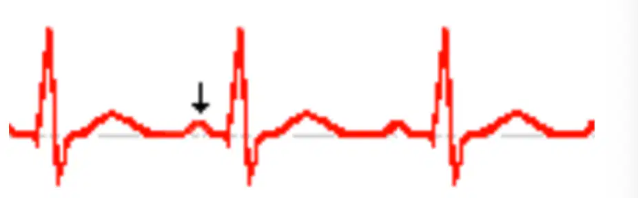
What part is this in an EKG Wave?
Q Wave
R Wave
S Wave
P Wave
How many leads does the EKG machine produce?
6
10
12
18
Which blood vessel comes out of the left ventricle?
The aorta
The vena cava
The pulmonary vein
The pulmonary artery
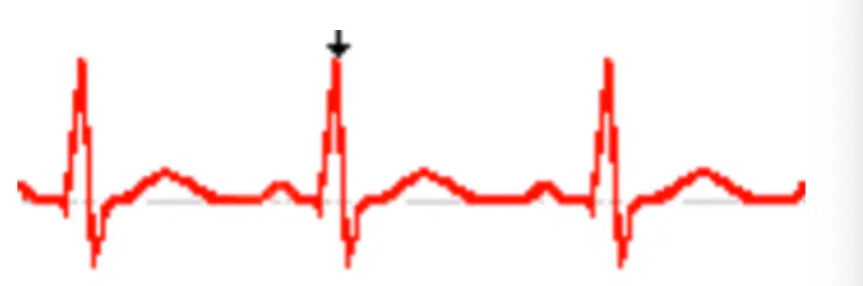
What part is this in an EKG wave?
Q Wave
R Wave
S Wave
P Wave
All of the following functions might be determined by the electrocardiogram
except...
Computes the duration of the repolarization
Length of time it takes to conduct the impluses
Measuring the patient's internal temperature
None of the above
Which of the following might NOT be shown by the ECG?
Duration of depolatization
Duration of repolarization
Data on the myocardium
All of the Above
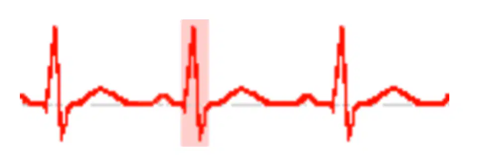
What is this part in an EKG Wave?
QR Segment
ST Interval
PR Segment
QRS Complex
When the oxygenated blood is being transported back to the left
atrium by the pulmonary veins, where is it coming from?
Right Atrium
Lungs
Kidneys
Left Ventricle
Which blood vessel comes out of the right ventricle?
The vena cava
The pulmonary vein
The pulmonary artery
The aorta
A cardiac cycle is the same as...
Blood pressure
One heart beat
Pulse
Oxygenation of the blood
The precordial V1 is placed on...
Fifth intercostal space to the right
Fourth intercostal space aligned with mid clavicle line
Fourth intercostal space to the left
Fourth intercostal space to the right
Which is the biggest blood vessel?
Aortic artery
Pulmonary Artery
Venules
Arteriole
The specialized blood cells in charge of defending the body from pathogens are called...
Erythrocytes
Leucocytes
Electrolytes
Osteoblast
The left atria receives blood from...
The vena cava
The pulmonary arteries
The pulmonary veins
The aorta
Which of the following are bipolar leads?
AVR, AVL, AVF
DI, DII, DIII
V1, V2, V3, V4, V5, V6
DI, DII, DIII, AVF, AVL, AVR
The ventricles are considered...
Low pressure chambers
High pressure chambers
Electrical chambers
Isoelectric chambers
The AV node is also known as...
The Gate Keeper
Natural Pacemaker of the heart
Natural Producer of electricity
Natural defibrillator of the heart
Which of the following would be considered correct for the layer of the heart which produces the contraction?
Epicardium
Subcutaneous
Myocardium
Endocardium
What does an EKG Machine records?
The circulation of the heart
The electrical conduction of the heart
The pulse of the heart
The patient's heart rate
Fifth inercostal space to the left aligned with mid clavicular line corresponds to which lead?
V4
V2
V3
V6
The Tunica _________ is the layer of the blood vessel that allows for them to attach to the muscles.
Media
Adventitia
Interna
Endothelium
Which are the types of circulation?
Tachycardic and Bradycardic
Dyastollic and Systolic
Systemic and Pulmonary
Latrogenic and Ferropenic
Which of the following does standard EKG paper becomes sensitive to damages?
Artificial light
Heat
Darkness
Cold
The intrinsic electricity refers to...
The external simulations of the heart
The conduction between electrodes
The electricity produced by the heart of its own
The electrical current between two ions
Where is The Bundle of His is located?
The Myocardial Wall
The Septal Wall
The AV Node
The SA Node
If an EKG tech has a patient with an amputated left arm, where would the LA electrode be placed?
On the left leg
On the left abdomen
On the superior suprasternal notch
On the right arm
Where in the heart does the right atria receives blood from?
The pulmonary vein
The pulmonary artery
The vena cava
The aorta
Which of the following are a part of a MINOR circulation?
Right ventricle, lungs, left atria
Left ventricle, lungs, right atria
Right atria, lungs, left atria
Left ventricle, systematic circulations, right atria
Which of the following structures is in charge of electrifying the Apex of the heart?
Purkenjie Branches
Bundles of His Branches
Bundle of His
Purkenjie Fibers
Which of the following is the 5th intercostal space, left midaxillary line is located?
V5
V6
V4
V3
Where would the posterior V leads V7-V9 be placed?
Below the scapula on the left side
Above the scapula on the right side
Below the scapula on the right side
All are correct
In a Limb Lead, which of the following is the color of the ground electrode?
Red
Green
White
Black
Which of the following are semilunar valves?
Aortic and Pulmonary
Mitral and Aortic
Tricuspid and Bicuspid
Biscuspid and Pulmonary
Which of the following are monopolar leads?
DI, DII, DIII, AVL, AVF, AVR
V1-6, AVR, AVL, AVF
AVR, AVL, AVF
DI, DII, DIII
Cardiovascular would be defined as...
The Heart
Blood
Blood Vessels
All the above
The blue lead corresponds to which electrode?
V1
V3
V4
V6
Which is the smallest vein?
Vena Cava
Arteriole
Venules
Median Cubital Vein
What are Electrolytes?
The unit of measurements of electric impulses of the heart
Chemical elements capable of producing electricity
Biological components of the heart
An electric reaction produce when the heart beats
What does depolarization mean?
Activation of the myocardial cells
The process in which a cell transfers ions to another
An augmentation of calcium in a cell
An irregular QRS complex reading
Clotting appears when which of the following stick to each other on a wound or lesion?
Erythrocytes
Leucocytes
Platelets
Plasma
Which of the following is a false statement?
The P wave represents atrial depolarization
The RR interval represents atrial repolarization
The QRS complex represents ventricular depolarization
The T wave represents ventricular repolarization
When preparing a patient for an EKG in the supine position, where should a technician stand to apply the electrodes?
The head of the bed
The foot of the bed
Patient's right side
Patient's left side
The pericardium is...
The protective sac of the heart
The protective layer of the ventricles
The protective layer of the septum
The most outer layer of the veins
Which of the following has a pulse?
Vein
Arteries
Capillaries
Valves
The deflections on an the Isoelectric Line/Baseline are from what field?
The negative field
The positive field
The neutral field
None of the above
Which of the following are precordial leads?
DI, DII, DIII, AVL, AVF, AVR
V1, V2, V3, V4, V5, V6
AVR, AVL, AVF
DI, DII, DIII
A slow respiration rate is also known as...
Hypotension
Bradycardia
Hypothermia
Bradypnea
The purple lead corresponds to which electrode?
V1
V3
V6
V5
Which blood vessel are capable of transporting O2 and CO2?
Arteries
Arterioles
Venules
Capillaries
Where is the heart located?
Behind the sternum
Between the 2nd and 6th ribs
In the thorax
All of the above
How does a pacemaker help Cardiac Conduction?
Checks blood pressure
Controls abnormal heart rhythms
Checks blood sugar
Controls blood pressure
Which of the following blood component brings nutrition and serves as a transportation method?
Water
Blood
Plasma
Hemoglobin
On a standard/common Telemetry monitoring, how many leads are used?
3 Leads
5 Leads
12 Leads
10 Leads
Which of these is NOT a type of blood vessel?
Lymph
Artery
Vein
Capillary
Gas exchange happens inside the...
Stomach
Brain
Lungs
Diaphragm
The specialized cell of the cardiovascular system is known as...
The Myocardial Cell
The Mitochondrial Cell
The Musculoskeletal Cell
The Myocytes Cell
Which component of the blood transports oxygen?
Plasma
Platelets
Red Blood Cells
White Blood Cells
A patient experiencing a body temperature below 95 degrees F is
Hyperthermic
Febrile
Hypovolemic
Hypothermic
When taking a radial pulse, the medical assistant should do all of the following, EXCEPT...
Point the patient's palms downard
Use the thumb to palpate the artery
Assess the pulse rate, rhythm, and volume
Use two or three fingertips to palpate the pulse at the inner wrist
The pulse point on the top of the foot is the...
Femoral
Dorsalis pedis
Temporal
Popliteral
When performing the palpatory method, which of the following indicates the level the medical assistant should inflate the cuff?
30mm above the radial pulse cessation
30mm above the brachial pulse cession
20mm above the reappaerance of the branchial pulse
30mm above the reappearance of the radial pulse
Faint tapping sounds as the blood pressure cuff initially deflates are recorded as the...
Pulse pressure
Pulse deficit
Systolic pressure
Diastolic pressure
To take the vital signs of an infant, the medical assistant should do what?
Have the parent leave the room
Have the parent sit the infant on the exam table
Use the stethoscope to listen to the heart and lungs while the parents hold the child
Take the pulse using the radial artery, and place your hand on the infant's child to feel respirations
What's the normal respiration rate for a newborn infant?
15 breaths/min.
35 breaths/min.
45 breaths/min.
55 breaths/min.
If a patient has just finished a hot beverage, wait 10-15 minutes before taking the temperature
True
False
Infants have an average pulse rate between 120-160 beats/min.
True
False
Blood pressure readings are measured in milligrams.
True
False
If the vital signs are all within normal limits, it is not necessary to document results.
True
False

Which of the following is most likely the cause for this artifact?
Patient Respirations
Electrical Inteference
Somatic Tremor
Pacemaker Spikes
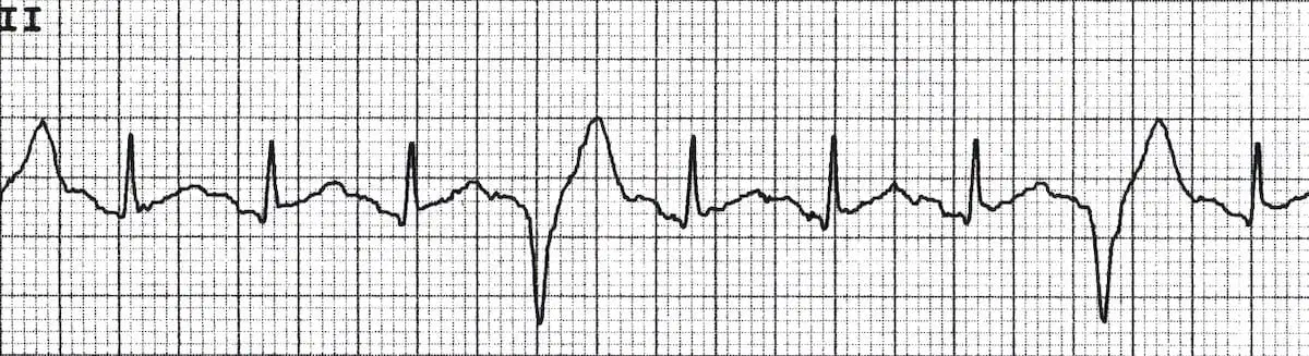
Identify the graph.
Premature atrial complex
Premature ventricular complex Bigeminy
Premature ventricular complex Trigeminy
Premature ventricular complex Quadgeminy

Identify the graph.
Atrial tachycardia
Premature ventricular complex
Premature atrial complex
Sinus Bradycardia
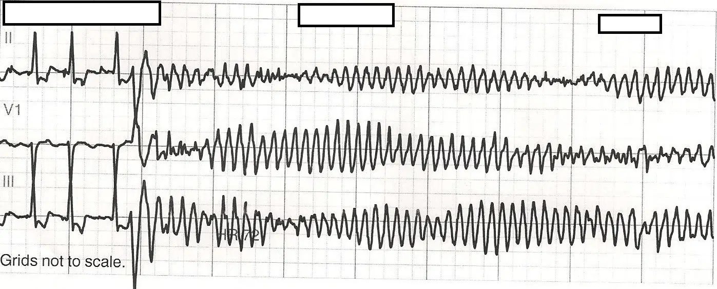
Identify the graph.
Idioventricular Rhythm
Torsades de Pointes
Sinus Rhythm
Ventricular Asystole

Identify the graph.
Atrial fibrillation
Third degree VA block
Classic Wenckebach
Multifocal Atrial Tachycardia
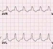
Identify the graph.
Accelerated Idioventricular Rhythm
Ventricular Tachycardia
Sinus Arrest
Atrial Paroxysmal Tachycardia
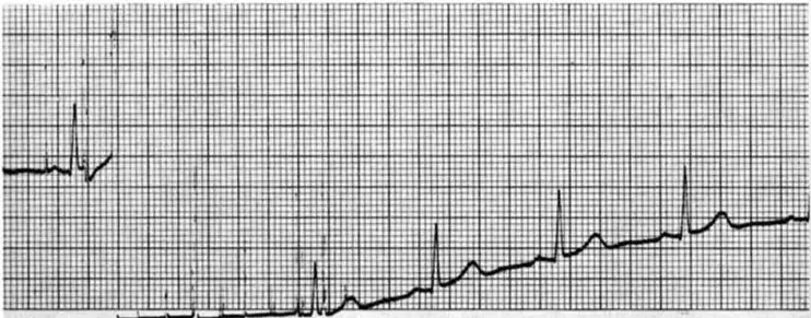
Which Artifact would this be?
Alternating Current (AC)
Muscle Tremor
Wandering Baseline
Interrupted Baseline
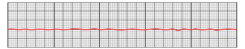
Identify the graph.
Cardiac Arrest
Premature Ventricular Complex
Asystole
Multifunctional Atrial Tachycardia
The AV Node may present:
Temporary Blockages
Regular Delay of the Electrical Current
Permanent Blockages
All of the above
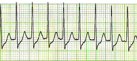
What is an atrial pacing caused by multiple ectopic focuses on top of the ventricles is called?
Supraventricular Tachycardia
Atrial Tachycardia
Atrial Flutter
Ventricular Tachycardia
What is the typical BPM of Sinus Tachycardia?
59 or less BPM
250 - 450 BPM
450 - 600 BPM
101 - 150 BPM
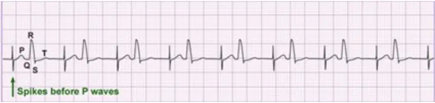
Identify the graph.
Pacemaker Pacing Artifact
Somatic Tremor Artifact
First Degree AV Block
Atrial Tachycardia
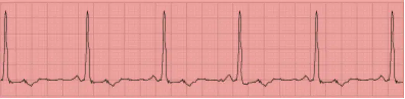
Identify the graph.
Atrial Ischemia
Ventricular Ischemia
Sinus Rhythm
Sinus Tachycardia
What is typical BPM for Accelerated Idioventricular?
50 - 100 BPM
250 - 400 BPM
61 - 100 BPM
40 - 60 BPM
What is a slow chain of PVC's called?
Sinus Rhythm
Idioventricular Rhythm
Atrial Flutter
Third Degree AV Block
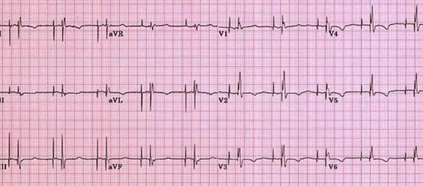
Identify the graph.
Dual Chamber Pacemaker
Bi Ventricular Pacemaker
Implantable Cardioverter Defibrillator (ICD)
Somatic Tremor
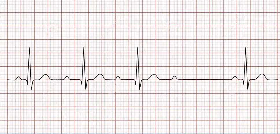
Identify the graph.
Second Degree AV Block Type I Mobitz
Second Degree AV Block Type II Mobitz
Sinus Arrest
Sinus Arrythmia
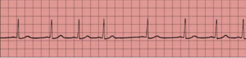
Identify the graph.
Sinus Arrythmia
Sinus Bradycardia
Second Degree AV Block
Sinus Rhythm
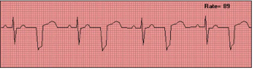
Identify the graph.
Ventricular Tachycardia
Idioventricular Rhythm
Ventricular Bigeminy
Ventricular Trigeminy
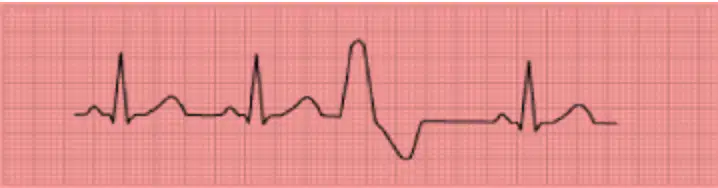
Identify the graph.
Ventricular Quadgeminy
Ventricular Fibrillation
Ventricular Tachycardia
Ventricular Trigeminy
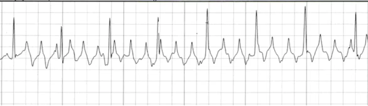
Identify the graph.
Atrial Tachycardia
Ventricular Fibrillation
Multifocal Atrial Tachycardia (MAT)
Atrial Flutter
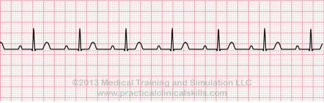
Identify the graph.
Sinus Rhythm
First Degree AV Block
Sinus Arrythmia
Sinus Arrest
What is the typical BPM for Atrial Fibrillation (A. FIB)?
401 - 650 BPM
150 - 300 BPM
60 - 450 BPM
Above 600 BPM
How do you define Ventricular Tachycardia?
Chain of PVC with below 100 BPM
Chain of PAC with above 100 BPM
Chain of PVC with above 100 BPM
Chain of PAC with below 100BPM
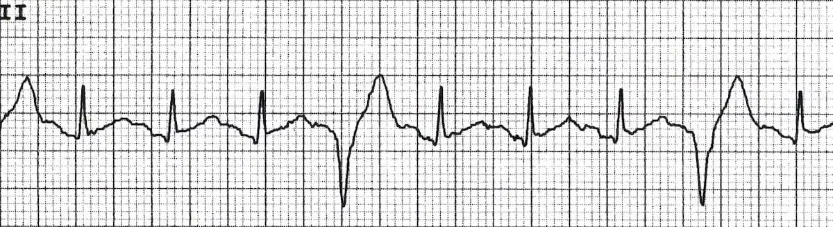
Identify the graph.
Third Degree AV Block
Ventricular Quadgeminy
Ventricular Tachycardia
Ventricular Trigeminy
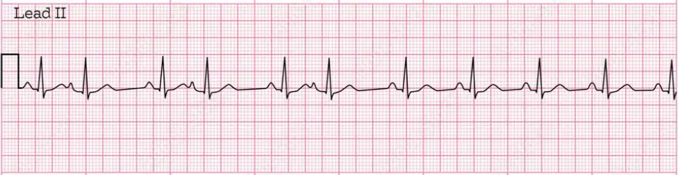
Identify the graph.
Multifocal Atrial Tachycardia
Premature Atrial Contraction
Wandering Atrial Pacemaker
Sinus Arrythmia
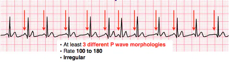
Identify the graph.
Multifocal Atrial Tachycardia
Atrial Tachycardia
Ventricular Tacycardia
Wandering Atrial Pacemaker
A Normal Sinus Rhythm strip would have....
The QRS complex missing
The P Wave inverted
All waves
A missing cardiac cycle
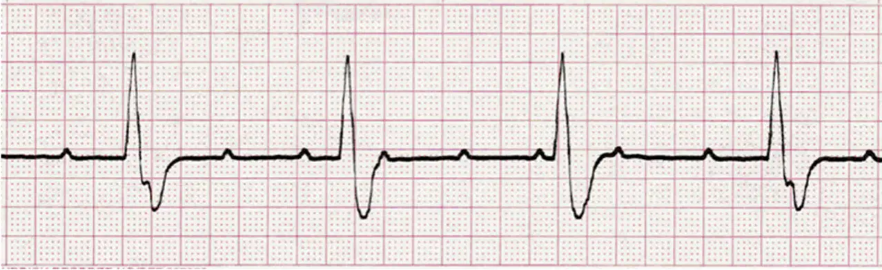
Identify the graph.
Sinus Arrest
Ventricular Tachycardia
Second Degree AV Block
Third Degree AV Block
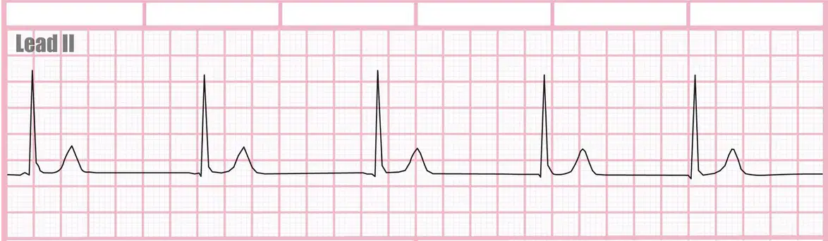
Identify the graph.
Junctional Rhythm
Idioventricular Rhythm
Accelarated Idioventricular Rhythm
V. Tachycardia Monomorphic

