
 Worksheets
A&P 1 2113 - Chapter 6/7/8 Review
Total questions: 79
Worksheet time: 42mins

These type of bones are typically longer than they are wide.
Irregular
Flat
Short
Long
Either end of a long bone.
Diaphysis
Periosteum
Spongy Bone
Epiphyses
Shaft of a long bone.
Epiphysis
Compact Bone
Sternum
Diaphysis
Hyaline cartilage that covers the ends of bones in synovial joints.
Endosteum
Periosteum
Articular cartilage
Synovial cartilage
Dense bone tissue that have a continuous extracellular matrix with no gaps.
Spongy Bone
Endosteum
Compact Bone
Periosteum
This bone has irregular interconnecting spaces between bony plates.
Spongy bone
Compact Bone
Irregular Bone
Wish Bone
Cavity in the diaphysis that contain red or yellow marrow.
Medullary Cavity
Marrow cavity
Nerve cavity
All of the above

What is represented by letter B in the diagram? This structure forms concentric rings around the central canal.
Lacunae
Lamellae
Canaliculi
Osteon
What can be found in the central canal of the haversian canal system?
osteocytes
lamella
blood vessels and nerves
red blood cells
Which part of the osteon contains the osteocytes?
Lacunae
Lamellae
Canaliculi
Central Canal
Tiny, hairlike canals, that transport blood, nutrients and wastes, connecting lacunae to each other and to the Haversian canal
Concentric Lamella
Volkmann's Canal
Lacunae
Canaliculli
Osteon
Osteoblasts are
bone building cells
bone destroying cells
mature bone cells
blood producing cells
Cells that break down bone are called
osteoclasts
osteoblasts
osteocytes
platelets
Type of ossification that uses a cartilage template which is gradually replaced by bone:
Endochondrial ossification
Intramembranous ossification
The bones in the skull are called
intramembraneous bones
endochondral bones
sesamoid bones
appendicular bones
Imagine you are studying the development of the human skull. Which process would you examine to understand how the flat bones of the skull are formed?
During intramembranous ossification, osteoclasts break down bone tissue to release calcium and phosphate.
Intramembranous ossification is the process of bone formation within the bone marrow cavity.
The process of intramembranous ossification involves the differentiation of mesenchymal cells into osteoblasts, which then deposit bone matrix to form trabeculae.
The process of intramembranous ossification involves the differentiation of mesenchymal cells into chondrocytes, which then form cartilage.

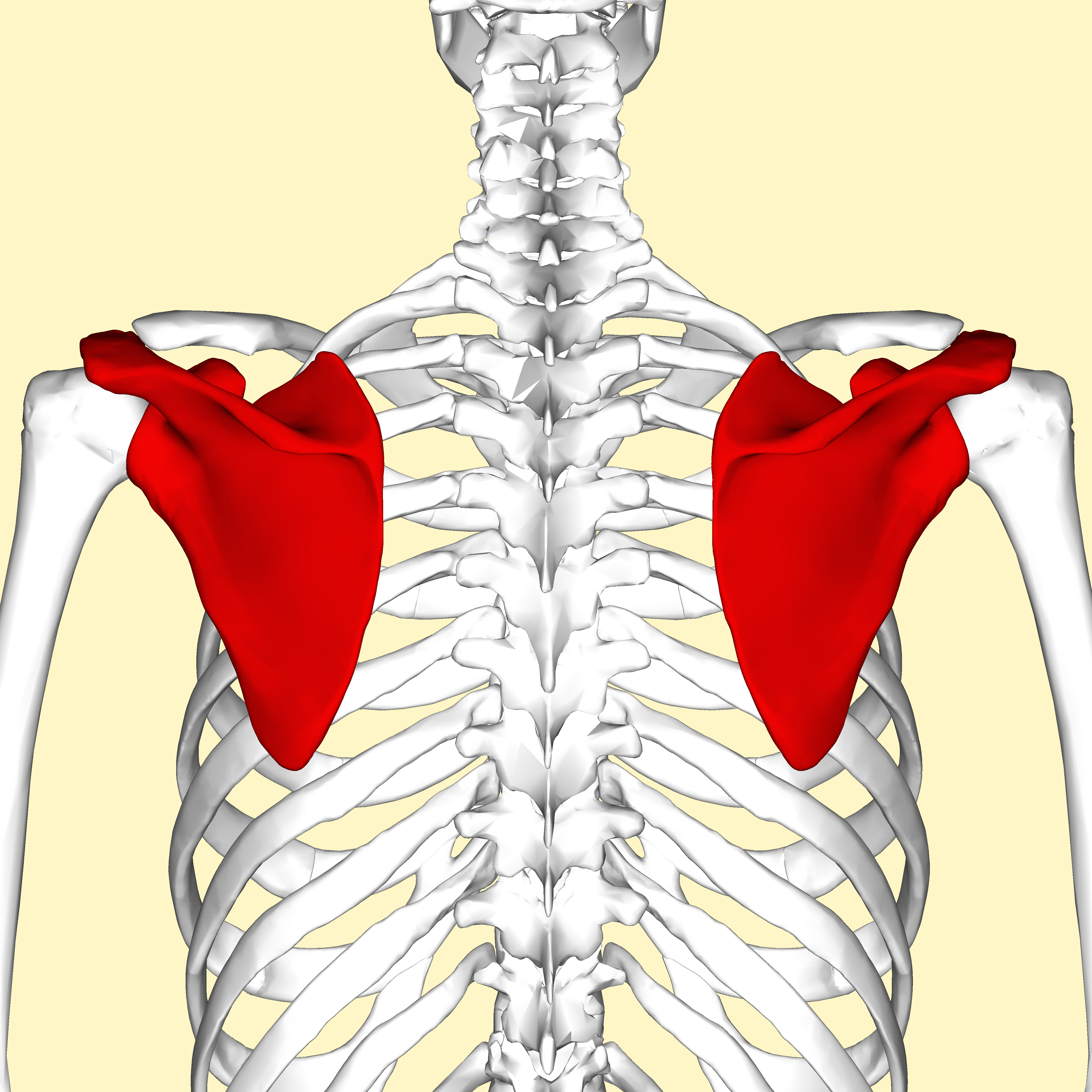
What bone is the arrow pointing to?
rib
spine
scapula
clavicle

What bone is the arrow pointing to (the bone closest to the thumb)?
ulna
humerus
radius
vertebrae



Accurate facts about the bone(s) are
Zygomatic, 2 irregular
Lacrimal, 2, flat
Nasal bone, 1 irregular bone
Nasal bones, 2 flat bones

Zygomatic
Temporal
Parietal
Maxilla

Mandible
Zygomatic
Temporal
Maxilla

Maxilla
lacrimal
Mandible
zygomatic

Two bones that make up the lateral parts of the nasal cavity; lined with membranes that help to filter warm air. It is represented by #2 in the image.
inferior nasal concha
nasal bones
zygomatic bones
sphenoid bone

How is this bone connected to your head?
frontal bone
zygomatic bones
temporomandibular joint
maxilla bones

This bone along with the nasal bones forms the ____________ canal which collects __________.
nasolacrimal, nasal discharge
nasolacrimal, tears
ear, ear wax
nasal, boogers

Zygomatic
Vomer
Ethmoid
Lacrimal
Which two bones make up the bony nasal septum?
Frontal and the vomer
Sphenoid and the nasal bones
Perpendicular plate of the ethmoid and the vomer
Perpendicular plate of the ethmoid and the lacrimal
The palatine process is an aspect of the ____ bone.
Maxillary
Lacrimal
Palatine
Zygomatic
Which bone holds the maxillary sinuses?
Zygoma
Lacrimal
Nasal
Maxilla

Which is the highlighted bone?
frontal
parietal
temporal
occipital

Which bone is highlighted
frontal
parietal
temporal
sphenoid

The highlighted area on the skull is part of the
occipital bone
parietal bone
temporal bone
frontal bone

Which is the highlighted bone
temporal
occipital
frontal
parietal

Which is the highlighted bone
sphenoid
temporal
ethmoid
lacrimal

The highlighted bone is called the
palatine
vomer
ethmoid
maxilla

The circled portion of the image is called the
external auditory meatus
coronoid process
mastoid process
styloid process

Which suture is highlighted on the skull?
coronal suture
squamous suture
sagittal suture
lambdoidal suture

Which suture is highlighted in the image?
squamous suture
coronal suture
sagittal suture
lambdoidal suture

The highlighted suture is called the
lambdoid suture
coronal suture
sagittal suture
squamous suture

The highlighted suture is called the
sagittal suture
coronal suture
squamous suture
lambdoidal suture

Which sinuses are green?
occipital
frontal
temporal
maxillary

Which sinuses are pictured in blue?
frontal
temporal
maxillary
occipital

Which bone represents the sphenoid bone?
blue section
yellow section
purple section
brown section

This bone is called the
hyoid
mastoid
styloid
mandible

The highlighted portion of the skeleton is called the
axial skeleton
appendicular skeleton
rib cage
cranial skeleton

The highlighted portion of the image is called the
appendicular skeleton
axial skeleton
arms and legs
lateral skeleton
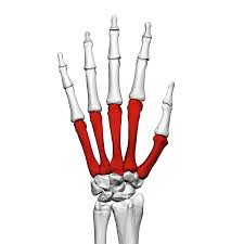
Which bones are highlighted
metacarpals
metatarsals
carpals
tarsals
Using your acronym determine the correct order of the wrist bones (lateral to medial, proximal to distal).
Hamate, trapezium, trapezoid, capitate, lunate, triquetrum, pisiform, scaphoid
Scaphoid, pisiform, hamate, capitate, lunate, triquetrum, trapezium, trapezoid
Scaphoid, lunate triquetrum, pisiform, trapezium, trapezoid, capitate, hamate
None of the above are in correct order
Which of the following is the correct order (proximal to distal, medial to lateral) of the ankle bones.
Cuboid, Talus, Navicular, Calcaneus
Calcaneus, Talus, Navicular, Cuboid
Talus, Cuboid, Navicular, Calcaneus
Cuboid, Navicular, Talus, Calcaneus
The head of the humerus articulates with the
Tarsals
Acetabulum
Glenoid cavity
Trochlear notch
What structure on the femur can be found on the center portion of the shaft?
Popliteal surface
Tibial tuberosity
Deltoid tuberosity
Linea aspera
The head of the radius articulates with the
Carpals
Capitulum
Trochlea
Styloid process

Name the outside portion of the tibia shown in blue.
Medial malleolus
Lateral malleolus
Acetabulum
Talus

Label G (anterior view of the left forearm)
Styloid process of ulna
Styloid process of radius
Radial tuberosity
Olecranon process
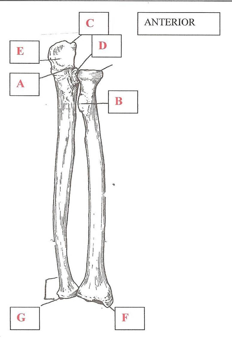
Label C (anterior view of the left forearm)
Semilunar Notch
Coronoid process
Radial tuberosity
Olecranon process

Label D (anterior view of the left arm)
Medial Malleolus
Lateral Malleolus
Medial condyle
Lateral condyle
The clavicle is composed of two ends, one is the sternal end and the other is
Acromial end
Coracoid end
Glenoid end
Medial end
The anatomical name for the big toe is the
Popliteal
Pollex
Hallux
Patella

What bone is labeled 2 and is shaded green?
ischium
pubis
ilium
acetabulum

What is the bone labeled 1?
ischium
pubis
ilium
acetabulum

What feature is labeled 6?
obturator foramen
acetabulum
greater sciatic notch
pubis symphysis
The appendicular skeleton is made up of...
Skull, Thoracic Cage, Vertebral Column
Pectoral Girdle, Thoracic Cage, Upper and Lower Limbs
Skull, Pelvic Girdle, Vertebral Column
Pectoral Girdle, Pelvic Girdle, Upper and Lower Limbs
The two bones that make up the pectoral girdle are...
clavicle and scapula
clavicle and spatula
clavitle and spatula
clavicle and sternum
The medial end of the clavicle articulates with which bone?
scapula
ribs
manubrium
body of the sternum

