
Font size
 Worksheets
microscopy and tissue
Total questions: 76
Worksheet time: 49mins
What is the part of the microscope used to look through?
Eyepiece
Revolving Nosepiece
Body Tube
Objective Lenses
What is the function of the stage clips in a microscope?
Hold the specimen slides in place
Focus images under the high-power objectives
Control the amount of light
Provide light for the specimen
Which part of the microscope provides light for the specimen?
Base
Fine Adjustment Knob
Light Source
Diaphragm
What determines how clearly a small object can be viewed in a microscope?
Stage Clips
Resolution
Light Source
Magnification
What is the function of the diaphragm in a microscope?
Adjust the amount of light passing through the specimen
Hold the specimen slides in place
Focus images under the high-power objectives
Provide light for the specimen
Who is known for the invention of the first practical compound microscope?
Hans Lippershey
Zacharias Jansen
Galileo Galilei
Antonie van Leeuwenhoek
What is the function of the condenser in a microscope?
Adjust the amount of light passing through the specimen
Hold the specimen slides in place
Focus images under the high-power objectives
Provide light for the specimen
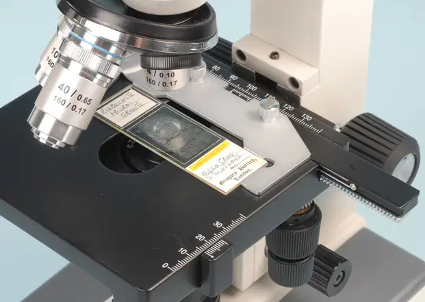
This is the rectangular solid part on which the specimen slide is placed. It is connected solidly to the lower side of the arm, and can move in both x and y directions.
(a)
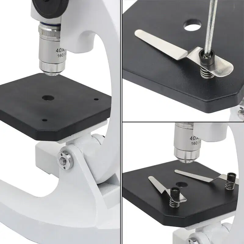
It is use to securely hold the slide in place so that it doesn’t accidentally move.
(a)
Used for precise focusing once coarse focusing has been completed. Use only this knob when on 40x or 100x.
Fine adjustment knob
Head or body tube:
Arm
Ocular (or eyepiece)
Vertical portion of the microscope connecting the base and the head..
Fine adjustment knob
Head or body tube:
Arm
Ocular (or eyepiece)
There are two lenses at the superior end of the head, through which observations are made. An ocular lens has a magnification of 10x. If your microscope has a pointer, it is attached to the right ocular and can be positioned by rotating the ocular lens
Fine adjustment knob
Head or body tube:
Arm
Ocular (or eyepiece)
Which of the following is used for magnification of extremely small specimens, such as bacterial cells.
100x OIO
40x HPO
10x LPO
4x scanner

Part of the microscope indicated by the red dot.
stage
illuminator
objectives
arm

Part of the microscope indicated by the red dot
stage
arm
objectives
illuminator

Part of the microscope indicated by the red dot
stage
arm
condenser
ocular lens

Part of the microscope indicated by the red dot
Stage
Condenser
Illuminator
Nosepiece

Label the part of the microscope. What is part A?
eyepiece lens
eyepiece tube
turret
objective lens

Label the part of the microscope. What is part D?
body
foot
objective lens
turret

Which objective should be used to first find an image?
Low power
Medium power
High power
Which part of the microscope should you use to sharpen the image?
Diaphragm
Coarse adjustment
Stage
Fine adjustment
We begin to look at our specimen using which combination?
high power and fine adjustment knob
high power and coarse adjustment knob
low power and coarse adjustment knob
low power and fine adjustment knob
Which scenario is correct?
Focusing a slide under high power with the coarse adjustment knob.
Focusing a slide under low power with the coarse adjustment knob
Carrying the microscope by the base and body tube
Focusing a slide by moving the diaphragm
Why is it important to choose the right stain for microscopic examination?
Staining has no impact on microscopic examination
It helps in highlighting specific structures within the specimen
Any stain can be used for microscopic examination
Staining is only for aesthetic purposes
What is the function of the diaphragm on a microscope?
To clean the microscope lens
To adjust the lighting
To hold the microscope steady
To control the amount of light reaching the specimen

What is J?
stage
Coarse Adjustment Knob
Fine Adjustment Knob
Base
What is K?
stage
Coarse Adjustment Knob
Fine Adjustment Knob
Base
Which of the following has 40x magnification?
low-power objective lens
eyepiece
high-power objective lens
oil-immersion objective lens


What is this part of the microscope called?
(a)
What is the color code of a 40X objective lens?
(a)
Site for aerobic respiration and the major site of ATP synthesis. It is commonly known as the POWERHOUSE OF THE CELL.
CILIA
FLAGELLA
LYSOSOME
MITOCHONDRIA
These are a GROUP OF CELLS with similar structure and function, along with extracellular substances located between cells.
ORGAN SYSTEM
ORGAN
TISSUES
ORGANISM
The following belong together except which one?
epithelium
bone
connective
nerve
The _____ tissue is considered a type of lining for body areas.
epithelial
connective
nervous
muscle
Tissues present in the brain and the spinal cord are known as
epithelial
muscle
nerve
connective
Tissues that provide movement of body are known as
nerve
epithelial
connective
muscle
Tissue means
Similar cells having common origin
Similar cells having common origin and functions
Cells having same metabolic activities
Similar cells with same metabolic activities
Smooth muscle is found mainly in
heart
stomach
brain
bone

Identify this tissue
simple squamous
simple columnar
stratified columnar

Identify this tissue.
simple cuboidal
adipose
stratified squamous

Identify this tissue.
simple columnar
stratified columnar
adipose

Identify this muscle tissue.
skeletal
cardiac
smooth

Identify this tissue.
blood
smooth muscle
bone

Identify this muscle tissue.
Skeletal
Smooth
Cardiac

Identify this muscle tissue.
smooth
cardiac
skeletal

Identify this tissue.
bone
cartilage
muscle
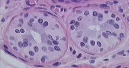
Identify the tissue
(a)
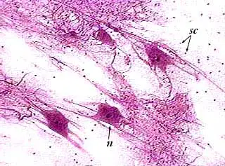
Name the tissue.
(a)
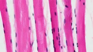
Name the tissue.
(a)
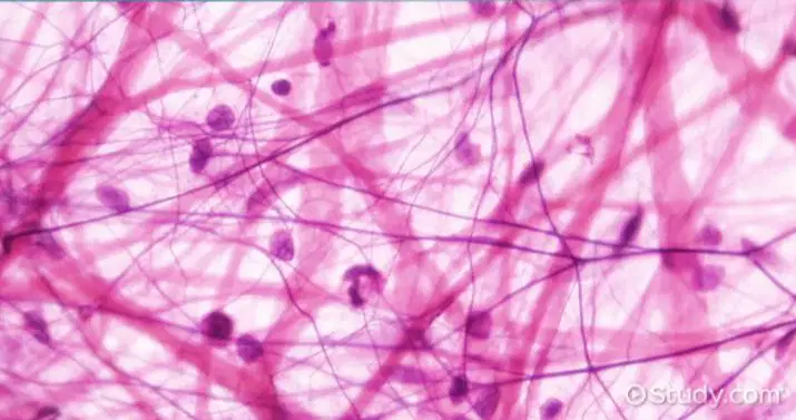
Name the tissue.
(a)
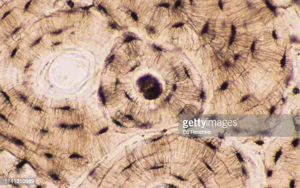
Name the tissue
(a)
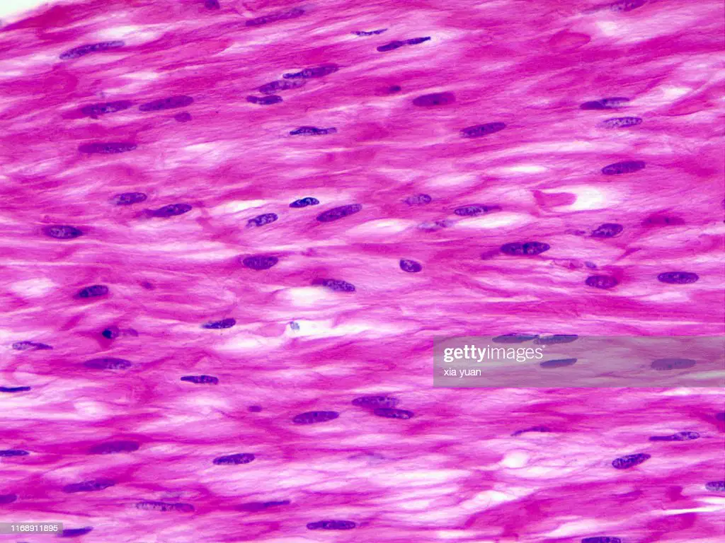
Name the tissue.
(a)
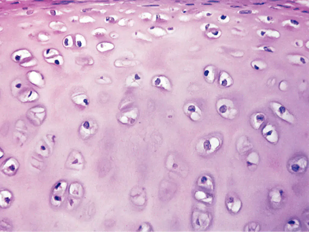
Name the tissue.
(a)
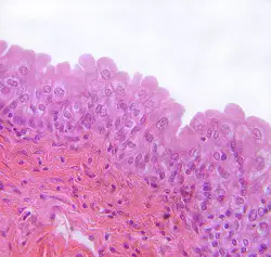
Name the tissue.
(a)
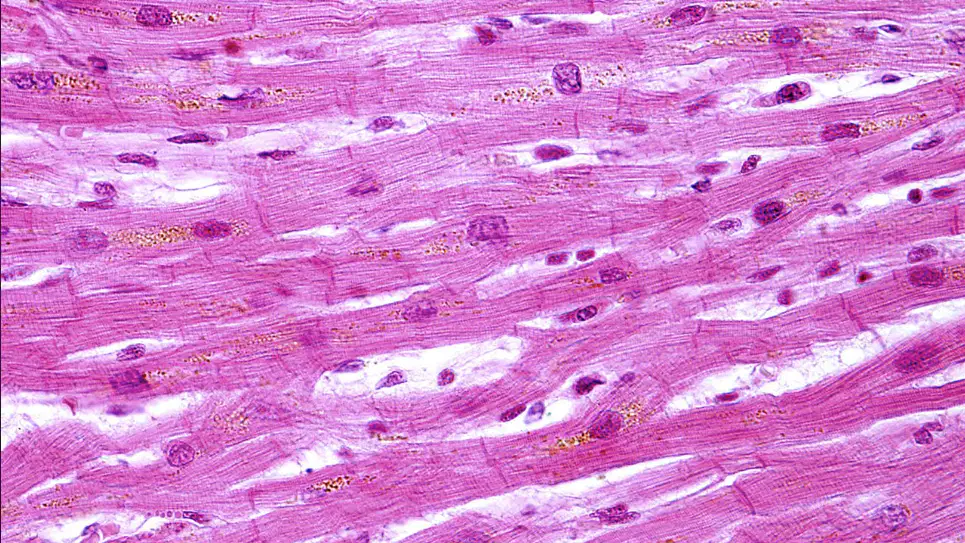
Name the tissue.
(a)
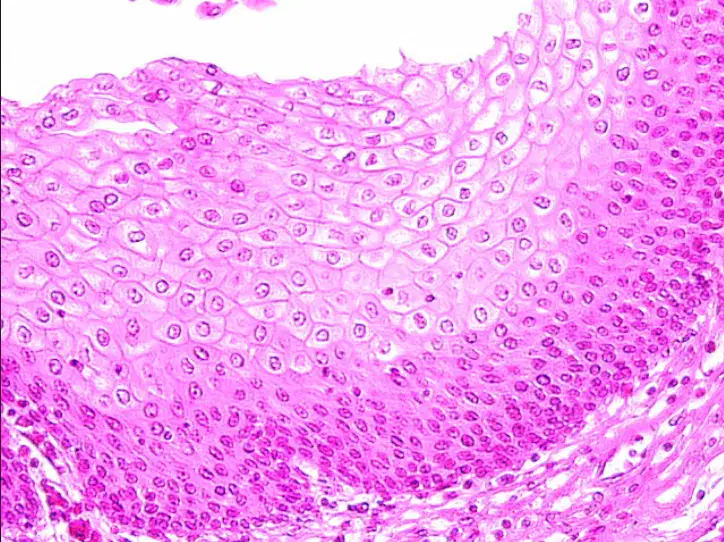
Name the tissue
(a)

Identify the tissue.
(a)

Identify the tissue.
(a)
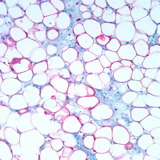
Identify the tissue.
(a)
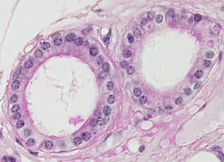
Identify the tissue.
(a)
How can you tell a pseudostratified columnar epithelium from a simple columnar epithelium or stratified columnar epithelium?
Look at the basal surface.
Look at the apical surface.
Check if its ciliated or not.
Look at the nuclei.
You are looking at a stratified epithelium and you notice that the cell shape at the basal surface is different than the cell shape at the apical surface. How do you name this epithelium?
Use the cell shape at the apical surface.
Use the cell shape at the basal surface.
Look in the middle and use that cell shape.
This will never happen.

What type of epithelium?
Non-Ciliated pseudostratified columnar epithelium.
Ciliated pseudostratified columnar epithelium.
Simple squamous epithelium
Stratified squamous epithelium.

Understanding tissue structure and function is important because
there is a relationship between the structure of each tissue type (how it is organized) and its function (what it does)
there is a relationship between tissue structure and organ function.
many diseases and pathologies are tissue and cell type specific.
changes at the tissue level affect the function of organs.
All of the choices are correct.
Which of the following is NOT a true statement about epithelial tissue?
Epithelial tissue is vascular.
Epithelial tissue provides physical protection for organs.
Epithelial tissue functions in absorption and secretion.
Epithelial tissue controls the permeability into and out of an area or organ.
Epithelial tissue consists almost entirely of cells, with very little extracellular material between them, thereby forming a barrier against entry into the body.
Epithelial cells
cover body surfaces or form glands.
usually have a free surface that is not in contact with other cells.
have a basement membrane that attaches to underlying tissues.
can be somewhat far away from any blood vessel.
All of the choices are correct.
The most correct classification of an epithelial tissue consisting of many layers of cells, in which the outer layers are flat and thin and the basal layers are cuboidal or columnar, is
simple columnar epithelium.
pseudostratified epithelium.
stratified squamous epithelium.
transitional epithelium.
simple squamous epithelium.
Epithelial cells that appear cube-like when an organ is relaxed, and flattened when the organ is distended by fluid, are classified as
simple columnar epithelium.
pseudostratified epithelium.
stratified squamous epithelium.
transitional epithelium.
simple squamous epithelium.
Connective tissue
joins together cells and tissues and transports substances.
provides a supporting framework for the body.
is characterized by large amounts of extracellular matrix.
Both "joins together cells and tissues and transports substances" and "is characterized by large amounts of extracellular matrix
All of the choices are correct.

