
 Worksheets
Exam 1
Total questions: 75
Worksheet time: 2hrs 58mins
Selenium is a component of the ____ a free radical scavenging system that limits the damage free radicals cause
Glutathione peroxidase
Selenium peroxidase
Cytosolic phospholipidase
Enzymatic redoxase
How is cell injury different from cell death?
Select all of the key mechanisms of cell injury or death
Decreased intracellular calcium
Permeabilization of cell membranes
Damage to DNA and proteins
Permeabilization of cell membranes
Disruption of biochemical pathways
Decreased intracellular calcium
ATP depletion
The major causes of ATP depletion are…
Hypoglycemia
Free radical damage
Hypoxia
Mitochondrial damage
Necrosis
Which of these are the correct sequence of events leading to cell injury or death due to ATP depletion?
Switch to anerobic glycolysis→Decrease O2→ Results in decreased ATP production→ Decreased function of Na+/K+ pumps → Increased influx of Ca2+, H20 and Na+ → Cellular swelling
Decreased function of Na+/K+ pumps→ Increased influx of Ca2+, H20 and Na+ → Cellular swelling→ Decrease O2→ switch to anerobic glycolysis→ Results in decreased ATP production
Cellular swelling→Increased influx of Ca2+, H20 and Na+ → Decrease O2→ Decreased function of Na+/K+ pumps→Switch to anerobic glycolysis→ Results in decreased ATP production
Decrease O2→ Switch to anerobic glycolysis→ Results in decreased ATP production→ Decreased function of Na+/K+ pumps→ Increased influx of Ca2+, H20 and Na+ → Cellular swelling
Which of these does mitochondrial damage NOT lead to?
Formation of mitochondrial permeability transition pore
Abnormal oxidative phosphorylation leading to the formation of ROS
Phospholipase activation
Leakage of pro-apoptotic proteins from the mitochondiral membrane leading ot cellular death by apoptosis
Increased Cytosolic Ca2+ activates what?
Select all the ways there can be an increase in cytosolic Ca2+ as a response to ischemia and toxins
Release from intracellular stores in mitochondrial and ER membranes
Anerobic glycolysis causing the release of Ca2+ intracellularly
Formation of the mitochondrial permeability transition pore
Increased influx of Ca2+ across the plasma membrane late in cell injury
What are free radicals?
Which of these are not sources of free radicals?
Redox reactions that occur during ATP production
Bursts produced in activated leukocytes during inflammation
Nitric Oxide
Glutathione peroxidase
DNA damage, Protein modification and lipid peroxidation all occur as a result of what?
(a)
What is the most common cause of cell swelling?
Decreased oxidation and use of FFAs
Hypoxia
Excess deposition of glycogen
What is the pathogenesis of cell swelling?
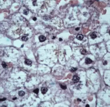
What changes can you observe in this histologic slide? What changes do you expect to see in the gross organ?
Select the main categories of the morphological manifestations of injury
Degeneration
Intracellular accumulation
Necrosis
Extracellular Alterations
Hyaline changes
Is cell swelling a reversible or irreversible morphological alteration during cell injury?
Reversible
Irreversible
What are some other names for acute cell swelling?
Hydropic degeneration
Cellular hypertrophy
Ballooning degeneration
What is a possible sequelae if there was hepatocellular hydropic degeneration occuring?
Obstruction of a bile duct
Hepatocellular necrosis
Increased bile flow

Given these gross and histological images what type of cellular injury is occurring? Why is this occurring?
What are three conditions that there is a pathologic alteration in glucose metabolism leading to glycogen accumulation?
Diabetes Insipidus
Diabetes Mellitus
Steroid hepatopathy
Glycogen storage disease
You have two slides. You stain one slide with PAS stain and it turns pink. Is it glycogen?
Describe the pathogenesis of glycogen accumulation starting with diabetes mellitus or steroid hepatopathy.
Is glycogen accumulation able to be seen grossly?
Yes
No
Which of the following are intracellular hyaline changes?
Hyaline droplets
Amyloid
Fibrinoid change
Deposition of collagen
What is hyaline?
Is glycogen accumulation reversible?
Yes
No
In what types of cells are hyaline droplets more prominent?
renal tubular epithelium
Liver
Brain
Enterocytes
epithelium
What is the pathogenesis of hyaline droplets?
What inclusions may look similar to hyaline droplets?
Viral inclusions
Acidophilic crystalline intranuclear inclusions
glycogen accumulation
Lead inclusions
Bacterial endotoxins
What stain causes amyloid to glow apple green under polarized light? What stain turns the amyloid deposits blue?
Hematoxylin and eosin (H&E) staining/Lugols iodine
Lugols iodine/PAS
Congo red/PAS
Lugols iodine/Wrights stain
Condo red/Lugols iodine
What is the pathogenesis of amyloid deposition through reactive systemic amyloidosis?
What is the difference between systemic familial amyloidosis and reactive systemic amyloidosis?
If there was an inciting cause vs genetic predisposition
If there was inflammation vs individual mutation
What are the two more common location for localized amyloidosis?
What sequelae can occur due to amyloidosis?
Pressure atrophy of cells
Proteinuria leading to kidney failure
Diabetes mellitus
Hepatic failure
Clotting disorders
What is fibrinoid change?
Deposition of fibrin in any tissue
Fibrous changes in a tissue
Necrosis of a blood vessel
What is the pathogenesis of fibrinoid change?
What are some components of fibrinoid change?
fibrin
immunoglobulins
epithelium
Platelets
complement
What are causes of extracellular hyaline deposition? What would it look like grossly (just think about this one, no answer in the choices)
Aging change
Acute injury
Ischemia
Chronic injury
What is fatty infiltration
Accumulation of fat in extracellular stromal tissue
Accumulation of fat in tissues that dont normally have adipocytes
Intracellular accumulation of fat droplets
hypertrophy of adipocytes
What is the difference between dystrophic and metastatic mineralization?
What are some examples of diseases that lead to metastatic calcification
Humoral hypercalcemia of malignancy
Secondary renal hyperparathyroidism
Chronic granulomatous disease
Vitamin D toxicosis
Necrosis of myocardium
What is coagulative necrosis?
Which of the following pathogenesis could occur and result in coagulative necrosis?
Fumonisin B1 ingested-->inhibits sphingosine N-acetyltransferase-->inhibits production of sphingolipids which are important in protecting the cell surface
Downer cow-->weight of body occludes vessels -->ischemia-->necrosis of thigh muscle
Diets high in fat-->Increase in ROS-->Free radical damage can lead to fat necrosis
Anemia-->hypoxia--> central lobular hepatic necrosis
What microscopic changes will you see with coagulative necrosis?
Nuclear pyknosis
inflammation at the center of the lesion
Increased basophilia
Karyorrhexis or karrylolysis
What is a cause of coagulative necrosis?
What is an infarct?
What is liquefactive necrosis?
What does liquefactive necrosis look like?
Abscesses
dry crumbly and gritty lesion
ischemic necrosis in the CNS
What is the cause of leukoencephalomalacia? What species is affected? What is the pathogenesis?
What is the suspected cause of polioencephalomalacia? What species is affected? What is the pathogenesis?
What lesions would you not see grossly if there is polioencephalomalacia?
Swelling of the brains gyri
necrosis of the white matter
cerebral laminar necrosis
Florescence under UV light
What are some causes of gas gangrene?
Aspiration pneumonia
ischemia
Viruses
Bacterial toxins
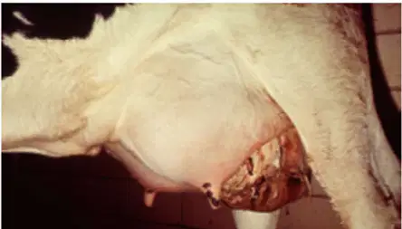
Is this wet or dry gangrene?
Dark red to black tissue
Soft moist and friable tissue
Foul odor with a line of demarcation where the necrosis is occurring
Wet gangrene
Dry gangrene
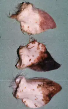
Is this wet or dry gangrene?
Dry shriveled leathery tissue
Cold tissue
Not smell or gas present
Wet gangrene
Dry gangrene
Whats the difference between wet and dry gangrene?
What is the pathogenesis of ergot poisoning resulting in wet gangrene?
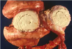
What are some causes of caseous necrosis?
Corynebacterium pseudotuberculosis
Salmonella
Mycobacterium bovis
Ecoli
What is fat necrosis and what are some causes?
What is the term for death of a cell at the end of its normal lifespan as seen in embryonic cells
that are no longer needed in development
(a)
What is the term for the death of a cell before the end of its normal life span, result of irreversible cell injury
(a)
What is the morphologic changes that follow cell death in a living animal, resulting from progressive, degradative action of enzymes on the cell
(a)
What is cell death directed by cellular signaling cascades and typically affects individual cells. Could be physiologic or pathologic
(a)
What is the term for the degradative changes in a cell due to action of endogenous enzymes, primarily from lysosomes. See in postmortem decomposition
(a)
What is it called when post mortem bacterial metablism and dissolution of host tissues result in the production of color and texture changes, gas production and odors?
(a)
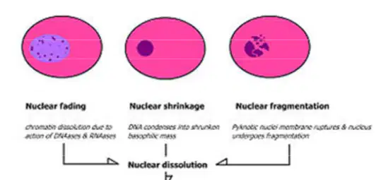
From left to right what nuclear patterns are being shown? What process are these cells going through?
List three potential sequelae to necrosis
What are some characteristic of apoptotic cells?
Cell swelling
cell shrinkage
Cytoplasmic blebs
Phagocytosis of apoptotic bodies
Inflammation
What are the two mechanisms of apoptosis?
Describe the mitochondrial pathway of apoptosis.
Describe the death receptor pathway of apoptosis
What post mortem change is present?
(a)
The cooling of the body after death
(a)
The stiffening of muscles after death
(a)
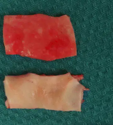
What postmortem change occured in the top section of a vessel compared to the bottom section
(a)
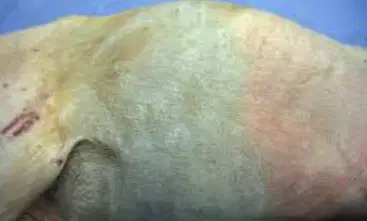
What postmortem change is occurring here
(a)

