
 Worksheets
Bio-107-Lab First Lab Practical
Total questions: 99
Worksheet time: 52mins
how would you describe the location of a bruise on the front right lower leg?
anterior, proximal to knee
posterior, distal to hip
anterior, distal to knee
posterior, proximal to hip
What directional term would be used to indicate sunburn on a patient’s back?
proximal
posterior
anterior
distal
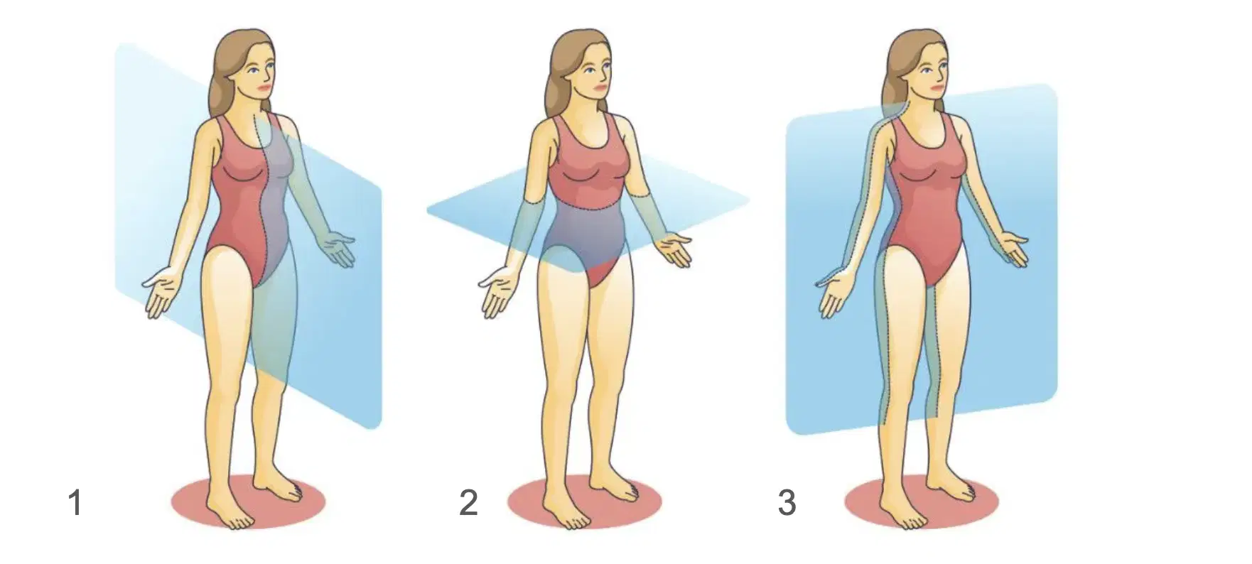
Label the planes of the body. In order of 1-3 Ex. The, Cat, Hat
Word Bank: Frontal, Median, Transverse
(a)
Which directional term is used to describe the relationship of the skin to the skeletal muscles?
superficial
lateral
deep
medial
The knee is proximal to the thigh.
True
False
Which of the following is the proper description of the human body in the anatomical position?
The body is erect with feet slightly apart, palms facing forward, and thumbs pointing away from the body.
The body is erect with feet together, palms facing the thighs, and thumbs pointing forward.
The body is erect with feet slightly apart and arms extending perpendicular to the torso along a straight line with the clavicle.
The body is erect with feet together, palms facing backward, and thumbs pointing toward the body.
Which of the following statements is correct?
The heart is posterior to the spine.
The heart is dorsal to the sternum.
The sternum is posterior to the spine.
The sternum is dorsal to the spine.
Which of the following statements regarding the umbilicus and the chin is true?
The umbilicus is caudal to the chin.
The umbilicus is superior to the chin.
The chin is caudal to the umbilicus.
The umbilicus is medial to the chin.
The anatomical position is characterized by all of the following EXCEPT ________.
body erect
thumbs pointed laterally
palms turned posteriorly
arms at sides
Which of the following describes a parasagittal plane?
a transverse cut just above the knees
two cuts dividing the body into left and right halves
any cut dividing the body into anterior and posterior portions
any sagittal plane except in the midline
What is a vertical section through the body, dividing it into anterior and posterior regions called?
transverse
frontal
median
sagittal
The elbow is proximal to the shoulder.
True
False
Which of the following is NOT correct for anatomical position?
Nose facing forward
Legs are straight with toes aimed forward
Arms fully extended
Palms facing inward towards the thighs
Which of the following is located in a median position?
Right foot
lung
mouth
shoulder
The knee is proximal to the ankle. Which of these other terms could also be used to correctly describe the relationship between the knee and the ankle?
The knee is superficial to the ankle.
The knee is medial to the ankle.
The knee is contralateral to the ankle.
The knee is superior to the ankle.
The cranial bones almost entirely surround the brain and protect it. Which of the following terms best describes the relationship between the cranial bones and the brain?
Cranial bones are anterior to the brain.
The cranial bones are superficial to the brain.
The cranial bones are proximal to the brain.
The cranial bones are lateral to the brain.
Which of the following is a term that can be used instead of dorsal?
lateral
posterior
ventral
distal
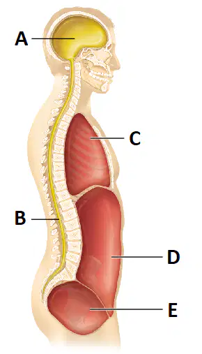
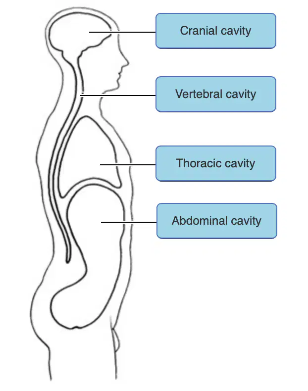
Which of the following is a correct pairing of a body cavity with its contents?
The cavity at C contains the lungs.
The cavity at E contains most of the digestive organs.
The cavity at D contains the reproductive organs.
The cavity at D contains the urinary bladder.
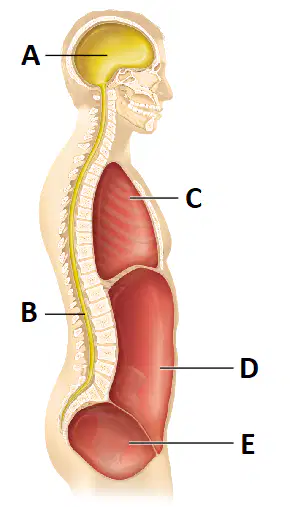
The ventral cavity includes which of the indicated subdivisions in the figure?
A and B
C and D
D and E
C, D, and E
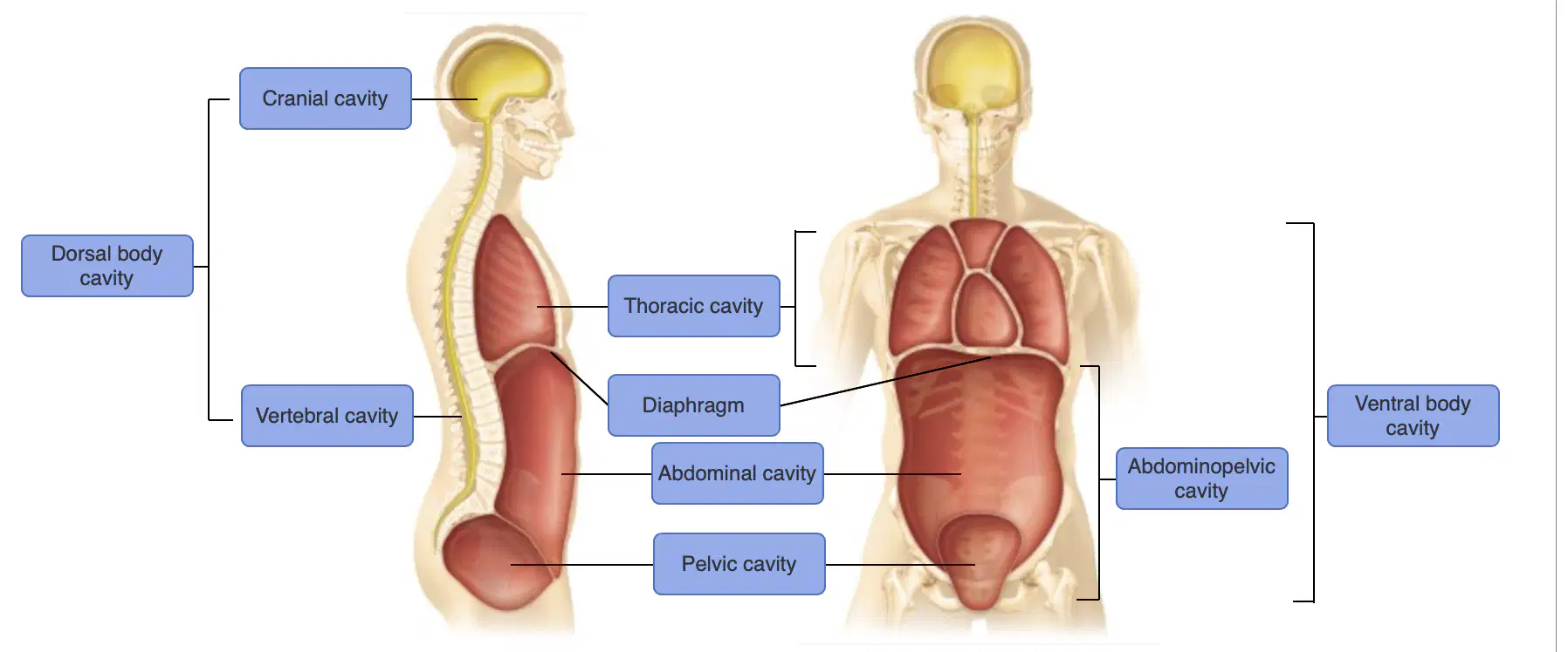
try to memorize
I sorta have it maybe ill get a 75?
I got this definitely gonna get at least an 85 :D
im going to fail :(
60 and below is what ill be getting :(
Easy 95-100 >:)
The dorsal body cavity is divided into which of the following subdivisions?
the vertebral/spinal and cranial cavities
the vertebral/spinal, cranial, and pleural cavities
the thoracic, pleural, and abdominopelvic cavities
the vertebral/spinal and thoracic cavities
Memories, please
This is the right answer
wrong
wrong
wrong
In which quadrant of the abdominopelvic cavity is the stomach located?
left lower quadrant
left upper quadrant
right lower quadrant
right upper quadrant
The thoracic cavity contains the ________. It is found ________ to the vertebral cavity.
digestive viscera; inferior
kidneys and spleen; deep
heart and lungs; anterior
stomach and liver; superficial
Which of the following epithelial tissue types is NOT correctly matched to its function?
stratified squamous epithelium; absorption
simple columnar epithelium with microvilli; absorption
simple cuboidal epithelium; secretion
simple squamous epithelium; filtration
Pseudostratified epithelia are well adapted to the digestive system because the cilia increase the surface area for absorption.
True
False
What function do transitional epithelia have?
stretching
absorption
filtration
resistance to friction
You observe a tissue under a microscope. There appears to be a lumen on one side of the tissue. Lining this lumen is a single layer of epithelial cells that seem to have long, fingerlike projections. Beneath the projections, the cells seem to have the shape of shoeboxes and are packed tightly together. What type of tissue are you looking at?
simple columnar epithelium
transitional epithelium
simple cuboidal epithelium
stratified squamous epithelium
Which of the following is a modification of the simple columnar epithelium that allows for efficient absorption along portions of the digestive tract?
thin and permeable
fibroblasts
cilia
dense microvilli
A ciliated version of pseudostratified columnar epithelium containing mucus-secreting goblet cells ________.
is more durable than all other epithelia
is found in some of the larger glands
lines most of the respiratory tract
aids in digestion
Which of the following epithelia forms the walls of the air sacs across which gas exchange occurs in the lungs?
pseudostratified columnar
simple columnar
simple cuboidal
simple squamous
________ epithelium appears to have two or three layers of cells, but all the cells are in contact with the basement membrane.
Stratified columnar
Transitional
Stratified cuboidal
Pseudostratified columnar
A many-layered epithelium with cuboidal basal cells and flat cells at its surface would be classified as ________.
stratified squamous
transitional
simple cuboidal
simple squamous
Select the correct statement regarding epithelia.
Stratified epithelia are tall, narrow cells.
Simple epithelia form impermeable barriers.
Stratified epithelia are present where protection from abrasion is important.
Pseudostratified epithelia consist of at least two layers of cells stacked on top of one another.
Simple cuboidal epithelia are usually found in areas where secretion and absorption occur.
True
False
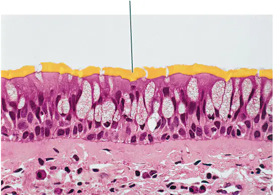
Identify the highlighted structures.
(a)
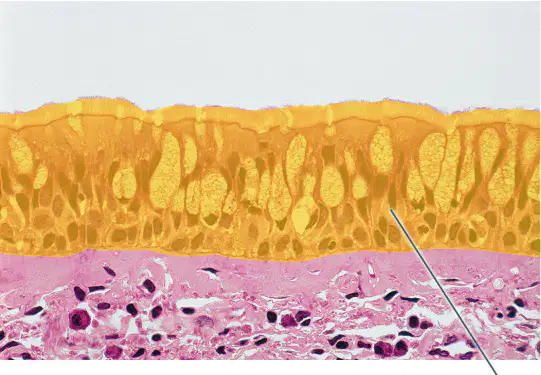
Identify the highlighted type of epithelial tissue.
(a)
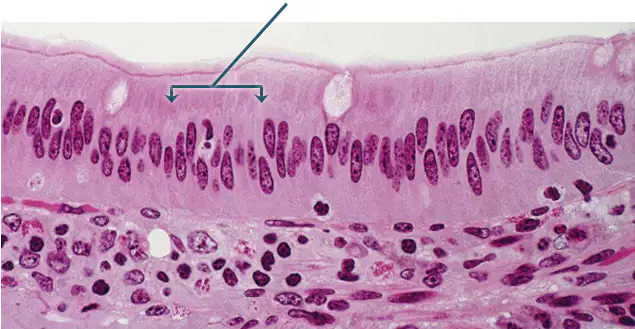
Identify the epithelium indicated by the arrows.
(a)
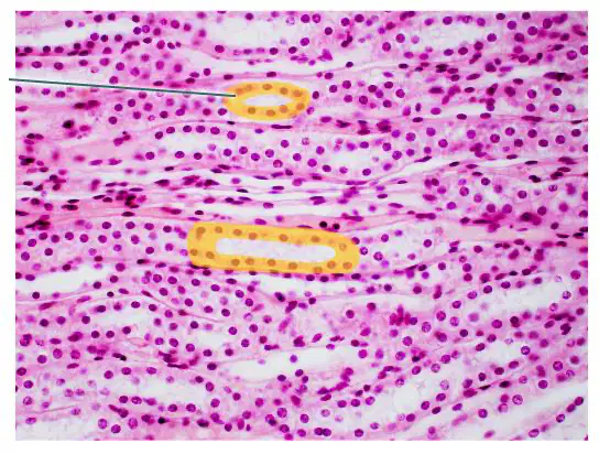
Identify the highlighted epithelium.
(a)
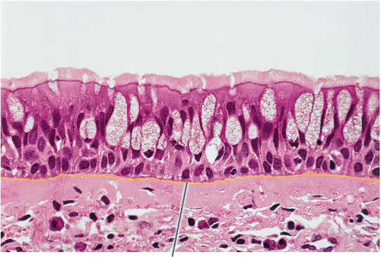
Which structure is highlighted?
basement membrane
goblet cell
nucleus
pseudostratified columnar epithelium
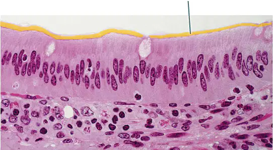
Which structures are highlighted?
goblet cells
nuclei
cilia
microvilli

Which structures are highlighted?
simple columnar cells
microvilli
nuclei
goblet cells
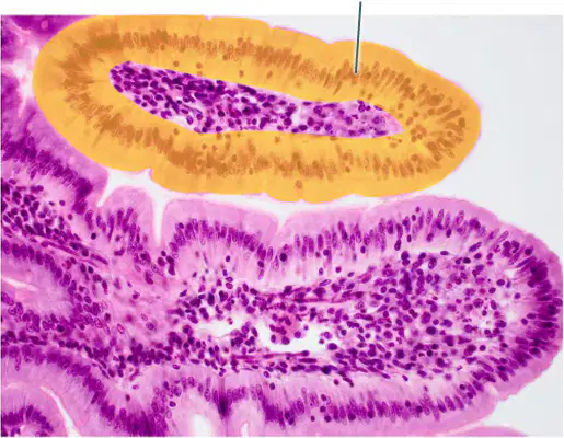
Which epithelial type is highlighted?
simple squamous epithelium
simple cuboidal epithelium
pseudostratified columnar epithelium
simple columnar epithelium
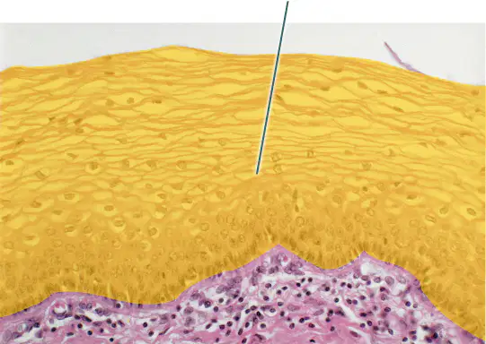
Which epithelial type is highlighted?
transitional epithelium
simple cuboidal epithelium
stratified squamous epithelium
pseudostratified columnar epithelium
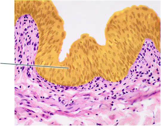
Which epithelial type is highlighted?
stratified cuboidal epithelium
stratified columnar epithelium
transitional epithelium
stratified squamous epithelium
Squamous epithelial cells ________.
are taller than they are wide, like columns
have no nuclei
are thin and flat, like fish scales
are as tall as they are wide, like cubes
Simple epithelia have ________.
all three types of epithelial cells arranged in one layer
multiple layers of just one type of epithelial cells
two types of epithelial cells, arranged in one layer
one type of epithelial cell, arranged in a single layer
Which of the following best describes pseudostratified epithelium?
Multiple layers of cells with various heights
Multiple layers of ciliated cells
Single layer of columnar cells
Single layer of cells with various heights
Multiple layers of cuboidal and columnar cells
What body system contains pseudostratified columnar epithelium?
Cardiovascular system
Endocrine system
Respiratory system
Digestive system
Urinary system
A single layer of tall rectangular cells best describes which of the following?
Simple squamous epithelium
Simple columnar epithelium
Simple cuboidal epithelium
Goblet cells
Stratified columnar epithelium
Identify the one characteristic that does NOT describe a goblet cell.
Secretes mucus onto the luminal surface
Columnar epithelial cell
Unicellular gland
Produces mucus
Component of a mucous gland
What is the function of cilia?
Movement of something across the cell’s surface
Circulation of cytoplasm within the cell
Increasing of surface area
Movement of the cell
Absorption
Identify the best description for the shape of squamous cells.
Spherical
Plate-like
Cube-like
Pyramidal
Cylindrical
Having multiple layers of flat cells against a free surface describes which of the following?
Stratified cuboidal epithelium
Pseudostratified squamous epithelium
Simple cuboidal epithelium
Simple squamous epithelium
Stratified squamous epithelium
Where is the basement membrane located?
Between neighboring epithelial cells
At the boundary between connective tissue and epithelium
At the basal surface of each epithelial cell
Across the apical surfaces of the epithelial cells
At the boundary between the lumen and connective tissue
What is the function of stratified squamous epithelium?
Protection
Diffusion
Secretion
Absorption
Filtration
Which description is best for transitional epithelium?
Cells that are more squamous in a relaxed state but more rounded when stretched.
Cells that can randomly exist in two different states.
Tissue that contains elastic fibers, permitting the tissue to stretch when necessary.
Cells that are more cuboidal in a relaxed state but more squamous when stretched.
Cells that transition from round to flat based on the age of the cell.
Identify the function of transitional epithelium.
Filtration
Absorption
Movement
Flexibility
Diffusion
What is the only body system that contains transitional epithelium?
Cardiovascular system
Respiratory system
Digestive system
Urinary system
Endocrine system
Epithelial tissue is typically localized to the following region(s) of the body.
only on the outermost surface of an organ
on the surfaces of organs, on the walls of cavities, on the outer surface of the body, and in glands of the body
only on the inner surfaces of body cavities
between other layers of tissue in many of the organs of the body
Based on its function, where would you likely find simple squamous epithelium?
in places that experience abrasion or friction
on the surface of the skin
along the digestive tract
in places where materials need to pass through a thin sheet of cells
What is the function of ciliated epithelium?
to move the epithelial cells from one area of the body to another
to absorb materials from the extracellular environment
to protect the underlying tissues
to move fluids across the tissue surface
Which of the following is an example of pseudostratified epithelium?
lining of the trachea
endocrine glands
kidney tubules
lung alveoli
Why are adipose tissue, blood, and bone all considered to be connective tissues?
They all have collagen and elastic fibers in their extracellular matrix.
They connect to each other.
They have a common origin: the embryonic tissue mesenchyme.
They have the same types of cells.
Connective tissues are made of different combinations of ground substance and fibers. As you compare slides of connective tissues, which would most likely be the strongest (toughest) type of connective tissue based on the composition of ground substance and fibers?
connective tissue high in reticular fibers
connective tissues high in collagen fibers
connective tissue high in elastic fibers
connective tissue high in ground substance
Which is the most atypical connective tissue since it neither connects structures nor provides structural support?
cartilage
osseous
areolar connective tissue
blood
Which tissue is correctly paired with its primary immature cell type?
bone; osteoblast
connective tissue proper; chondrocyte
cartilage; fibroblast
bone; fibrocyte
Which of the following statements is true?
Dense irregular connective tissue has a high proportion of elastic fibers.
Dense connective tissue is dominated by collagen fibers.
Loose connective tissue includes cartilage.
Closely packed bundles of collagen fibers are characteristic of loose connective tissues.
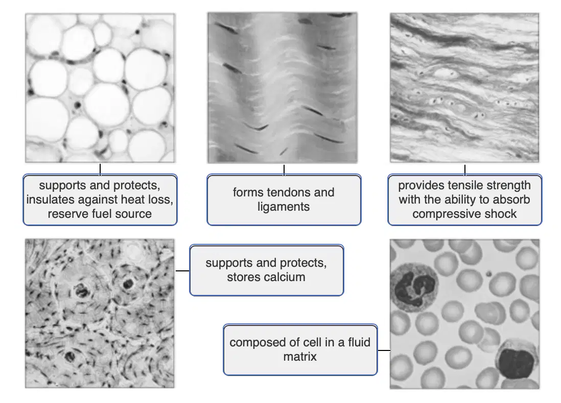
Memorize please
Pick This
dont pick this
The reason that intervertebral discs exhibit a large amount of tensile strength, which allows them to absorb shock, is because they possess ________.
hydroxyapatite crystals
reticular fibers
collagen fibers
elastic fibers
What tissue has lacunae, calcium salts, and blood vessels?
cartilage tissue
fibrocartilaginous tissue
areolar tissue
bone (osseous tissue)
The primary blast cell for bone is the ________.
osteoblast
fibroblast
hemocytoblast
chondroblast
How is hyaline cartilage different from elastic cartilage or fibrocartilage?
It contains more nuclei.
It is more vascularized.
Fibers are not normally visible.
It allows great flexibility.
Which of the following is found in hyaline cartilage?
fibroblast
mast cell
chondrocyte
macrophage
The shape of the external ear is maintained by ________.
fibrocartilage
hyaline cartilage
elastic cartilage
adipose tissue
Select the correct statement regarding adipose tissue.
Most of the cell volume is occupied by the nucleus.
It is composed mostly of extracellular matrix.
Its primary function is nutrient storage.
Mature adipose cells are highly mitotic.
Tendons and ligaments are composed mainly of dense irregular connective tissue.
True
False
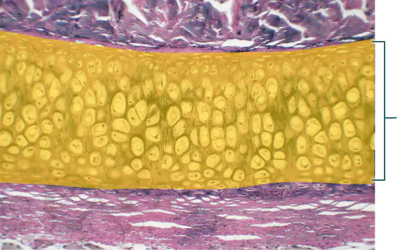
Which tissue is highlighted?
hyaline cartilage
dense regular elastic tissue
elastic cartilage
areolar connective tissue
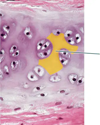
Which component of the connective tissue in this field of view is highlighted?
collagen fibers
lacunae
chondrocytes
extracellular matrix
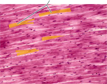
What structure connects the highlighted muscle cells to one another?
sarcomeres
Z discs
intercalated discs
tight junctions
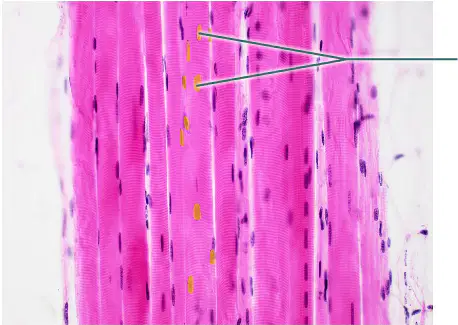
Which structures are highlighted?
fibers
nuclei
striations
fascicles
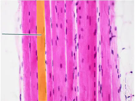
Which structure is highlighted?
fascicle
sarcomere
skeletal muscle fiber
perimysium

What is a characteristic of the highlighted muscle tissue?
striated
branching
voluntary
uni-nucleated
Under a microscope you observe a tissue that appears to have long fibers that appear striated. The nuclei are pushed off to the side of the fibers. The tissue looks very vascular. What type of tissue are you observing?
skeletal muscle tissue
dense irregular connective tissue
dense regular connective tissue
osseous tissue
Smooth muscle __________.
contains intercalated discs
has striations
has long, cylindrical cells
is under involuntary control
Which tissue in the wall of the uterus is required for labor contractions?
stratified squamous
skeletal muscle
smooth muscle
elastic cartilage
Intercalated discs and striations are both characteristics of skeletal muscle.
True
False
What visual feature sets hyaline cartilage apart from other types of cartilage?
Cells exist in spaces called lacunae.
Very few cells can be observed.
Matrix appears fibrous.
Matrix appears transparent.
Matrix does not contain fibers.
Where are chondrocytes located?
Chondrocytes aren’t found in cartilage.
Outside lacunae
Between collagen fibers
Inside lacunae
Outer periphery of the tissue
What does the term “regular” in “dense regular connective tissue” refer to?
Fibers are all the same type.
Fibers are evenly distributed throughout the tissue.
Fibers run in an interwoven web.
Fibers run parallel.
Fibers run perpendicular.
What are the fiber types found in the two forms of dense regular connective tissue?
Elastic and reticular
Reticular and collagen
Collagen and elastic
What does the term “dense” in “dense irregular connective tissue” refer to?
Density of the ground substance
Density of fibers
Density of cells
Density of fibroblast nuclei
What is the dominant fiber type found in dense irregular connective tissue?
Elastic
Collagen
Reticular
What type of connective tissue is areolar connective tissue?
Dense regular connective tissue
Dense connective tissue
Dense irregular connective tissue
Loose connective tissue
Cartilage
What fiber type permits areolar connective tissue to retain a degree of flexibility?
Reticular
Collagen
Elastic
Which description best identifies the unique attributes of connective tissue?
Connective tissues transmit signals from one part of the body to another.
Connective tissues are hard and inflexible, whereas soft, flexible structures of the body are made up of other tissue types.
Connective tissues, unlike other tissues, lack blood vessels and blood.
Connective tissue contains a large amount of an extracellular matrix.
A histologist examines a tissue slide and observes many fibers tightly packed together in a parallel arrangement. A few cells can be seen squeezed between the fibers. Which type of tissue is being studied?
dense irregular connective tissue
areolar connective tissue
adipose connective tissue
dense regular fibrous connective tissue
Why is blood classified as a connective tissue?
Since all connective tissues are highly vascularized, blood is included as a connective tissue.
Blood helps to connect body parts together.
Blood cells, and the cells of all other connective tissues, come from the same kind of embryonic stem cell.
Blood tissue contains a matrix that is very similar to that of other types of connective tissues.
What is the ground substance in typical connective tissue matrix?
interstitial fluid, proteoglycans, and cell adhesion proteins
dissolved bone salts
collagen, elastic, and reticular fibers
fibroblasts, chondroblasts, osteoblasts, and mesenchyme cells

