
 Worksheets
AP 2113 Lab Final Review
Total questions: 94
Worksheet time: 1hrs 16mins
Hinge joints allow What type of movement?
Flexion & Extension
Abduction & Adduction
Rotation & Extension
Flexion & Rotation

Name this joint
gliding
saddle
ball and socket
condyloid


When kicking a football, _____________________ joint is one of the main movements required.
Extension at the elbow
Extension at the Knee
Abduction at the hip
Circumduction at the hip

Bending your head back until it hurts is an example of ________.
A) flexion
B) extension
C) hyperextension
D) circumduction
The movement "adduction" involves:
A decrease in the joint angle
An increase in the joint angle
Taking the body part away from the midline of the body
Taking the body part towards the midline of the body

Extension of the ankle joint (toes moves inferiorly from the anatomical position)
plantar flexion
dorsiflexion
inversion
eversion

Lateral movement of the plantar surface of the foot
inversion
plantar flexion
dorsiflexion
eversion
Flexion that exceeds the normal range of motion
flexion
hyperflexion
dorsiflexion
plantar flexion
Motion that rotates the palm of the hand away from the anatomical position
supination
inversion
eversion
pronation

Flexion of the ankle joint (toes move superiorly from the anatomical position)
hyperflexion
flexion
dorsiflexion
plantar flexion
Medial movement of the plantar surface of the foot
plantar flexion
dorsiflexion
eversion
inversion
Which of the following is an example of a synarthrosis/synarthrotic joint?
knee
hip
neck
skull sutures

Ligaments that stabilize the knee by restricting anterior and posterior movements of the femur
medial & lateral menisci
anterior and posterior cruciate ligaments
anterior and posterior menisci
medial and later cruciate ligaments
What is the joint called where the tibia and femur articulate?
Tibiofibular
Tibiofemoral
Tibiocondylar
Tibiotubercle
Term of motion meaning away from the body.
abduction
Extension
flexion
adduction
Found at the posterior base of the cerebrum and is in charge of balance and coordinating movements:
Thalamus
Reticular Formation
Hypothalamus
Cerebellum


During sympathetic stimulation, the digestive secretion will be inhibited as the energy can be conserved to survive in the danger reaction
True
False
The "fight or flight" response during threatening situations in the role of the:
somatic nervous system
sympathetic nervous system
parasympathetic system
cerebellum
Parasympathetic system is dominates in “fight or flight” situation.
True
False

Oculomotor
Optic
Olfactory
Trochlear

Olfactory
Oculomotor
Trigeminal
Optic

Oculomotor
Optic
Trochlear
Trigeminal

Trochlear
Abducens
Trigeminal
Facial

Hypoglossal
Abducens
Trochlear
Trigeminal

Abducens
Accessory
Facial
Trochlear

Abducens
Vagus
Accessory
Facial

Vestibulocochlear
Vagus
Abducens
Glossopharyngeal

Hypoglossal
Glossopharyngeal
Vagus
Facial

Accessory
Abducens
Vestibulocochlear
Vagus

Hypoglossal
Accessory
Abducens
Vagus

Accessory
Hypoglossal
Vagus
Facial
Hearing and balance
Oculomotor
Accessory
Vestibuolocochlear
Hypoglossal
Smell
Optic
Glossopharyngeal
Hypoglossal
Olfactory
Eye movement
Eyelid opening
Pupillary Constriction
Visual focus
Optic
Trochlear
Oculomotor
Abducens
Facial sensation
Tongue sensation
Open/close jaw
Trochlear
Facial
Vagus
Trigeminal
Shoulder shrugging
Head turning
Accessory
Hypoglossal
Vagus
Abducens
Nerves that extend beyond the end of the spinal cord into the lower portion of the spinal column "horses tail"
Cauda Equina
Anterior Fissure
Ventral Horn
Sacral Plexus

The coloured circular part of the eye is the
Eyelashes
Iris
Pupil

Structure 4
Cornea
Lens
Pupil
Vitreous humor

Structure 4
Auditory Nerve
Cochlea
Eustachian Tube
Pinna

It is a hole in the middle of the iris that takes in light.
retina
iris
pupil
sclera
Which of the following is the correct order in which light travels through the eye?
Pupil, Optic Nerve,Cornea, Lens
Pupil, Lens, Retina, Optic Nerve
Optic Nerve,Cornea, Retina, Lens
Cornea, Lens, Retina, Optic Nerve


Area #1 is called the
Incus
Auricle
Auditory canal
Tympanic membrane

Area #1 is called -
Hammer (Malleus)
Anvil (Incus)
Stirrup (Stapes)
Ossicles

Area #3 is called the -
Malleus (Hammer)
Ossicles
Anvil (Incus)
Stapes (stirrup)
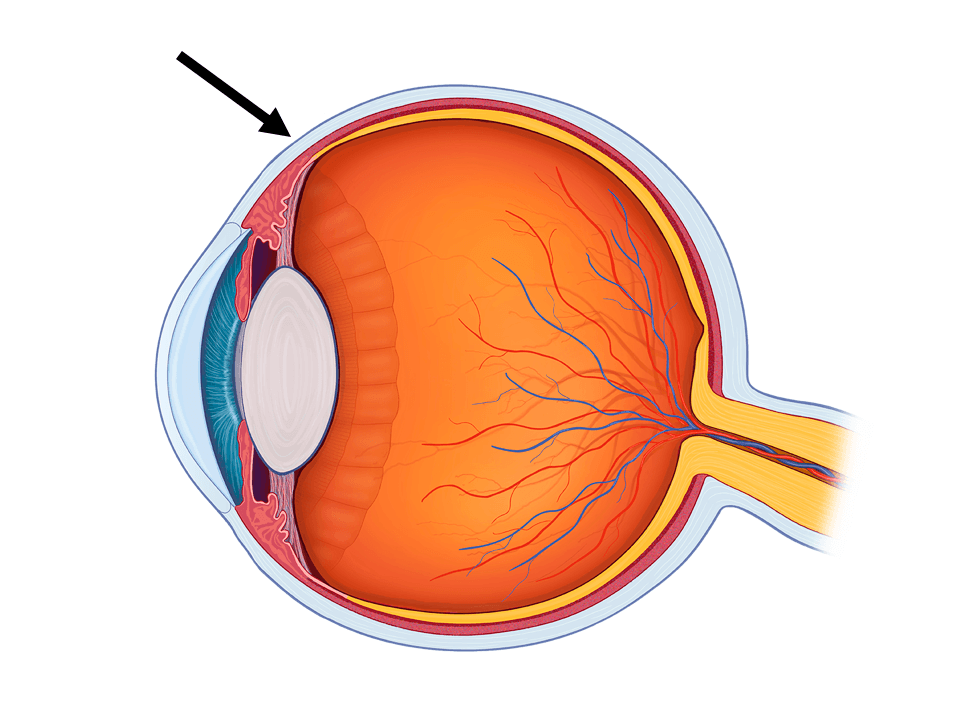
The white outer part of the eye.
Cornea
Lens
Sclera
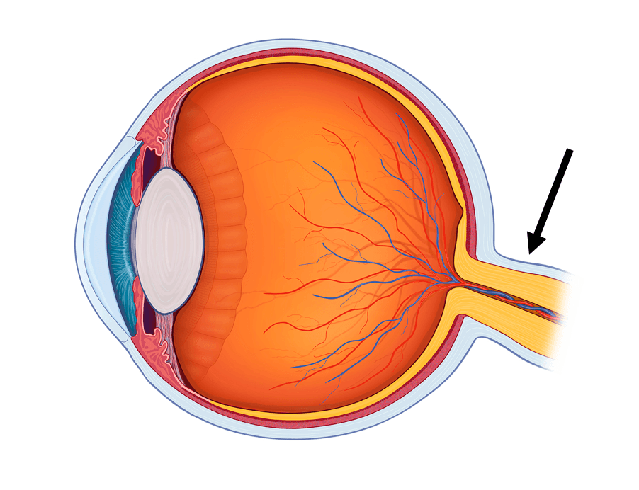
This part of the eye sends signals from the retina to the visual cortex.
Optic Nerve
Retina
Iris


Which part is #5?
pupil
iris
cornea
lens


Hey, wanna make eye contact? These muscles around your eye help you bat your eyes; closes the eyelids and enables you to wink at the special person!
Orbicularis Oris
Orbicularis Oculi
Corrugator
Frontalis

Contraction of this muscle enables you to kiss! #smooches
Orbicularis Oris
Orbicularis Oculi
Corrugator
Occipitals

This muscle pulls the corner of mouth laterally; presses the cheek against the teeth.
Masseter
Buccinator
Zygomaticus Major
Zygomaticus Minor

what muscle is this
pectoral
deltoid
triceps
biceps



