
 Worksheets
Respiratory System Review
Total questions: 96
Worksheet time: 24hrs 0mins
Which of the following best describes the anatomy of the respiratory tract?
The respiratory tract consists of only the lungs.
The respiratory tract includes both the upper and lower sections.
The respiratory tract is only made up of the trachea.
The respiratory tract is unrelated to breathing.
What is one of the main functions of the respiratory system?
To digest food
To pump blood
To facilitate gas exchange
To produce hormones
How are the respiratory and cardiovascular systems related?
They function independently of each other.
The respiratory system supplies oxygen, which the cardiovascular system transports.
The cardiovascular system controls breathing rate.
The respiratory system digests nutrients for the cardiovascular system.
Which process is directly controlled by the mechanisms of ventilation?
Blood clotting
Movement of air in and out of the lungs
Production of red blood cells
Digestion of proteins
How does ventilation impact blood pH?
It has no effect on blood pH.
It increases blood sugar levels.
It helps regulate blood pH by controlling carbon dioxide levels.
It only affects oxygen levels, not pH.
What is the main function of the respiratory system?
To digest food
To supply the body with oxygen and eliminate carbon dioxide
To produce hormones
To filter waste from the blood
Which of the following is NOT listed as a respiratory or cardiovascular process in the overview?
Ventilation
External Respiration
Digestion
Internal Respiration
Arrange the following processes in the correct order as they are listed in the overview: Ventilation, External Respiration, Transport, Internal Respiration.
Ventilation, Transport, External Respiration, Internal Respiration
Ventilation, External Respiration, Transport, Internal Respiration
External Respiration, Ventilation, Transport, Internal Respiration
Transport, Ventilation, Internal Respiration, External Respiration
Which of the following is part of the upper respiratory tract?
Trachea
Lungs
Nose and Nasal Cavity
Bronchi
Which structure is found in the lower respiratory tract?
Pharynx
Larynx
Trachea
Nasal Cavity
Which of the following lists only structures of the lower respiratory tract?
Nose, Pharynx, Larynx
Trachea, Bronchi, Lungs
Nasal Cavity, Larynx, Bronchi
Pharynx, Alveoli, Lungs
A patient has an infection in their pharynx. Which part of the respiratory tract is affected?
Upper respiratory tract
Lower respiratory tract
Both upper and lower respiratory tracts
Neither tract
Explain why the alveoli are classified as part of the lower respiratory tract and not the upper respiratory tract.
They are located in the lungs, which are part of the lower tract.
They are found in the nasal cavity.
They are part of the pharynx.
They are located in the larynx.

Which of the following structures is NOT part of the upper respiratory system?
Sphenoid sinus
Nasal cavity
Trachea
Lungs
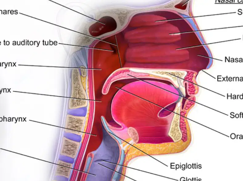
What is the function of the epiglottis in the upper respiratory system?
To produce sound
To prevent food from entering the trachea
To filter dust from the air
To warm the air
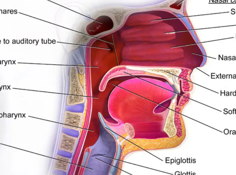
Which structure connects the nasal cavity to the pharynx?
Internal nares
Hard palate
Glottis
Vocal fold
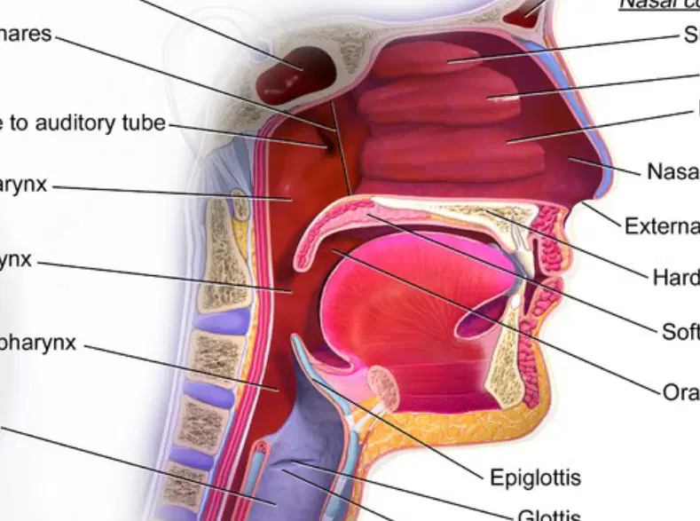
A patient has difficulty speaking and it is found that their vocal folds are not functioning properly. Which part of the upper respiratory system is most likely affected?
Oropharynx
Glottis
Sphenoid sinus
Hard palate
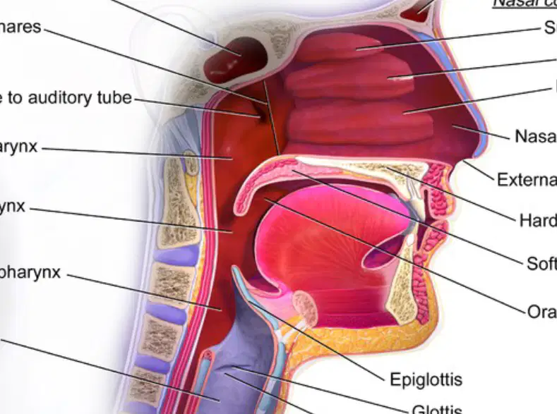
If the soft palate fails to function properly, which of the following processes would be most directly affected?
Swallowing and separating the nasal cavity from the oral cavity during swallowing
Producing mucus
Filtering air
Vibrating to produce sound
What is the primary function of nose hairs in the nasal cavity?
To produce mucus
To filter inspired air
To warm the air
To provide resonance for sound
Which structure in the nasal cavity is responsible for warming, cleansing, and humidifying inspired air?
Paranasal sinuses
Nasal mucosa
Nasal conchae
Nose hairs
What does the olfactory mucosa contain?
Goblet cells
Smell receptors
Cilia
Blood vessels
Which type of epithelium is found in the respiratory mucosa of the nasal cavity?
Simple squamous epithelium
Stratified squamous epithelium
Pseudostratified ciliated columnar epithelium
Transitional epithelium
What is one function of the paranasal sinuses?
To filter air
To produce smell receptors
To lighten the skull and provide resonance for sound production
To humidify air
Explain how the nasal conchae contribute to the process of respiration. (DoK Level 2)
They filter dust particles from the air.
They warm, cleanse, and humidify inspired air, making it suitable for the lungs.
They produce mucus to trap pathogens.
They provide resonance for sound production.
A patient has difficulty smelling and frequent respiratory infections. Based on your knowledge of nasal cavity structures, which two components might be malfunctioning and why? (DoK Level 3)
Nose hairs and paranasal sinuses, because they filter air and lighten the skull.
Olfactory mucosa and respiratory mucosa, because one contains smell receptors and the other traps pathogens.
Nasal conchae and goblet cells, because they both produce mucus.
Paranasal sinuses and nasal conchae, because they both provide resonance for sound.
Which portion of the pharynx is the uppermost and contains the pharyngeal tonsils (adenoids)?
Nasopharynx
Oropharynx
Laryngopharynx
Esophagus
What is the function of the uvula during swallowing?
It opens the Eustachian tube
It closes the passageway into the nasopharynx
It digests food
It produces saliva
Which region of the pharynx is located posterior to the oral cavity and is also part of the digestive system?
Nasopharynx
Oropharynx
Laryngopharynx
Trachea
Where are the palatine and lingual tonsils found?
In the nasopharynx
In the oropharynx
In the laryngopharynx
In the esophagus
The laryngopharynx is located:
Inferior to the esophagus
Superior to the esophagus
Posterior to the nasal cavity
Anterior to the oral cavity
Explain how the structure of the pharynx supports both respiratory and digestive functions.
The pharynx is divided into regions that separately handle air and food, with structures like the uvula and tonsils aiding in directing substances appropriately.
The pharynx only supports respiratory functions.
The pharynx only supports digestive functions.
The pharynx is not involved in either respiratory or digestive functions.
Which cartilage is responsible for producing the "Adam's Apple" in the larynx?
Cricoid Cartilage
Arytenoid Cartilage
Thyroid Cartilage
Epiglottis
What is one of the major functions of the larynx?
Absorbing nutrients
Providing a patent airway
Producing digestive enzymes
Filtering blood
Which cartilage anchors the vocal cords and pivots to change their length and tension during phonation?
Thyroid Cartilage
Arytenoid Cartilage
Cricoid Cartilage
Epiglottis
How does the epiglottis contribute to the function of the larynx during swallowing?
It produces sound
It diverts food away from the airway toward the esophagus
It increases abdominal pressure
It anchors the vocal cords
Which of the following is NOT a function of the larynx?
Voice production
Allowing for abdominal pressure to increase
Absorbing oxygen
Providing a patent airway
Explain how the arytenoid cartilage contributes to voice production.
It absorbs sound waves
It pivots to change the length and tension of the vocal cords during phonation
It produces mucus for lubrication
It diverts food away from the airway

Which structure is located at the topmost part of the larynx as shown in the diagram?
Hyoid Bone
Thyroid Cartilage
Cricoid Cartilage
Trachea

What is the function of the epiglottis as indicated in the diagram?
It produces sound.
It prevents food from entering the trachea during swallowing.
It supports the vocal cords.
It connects the larynx to the esophagus.

Which cartilage is located directly below the thyroid cartilage in the diagram?
Hyoid Bone
Cricoid Cartilage
Epiglottis
Trachea

If a person has difficulty producing high-pitched sounds, which muscle shown in the diagram might be involved?
Cricothyroid Muscles
Thyrohyoid Membrane
Trachea
Hyoid Bone
What happens as air rushes through the glottis and resonates in the oral, nasal, and sinus cavities?
Sounds are produced and modified.
The vocal cords stop vibrating.
The tongue becomes rigid.
The nasal cavity closes.
Which structures can modify the buzzing noises produced by the vocal cords?
Tongue, lips, pharyngeal muscles, and soft palate
Teeth and gums only
Only the nasal cavity
Only the glottis
What is true about the vocal cords during whispering?
The vocal cords do not vibrate.
The vocal cords vibrate rapidly.
The vocal cords produce loud sounds.
The vocal cords close completely.
Explain how the modification of sound occurs after the vocal cords produce buzzing noises. (DoK Level 2)
The buzzing noises are modified by the tongue, lips, pharyngeal muscles, and soft palate.
The buzzing noises are absorbed by the lungs.
The buzzing noises are stopped by the teeth.
The buzzing noises are amplified by the diaphragm.
A person is unable to produce sound while whispering. Using your understanding of sound production, explain why this occurs. (DoK Level 3)
The vocal cords do not vibrate during whispering, so no sound is produced.
The tongue blocks the glottis during whispering.
The nasal cavity is closed during whispering.
The air does not pass through the oral cavity during whispering.
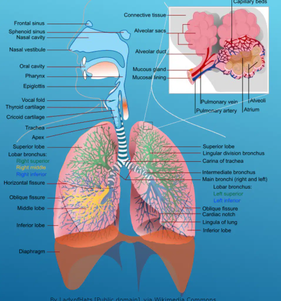
Which structure is responsible for the exchange of gases in the lungs?
Alveoli
Trachea
Diaphragm
Vocal fold

What is the function of the diaphragm in the respiratory system?
It contracts and relaxes to help move air in and out of the lungs.
It filters dust and microbes from inhaled air.
It produces mucus to trap particles.
It exchanges oxygen and carbon dioxide.

Which of the following is NOT a part of the upper respiratory tract?
Trachea
Nasal cavity
Pharynx
Frontal sinus

Compare the roles of the right and left lobar bronchi in the respiratory system.
Both direct air to their respective lung lobes, but the right has three branches and the left has two.
The right lobar bronchus only filters air, while the left exchanges gases.
The left lobar bronchus is responsible for vocalization, while the right is not.
Both are responsible for producing mucus in the lungs.

Why is the mucosal lining important in the respiratory system?
It traps dust and microbes, protecting the lungs.
It contracts to move air in and out.
It exchanges oxygen and carbon dioxide.
It produces sound for speech.
What is the primary function of the mucociliary escalator in the trachea?
It supports the trachea with cartilage rings.
It traps debris and moves it up and out of the respiratory tract.
It produces sound for speech.
It absorbs oxygen from the air.
What type of epithelium lines the trachea?
Simple squamous epithelium
Stratified squamous epithelium
Pseudostratified columnar epithelium
Transitional epithelium
Which structure does the trachea bifurcate into?
The left and right main stem bronchi
The alveoli
The nasal cavity
The diaphragm
What supports the trachea and keeps it open?
Flat plates of elastic cartilage
"C" shaped rings of hyaline cartilage
Muscle fibers
Ligaments
Explain how the structure of the trachea contributes to its function in the respiratory system.
The trachea is lined with smooth muscle to help with gas exchange.
The trachea is supported by "C" shaped rings of hyaline cartilage, which keep the airway open, and is lined with pseudostratified columnar epithelium that helps trap and remove debris.
The trachea is made of bone to provide maximum protection.
The trachea is covered in cilia to absorb nutrients.
Which of the following best describes the bronchi?
The smallest airways in the lungs
The largest airways distal to the trachea
Blood vessels that supply the lungs
Muscles that control breathing
What type of tissue supports the bronchi?
Elastic cartilage
Fibrous tissue
Hyaline cartilage
Smooth muscle
What is the function of the primary (main stem) bronchi?
Deliver air to each lobe
Deliver air to each segment
Deliver air to each lung
Filter dust from the air
Which type of bronchi delivers air to each lobe of the lung?
Primary bronchi
Secondary bronchi
Tertiary bronchi
Alveolar ducts
Explain how the structure of the bronchi supports their function in the respiratory system.
The bronchi are lined with smooth muscle to help move air quickly.
The bronchi are supported by hyaline cartilage and lined with pseudostratified columnar epithelium, which helps maintain open airways and filter air.
The bronchi are made of bone to provide maximum protection.
The bronchi are filled with fluid to trap dust particles.
Which of the following best describes bronchioles?
The largest airways in the respiratory system
The smallest airways leading eventually to the alveolar duct and alveoli
The main site of gas exchange in the lungs
The tubes that connect the nose to the throat
What happens to air flow when the diameter of the bronchiolar airway increases?
Air flow decreases
Air flow remains the same
Air flow increases
Air flow stops completely
Which muscle type is responsible for modifying the diameter of bronchioles?
Cardiac muscle
Skeletal muscle
Bronchiolar smooth muscle
Striated muscle
Asthma is best characterized by which of the following?
Permanent narrowing of the airways
Episodic, reversible bronchospasm due to exaggerated bronchoconstriction response to stimuli
Complete blockage of the trachea
Inflammation of the alveoli only
A patient experiences decreased air flow due to a reduced airway diameter. Which physiological change is most likely occurring?
The bronchiolar smooth muscle is relaxing
The bronchiolar smooth muscle is contracting
The alveoli are expanding
The trachea is dilating
What are alveoli?
Very small air sacs of the lungs
Large blood vessels in the lungs
Muscles that control breathing
Bones in the chest cavity
Which of the following is a major structure in the respiratory zone?
Alveoli
Trachea
Bronchi
Diaphragm
What is the primary function of Type 1 alveolar cells?
Line each alveolus and form a sheet of simple squamous epithelium specialized for diffusion
Produce surfactant to reduce surface tension
Roam the alveolar surface to keep the lungs clean
Deliver oxygen to the blood
How do Type 2 alveolar cells help prevent alveolar collapse?
By producing surfactant that reduces surface tension within each alveolus
By forming a barrier to pathogens
By increasing blood flow to the lungs
By transporting oxygen directly to the blood
What is the role of macrophages in the alveoli?
Roam the alveolar surface keeping the lungs clean
Produce surfactant
Line the alveolus for diffusion
Deliver blood to the alveolus
What is the term for the surface between each alveolus and the pulmonary capillaries delivering blood?
Respiratory membrane
Alveolar wall
Bronchial barrier
Pleural lining
Explain why the production of surfactant by Type 2 alveolar cells is critical for lung function. (DoK Level 3)
It reduces surface tension, preventing alveolar collapse and ensuring efficient gas exchange.
It increases the number of alveoli in the lungs.
It helps the lungs expand by increasing blood flow.
It removes pathogens from the alveolar surface.

What structure is depicted in the diagram shown?
Alveoli in the lungs
Nephrons in the kidney
Villi in the small intestine
Lobules in the liver

Which function is primarily associated with the structures shown in the diagram?
Gas exchange between air and blood
Filtration of blood
Absorption of nutrients
Production of bile

Why are the walls of the structures shown in the diagram thin and surrounded by capillaries?
To maximize the efficiency of gas exchange
To increase the rate of nutrient absorption
To filter waste products from the blood
To secrete digestive enzymes
Which of the following is the superior most portion of each lung that extends above the first rib?
Apex
Base
Hilum
Cardiac notch
What is the function of the hilum in the lungs?
It is where blood vessels, lymphatic vessels, bronchi, and nerves enter each lung.
It is the dome-shaped region that sits on the diaphragm.
It is the superior most portion of the lung.
It is the region that makes room for the heart.
Which lung is larger and how many lobes does it have?
The right lung is larger and has 3 lobes.
The left lung is larger and has 2 lobes.
The right lung is larger and has 2 lobes.
The left lung is larger and has 3 lobes.
Why does the left lung have a cardiac notch?
To make room for the heart, which favors the left side of the median plane.
To allow more air to enter the left lung.
To provide space for the diaphragm.
To separate the upper and lower lobes.
Which region of the lung sits on the diaphragm?
Base
Apex
Hilum
Cardiac notch
Which layer of the pleura covers each lung tightly?
Parietal pleura
Visceral pleura
Pleural space
Pericardial sac
What is the main function of the serous fluid in the pleural space?
To provide oxygen to the lungs
To lubricate between the pleural layers
To absorb carbon dioxide
To increase intrapleural pressure
Where does the parietal pleura line?
The surface of the heart
The thoracic cavity wall, diaphragm, and abuts the pericardial sac
The inside of the trachea
The alveoli
Why is intrapleural pressure negative throughout the respiratory cycle?
To allow air to escape from the lungs
To adhere the lungs to the body cavity wall and diaphragm
To increase the amount of serous fluid
To prevent the lungs from expanding
A patient has a condition where the pleural space lacks serous fluid. What is the most likely consequence?
Increased lubrication between pleural layers
Friction and possible pain during breathing
Enhanced oxygen exchange
Higher intrapleural pressure

Which structure separates the thoracic cavity from the abdominal cavity and plays a key role in breathing?
Diaphragm
Parietal pleura
Intercostal muscle
Pleural cavity

What is the name of the thin membrane that directly covers the surface of the lungs?
Parietal pleura
Visceral pleura
Pleural cavity
Intercostal muscle

Which of the following best describes the function of the pleural cavity?
It produces red blood cells.
It allows smooth movement of the lungs during breathing.
It contracts to help with exhalation.
It filters air entering the lungs.

If the parietal pleura is damaged, which of the following is most likely to occur?
The diaphragm will stop contracting.
The lungs may not move smoothly within the chest cavity.
The intercostal muscles will become paralyzed.
The rib cage will collapse.
According to Boyle’s law, what happens to the pressure of a gas when its volume increases?
Pressure increases
Pressure decreases
Pressure remains the same
Temperature increases
Which nerve innervates the diaphragm to cause it to contract and flatten during inspiration?
Vagus nerve
Phrenic nerve
Sciatic nerve
Trigeminal nerve
What is the effect of the contraction of the external intercostal muscles during inspiration?
Decreases the thoracic cavity size
Elevates the anterior margin of the ribs, enlarging the thoracic cavity
Relaxes the diaphragm
Reduces lung volume
During inspiration, what happens to the volume and pressure inside the lungs?
Volume decreases, pressure increases
Volume increases, pressure decreases
Volume and pressure both increase
Volume and pressure both decrease
Why does air move from the atmosphere into the lungs during inspiration?
Because the pressure inside the lungs is higher than atmospheric pressure
Because the pressure inside the lungs is lower than atmospheric pressure
Because the diaphragm relaxes
Because the thoracic cavity contracts
Explain how Boyle’s law is applied during the process of inspiration in the lungs.
As lung volume decreases, pressure increases, causing air to enter the lungs
As lung volume increases, pressure decreases, causing air to enter the lungs
As lung volume increases, pressure increases, causing air to leave the lungs
As lung volume decreases, pressure decreases, causing air to leave the lungs

