
 Worksheets
Exam 2 Prep (Lessons 8.1 and 8.2) NSCI 175 UNC
Total questions: 98
Worksheet time: 52mins
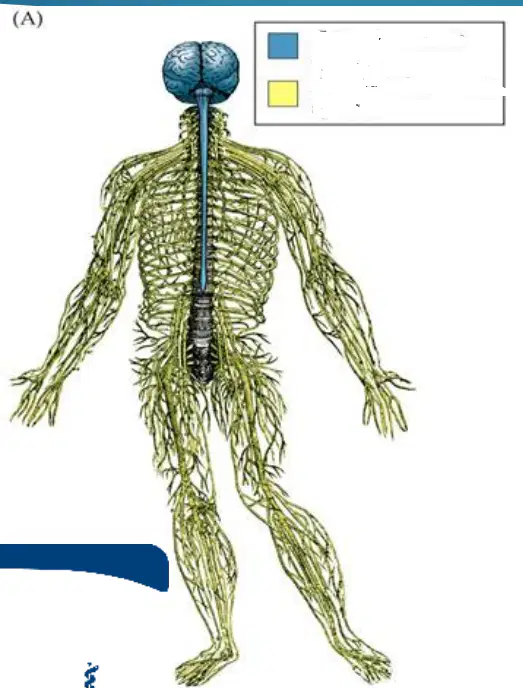
Blue is the ____ nervous system
Central
Peripheral
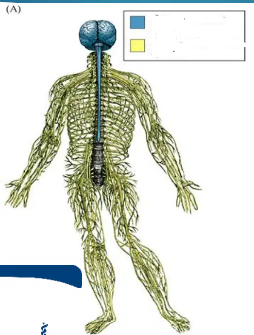
Yellow is the _____ nervous system
Central
Peripheral
The Peripheral Nervous system has three components; those three components are...
Cranial Nerves
Spinal Nerves
Autonomic nervous system
Trigeminal Nerves
Optic Nerves
Connected to the brain
Cranial Nerves
Spinal Nerves
Autonomic Nervous System
Also called somatic nerves, connected to spinal cord
Cranial nerves
Spinal nerves
Autonomic nervous system
Primarily controls glands and internal organs
Cranial nerves
Spinal nerves
Autonomic nervous system
Olfactory, optic, vagus,, trigeminal, facial, glossopharyngeal, and vesitbulochlear are...
Sensory
Motor
Oculomotor, trochlear, abducens, jaw muscles, facial muscles, salivary glands, tear glands, internal organs, throat muscles, and hypoglossal all deal with ...
Motor
Sensory
Olfactory I deal with...
Smell
Vision
Muscles that move the eyes
Tongue, soft palate
Optic II deal with..
Vision
Face, sinuses, teeth
Inner ear: hearing and balance
Neck muscles
Muscles that move the eyes are associated with the...
Oculomotor III
Trochlear IV
Abducens VI
Facial V
Olfactory I
Trigeminal V deals with
Jaws mucles
Face, sinuses, teeth
Tongue, soft palate
Tongue Muscles
Hypoglossal XII deals with..
Tongue muscles
Neck muscles
Throat muscles
Jaw muscles
Spinal accessory XI deals with
Neck muscles
Tongue muscles
Jaw muscles
Muscles that move the eyes
Vagus X deals with...
Information from internal organs
Internal organs
Tongue muscles
Smell
Vision
Glossopharyngeal IX deals with..
Taste and other mouth sensations
Throat muscles
Inner ear: hearing and balance
Face, sinuses teeth
Jaw muscles
Vestibulocochlear VIII deals with
Inner ear: hearing and balance
Tongue, soft palate
Vision
Smell
Facial VII deals with
Tongue, soft palate
Facial muscles, salivary glands, tear glands
Taste and other mouth sensations
Throat muscle
Conscious and subconscious control over skeletal muscles, skin, and joints
Somatic (Spinal nerves)
Autonomic
Autonomic truths and falses (Select the truths)
Control of viscera (body organs, smooth muscles, blood vessels, gland)
Control pressure of oxygen in the blood (heart rate, blood vessel diameter)
Conscious and subconscious control over skeletal muscles, skin and joints
The majority of the cranial nerves contain axon of the PNS. However two are actually parts of the central nervous system. Which two are they? Select all that apply.
Olfactory
Optic
Trigeminal
Vagas
Trochlear
____ can be used to describe a nerve that carries info from the body to the spinal cord
Afferent
Efferent
Sympathetic
Parasympathetic
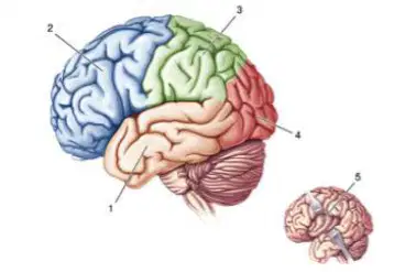
What is 1?
Temporal Lobe
Frontal Lobe
Parietal Lobe
Occipital Lobe
Insular Lobe
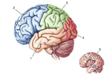
What is 2
Temporal Lobe
Frontal Lobe
Parietal Lobe
Occipital Lobe
Insular Lobe
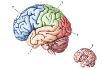
What is 3?
Temporal Love
Frontal Lobe
Parietal Lobe
Occipital Lobe
Insular Lobe
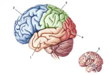
What is 4?
Temporal Lobe
Frontal Lobe
Parietal Lobe
Occipital Lobe
Insular Lobe
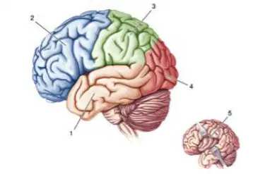
What is 5?
Temporal Lobe
Frontal Lobe
Parietal Lobe
Occipital Lobe
Insular Lobe
Primary auditory cortex, learning and memory
Temporal Lobe
Frontal Lobe
Parietal Lobe
Insular Lobe
Primary motor cortex, complex cognitive processes: language, executive function, attention, memory, impulsivity, planning, strategizing, inhibition to obtain goals/rewards (part of what makes humans unique, largest of all animals)
Temporal Lobe
Insular Lobe
Parietal Lobe
Frontal Lobe
Primary somatic sensory areas, visual processing
Parietal Lobe
Frontal Lobe
Temporal Lobe
Insular Lobe
Primary visual cortex, other visual areas
Occipital Lobe
Insular Lobe
Parietal Lobe
Frontal Lobe
Gustatory cortex (Sense of taste)
Occipital Lobe
Parietal Lobe
Temporal Lobe
Insular Lobe
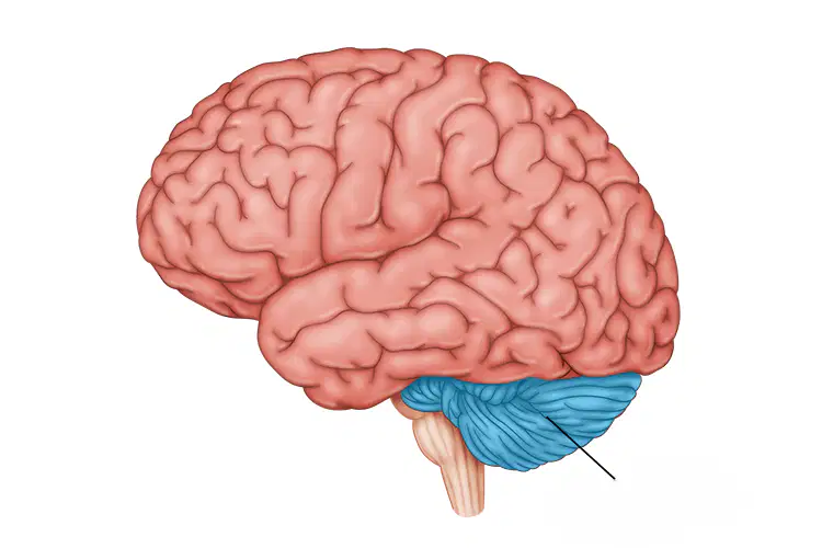
This is the?
Midbrain
Thalamus
Cerebellum
Medulla Oblongata
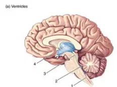
What is 1?
Spinal Canal
Fourth ventricle
Cerebral aqueduct
Third ventricle
Lateral ventricle
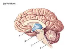
What is 2?
Spinal Canal
Cerebral aqueduct
Fourth ventricle
Third ventricle
Lateral ventricle
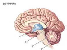
What is 3?
Spinal Canal
Third ventricle
Cerebral aqueduct
Fourth ventricle
Lateral ventricle
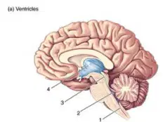
What is 4?
Fourth ventricle
Spinal Canal
Third ventricle
Cerebral aqueduct
Lateral ventricle
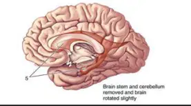
What is 5?
Spinal Canal
Fourth Ventricle
Lateral Ventricle
Third Ventricle
Cerebral aqueduct
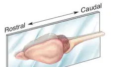
Relating to an imaginary line that runs through the middle of the body from top to bottom, dividing it into left and right halves
Midsagittal
Horizontal
Coronal
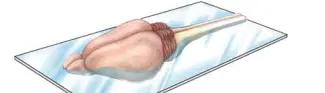
The transverse plane, which is a plane that divides the body into top and bottom halves
Midsagittal
Horizontal
Coronal
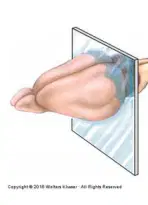
What kind of cut would you make to disconnect the right and left cerebral hemispheres?
Midsagittal
Horizontal
Coronal
Same side of the body
Ipsilateral
Contralateral
Opposite side of the body
Ipsilateral
Contralateral
The left and right portions of the body are divided by the ______ plane
frontal
sagittal
coronal
horizontal
Where is CSF produced?
ventricles of the paired cerebral hemispheres
Diencephalon
Subarachnoid space
Arachnoid villi
Axons of the nervous system are described as afferent and efferent according to the direction in which they carry information. Which of the following represents an efferent projection?
All of these
Interneurons in the spinal cord
Sensory input to the spinal cord
Motor output of the spinal cord
Which major white matter system bridge the cerebral hemispheres
dorsal columns
corpus callosum
posterior capsule
internal capsule
Which of the following mediates vital bodily functions?
Brain stem
Meninges
Cerebrum
Cerebellum
At what point do the somatic sensory axons enter the spinal cord?
Dorsal root ganglia
Ventral roots
Dorsal roots
Ventral root ganglia
The brain and spinal cord are wrapped in protective membranes known collectively as the
dura mater
myelin
meninges
pia mater
Which membrane lies closest to the brain
Pia mater
Dura mater
Meninges
Arcachnoid
What are the advantages of MRI over CT scan?
Does not use X-irradiation
Yields a more detailed map of the brain
Images of brain slices can be made in any plane desired
All of these
In which plane of section would it be easiest to see the corpus callosum?
Sagittal cut directly down the midline
Horizontal cut at the level of the brainstem
Coronal cut at the level of the cerebellum
Sagittal cut at the level of the temporal lobe
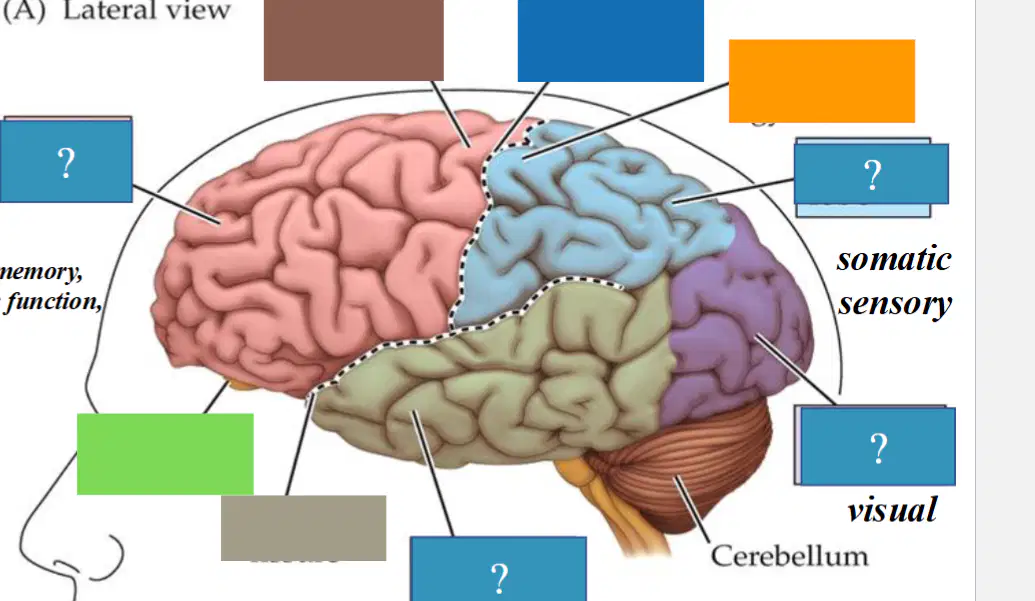
What is brown pointing to?
Precentral gyrus
Central sulcus
Postcentral gyrus
Sylvian fissure
Olfactory bulb
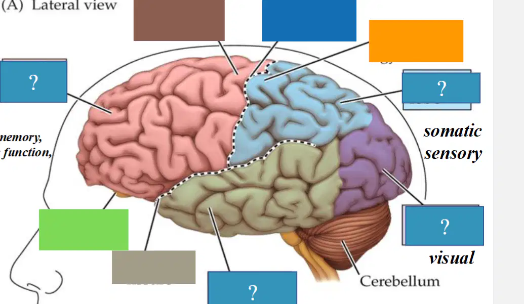
What is blue pointing to (THE BLUE WITHOUT A "?")
Central sulcus
Postcentral gyrus
Precentral gyrus
Olfactory bulb
Sylvian fissure
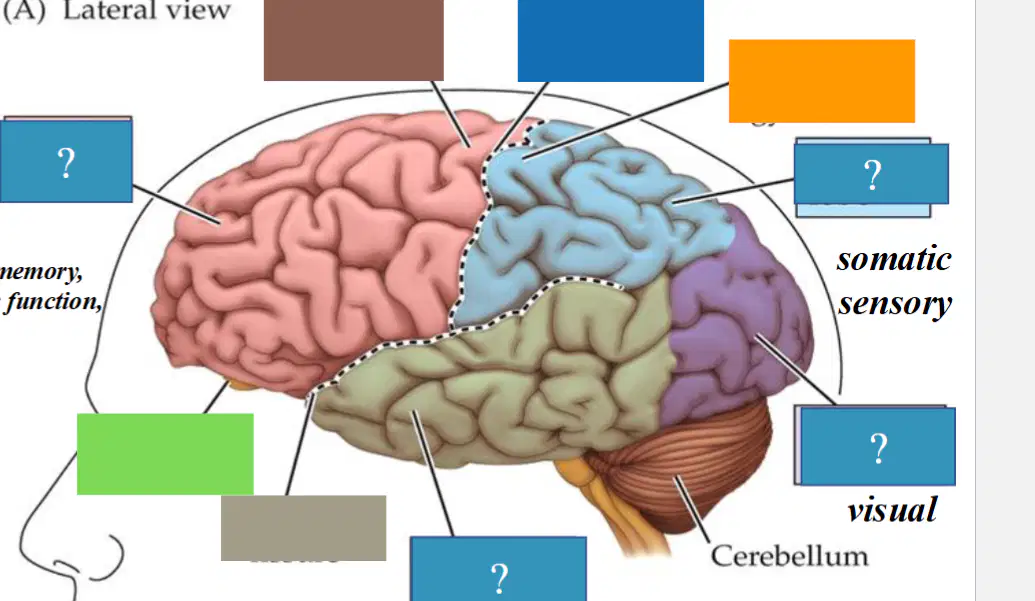
What is orange pointing to?
Postcentral gyrus
Central gyrus
Precentral gryrus
Olfactory bulb
Sylvian fissure
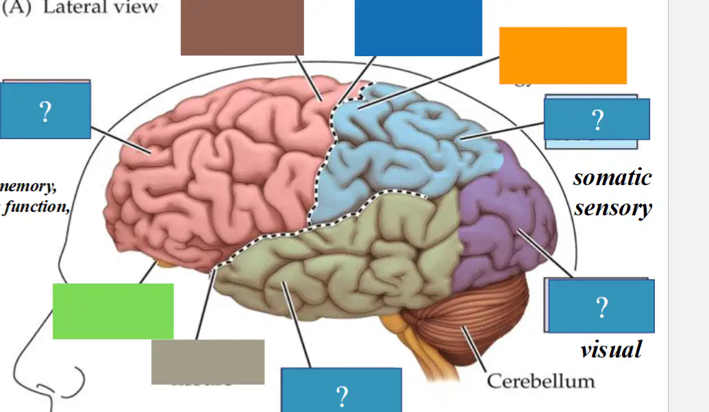
What is light green pointing to?
Sylvian fissure
Olfactory bulb
Postcentral gyrus
Central sulcus
Precentral gyrus
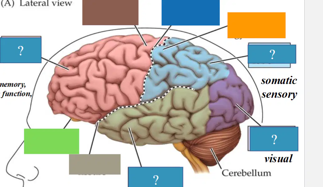
What is gray pointing to?
Olfactory bulb
Precentral gyrus
Central sulcus
Postcentral gyrus
Sylvian fissure
A rounded mass of tissue in the brain that receives and processes information about smells from the nose
Olfactory bulb
Cerebellum
Central sulcus
Postcentral gyrus
A part of the brain that controls voluntary movement and is located in the frontal lobe
Precentral gyrus
Sylvian fissure
Postcentral gyrus
Central sulcus
Is a key landmark that separates the insula medially and the frontal lobe superiorly from the temporal lobe. (also known as the lateral sulcus or lateral fissure)
Sylvian fissure
Postcentral fissure
Precentral gyrus
Central fissure
A deep groove in the brain that separates the frontal lobe from the parietal lobe
Central sulcus
Postcentral gyrus
Precentral gyrus
Olfactory bulb
A region of the brain that processes general somatic sensations, such as touch, pressure, temperature, and pain
Postcentral gyrus
Precentral gyrus
Central sulcus
Sylvian fissure
Movement of specific body parts is regulated by
distinct regions within the ______ cortex, and a similar
pattern of organization is present in the
________ cortex.
Motor cortex; Somatosensory cortex
Somatosensory cortex; Motor cortex
Primary motor & sensory
cortices are NOT separated by
the central sulcus
True
False
How cells of the cerebral cortex are organized
The cortex is organized into layers (Parallel to the surface of the brain)
Layer of neurons closest to the surface is Layer I
At least one cell layer contains pyramidal cells emit dendrite and extend to layer I
Layer of neurons furthest away from the surface is Layer I
The cortex is organized into layers (Perpendicular to the surface of the brain)
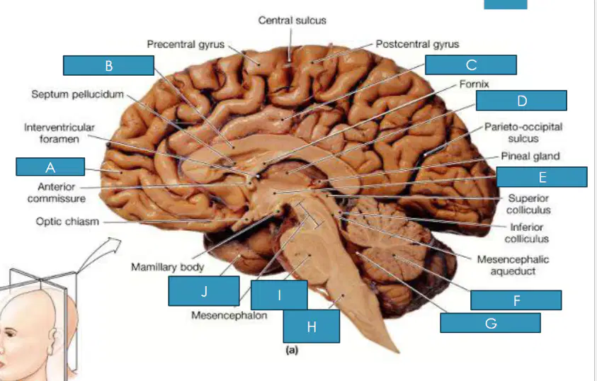
What is A?
Cingulate Gyrus
Septum Pellucidum
Fornix
Frontal Lobe
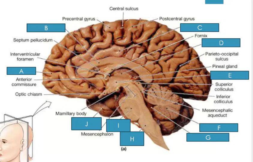
What is B?
Cingulate Gyrus
Thalamus
Corpus Callosum
Medulla
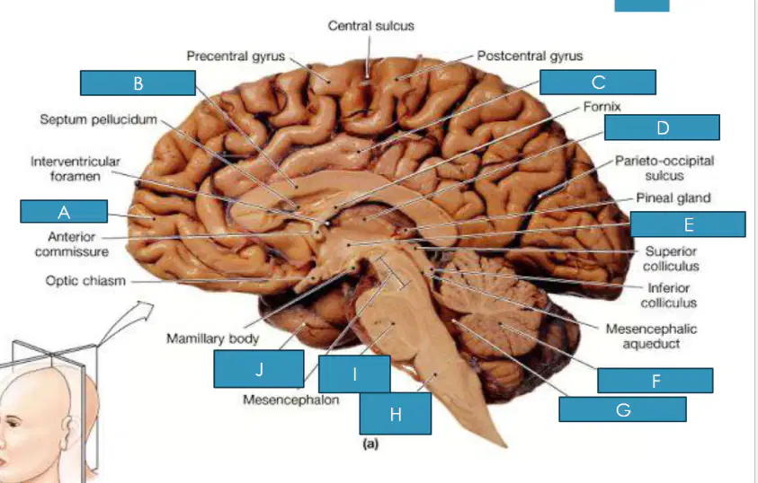
What is C?
Cingulate gyrus
Thalamus
Pineal Gland
Pons
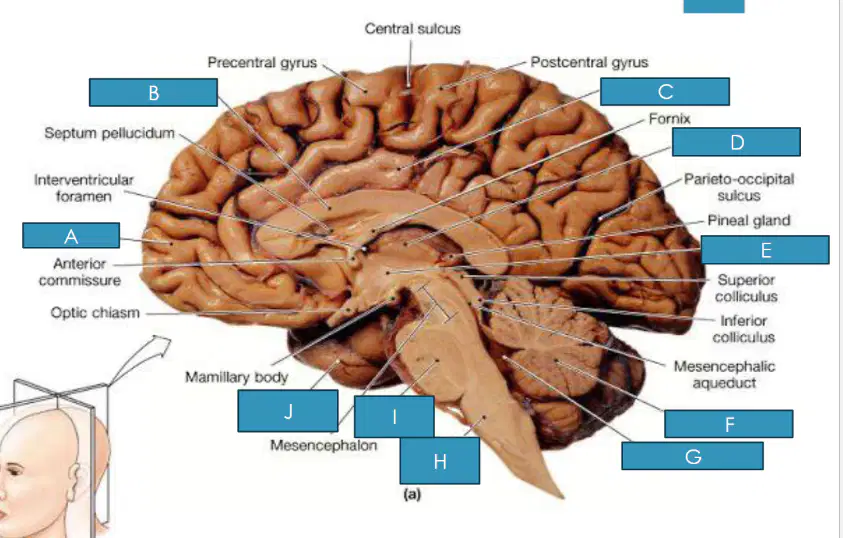
What is D?
Thalamus
Pineal Gland
Cerebellum
Fourth Ventricle
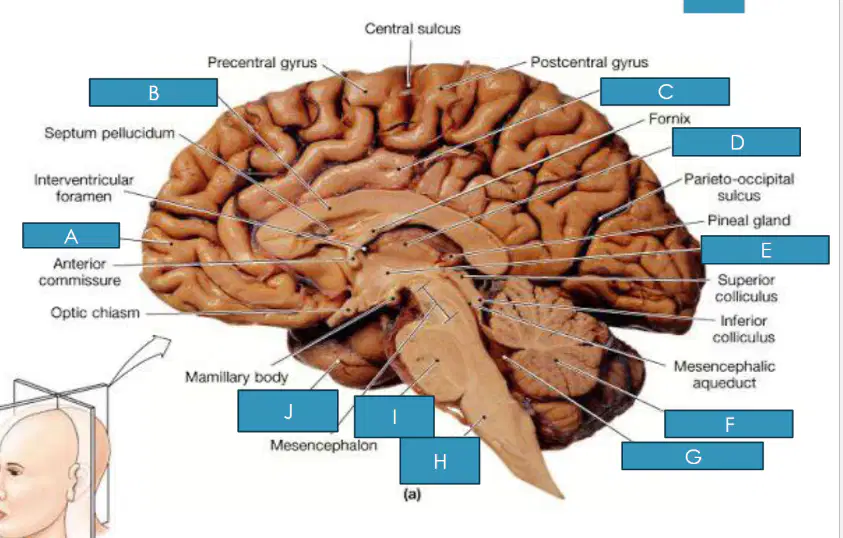
What is E?
Pons
Pineal Gland
Hypothalamus
Medulla
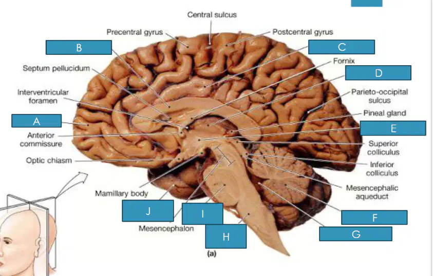
What is F?
Pons
Cerebellum
Corpus Callosum
Frontal Lobe
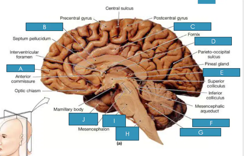
What is G?
Cerebellum
Medulla
Fourth Ventricle
Hypothalamus
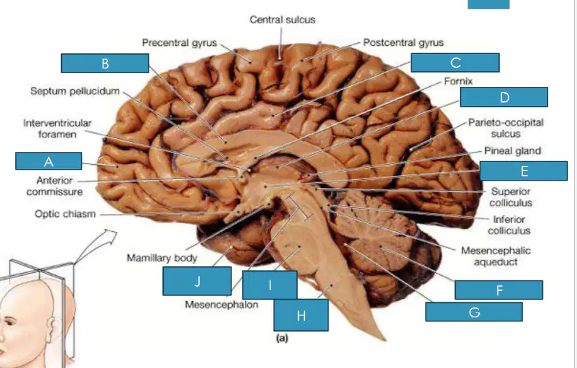
What is H?
Medulla
Cerebellum
Pons
Pineal Gland
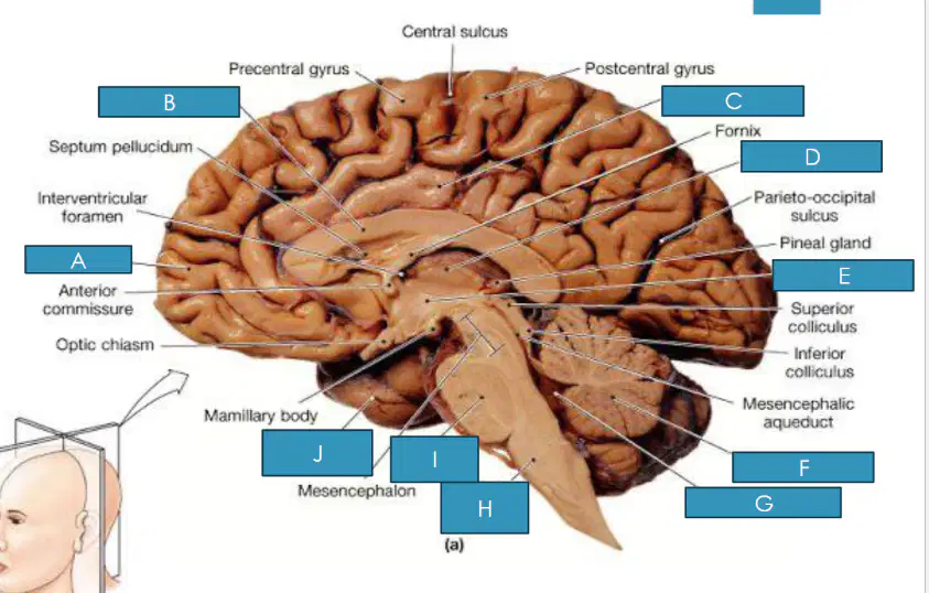
What is I?
Fourth Ventricle
Cingulate Gyrus
Pons
Hypothalamus
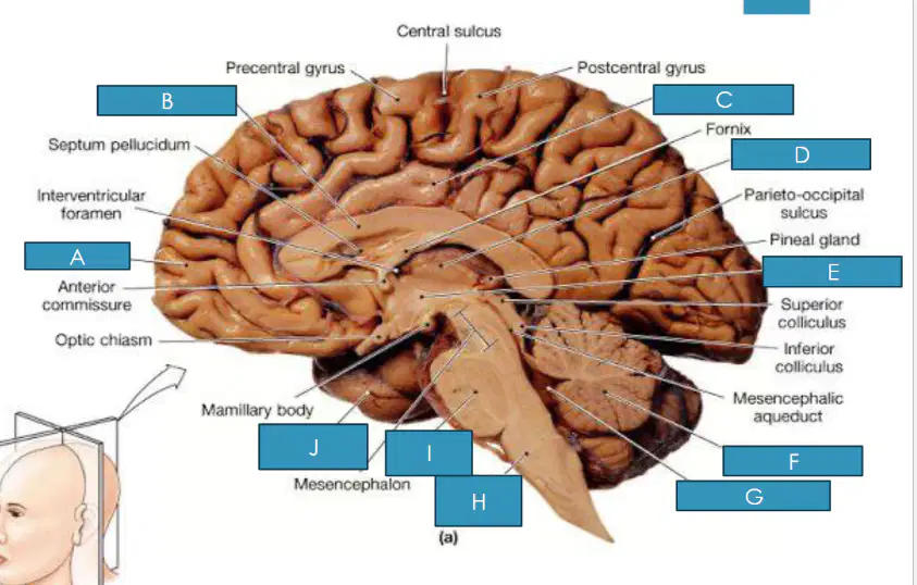
What is J?
Thalamus
Hypothalamus
Temporal Lobe
Pineal Gland
Similarities between Mammalian brains and Human Brain
Three trispheres
Two hemispheres
Ventricles and surrounding brain regions (midbrain)
Cerebellum, pons, and medulla
Convolution
Differences between Mammalian and Human brains
Two Hemispheres
Convolutions
Cerebral hemisphere
Olfactory bulb
Pre-frontal cortex
Utilizes X-ray absorption to generate an image of a slice of the brain, allowing the view of the gross organization of gray and white matter, and the position of ventricles
Computed tomography (CT)
Positron emission tomography (PET)
Magnetic Resonance Imaging (MRI)
Functional MRI
Utilizes radioactivity to measure metabolism in the brain. Brain activity stimulated light signals generate an image of the brain activity
Positron emission tomography (PET)
Computed tomography (CT)
Magnetic Resonance Imaging (MRI)(
Functional MRI
Utilizes a magnetic field to generate an image of the brain. Activity of hydrogen atoms provide a more detailed image in the absence of radiation.
Magnetic Resonance Imaging (MRI)
Functional MRI
Computed tomography (CT)
Positron emission tomography (PET)
Utilizes a magnetic field to generate an image of the brain and overlays it with an image highlighting the effects of the magnetic field on oxyhemoglobin (measure of metabolism).
Magnetic Resonance Imaging
Functional MRI
Positron emission tomography (PET)
Computed tomography (CT)
The following are major cell groups in the brain
corpus collosum
fornix
caudate nucleus
putamen
septal area
The ridges of tissue on the convoluted surface of the cortex are called...
sulci
nuclei
ganglia
gyri
Neurons in the __________ are often referred to as the gateway to the cerebral cortex
thalamus
olfactory bulb
brain stem
cerebellum
The brainstem contains which of the following structures
central sulcus
cerebellum
medulla
corpus callosum
Which region of the cortex is necessary for vision?
temporal cortex
frontal cortex
occipital cortex
postcentral gyrus
Which of the following is the most posterior cerebral lobe in the brain?
temporal lobe
occipital lobe
frontal lobe
parietal lobe
In the cerebral cortex, layer 5 is especially noticeable because it
is a different color from the rest
contains many fibers
contains many large cell bodies
is quite variable from region to region
Which region of the cortex is crucial for motor control?
precentral gyrus
postcentral gyrus
prefrontal cortex
parietal lobe
How many distinct layers are observed in the human neocortex?
6
1
3
over 1 million
Relay station, breathing regulation, sleep-wake cycle, facial movements, sensory and motor pathways, it is essential for many unconscious processes and helps integrate sensory and motor functions
Pons
Hypothalamus
Thalamus
Temporal Lobe
Autonomic Functions, reflex actions, nerve signal conduit, maintaining vital bodily functions and ensuring basic survival mechanisms are in places
Medulla
Temporal Lobe
Pons
Cerebellum
Bundle of axons that physically connects two hemispheres
Corpus Callosum
Thalamus
Hypothalamus
Cerebellum
Relays info from the cerebrum and cerebellum to the spinal cord and regulates consciousness and body temperature
Brain stem
Cingulate gyrus
Corpus Callosum
Hypothalamus
Which of the following statements correctly identifies a key difference between the human brain and other mammalian brains?
Both human and other mammalian brains have two hemispheres
The olfactory bulb is larger in humans compared to other mammals
The human brain has more convolutions (sulci and gyri), leading to an increased s.....
All mammals have a prefrontal cortex of equal size
None of the above
A famous study in London demonstrated Taxi drivers had a larger hippocampus than bus drivers, indicating that the hippocampus may be responsible for spatial navigation. Which methodology did they use to come to these conclusions?
Positron emission tomography (PET)
Magnetic Resonance Imaging (MRI)
functional MRI (fMRI)
Immunohistochemistry
None of the above

