
 Worksheets
Advanced Biochemistry - 2022 MCQs
Total questions: 83
Worksheet time: 42mins

What is the correct order of events during an action potential?
(A) -> (B) -> (C) -> (D)
(D) -> (A) -> (B) -> (C)
(D) -> (A) -> (C) -> (B)
(C) -> (A) -> (B) -> (D)
(A) -> (C) -> (B) -> (D)
What is the biochemical mechanism behind the refractory period of electrically excitable cells?
The cell needs time to re-establish Na+ balance.
The Na+ channel voltage sensor blocks the channel.
The Na+ channel voltage sensor alters the selectivity filter characteristics.
The Ca2+ channel is activated, flooding the cytoplasm with more positive charge.
The Na+ channel’s gate closes for a specific period of time based on the duration that it was previously open.
Solute binding in transporter mediated diffusion can be blocked by ______.
Inhibitors
GTP
Divalent cations
Divalent anions
ATP
______ transporters constitute the largest family of membrane transport protein.
V-type proton pump
K+ ion
Ca2+ ion
P-type pump
ABC
What is the primary characteristic of plants that are defective in chlorophyll synthesis?
They are light sensitive.
They are yellow and are unable to develop into mature plants.
They accumulate aminolevulinate.
They have abnormal leaf morphology.
All of the above.
Mutant analysis is one method of providing information about biosynthetic pathways. Which of the following characteristics of a mutant would be most helpful in elucidating the pathway?
The mutant accumulates a substrate in the pathway.
Growth of the mutant on a wide range of medias.
Sequence of the mutant’s 16S rRNA gene.
The mutant is able to incorporate radiolabelled S35-Methionine.
None of the above.
Which of the following best describes the cause of the porphyria disease group?
Light sensitivity.
Propensity to develop dementia.
Defect in chlorophyll biosynthesis.
Defect in phytochrome biosynthesis.
Defect in heme biosynthesis.
Scientists can determine if a biosynthetic pathway is present in an organism with a sequenced genome by …
Scanning for orthologous proteins based on known gene sequences.
Scanning for synonymous proteins based on known gene sequences.
Growing the organism on an alternative carbon source.
Growing the organism on an alternative sugar source.
None of the above.
Aminolevulinic acid synthesis can proceed by two pathways, the …
Shemin pathway using acetyl-CoA and glycine substrates and the C5 pathway using glutamate substrate.
Shemin pathway using succinyl-CoA and proline substrates and the C5 pathway using aspartate substrate.
Shemin pathway using acetyl-CoA and glycine substrates and the C5 pathway using aspartate substrate.
Shemin pathway using acetyl-CoA and glycine substrates and the C5 pathway using glutamate substrate.
Shemin pathway using succinyl-CoA and glycine substrates and the C5 pathway using glutamate substrate.
What technique is used to elucidate steps within biosynthetic pathways?
Cloning whole organisms.
Enzyme degradation.
Mutant complementation.
Isotonic tracers.
None of the above.
Which of the following is required for protein import into mitochondria?
GTP hydrolysis.
ATP hydrolysis.
Signal sequence with many acid residues.
A protein that is fully folded.
Ca²⁺ gradient to power the membrane translocator.
What is the dominant method of protein import into the mitochondria?
Co-translational translocation.
Co-translational insertion.
Post-translational translocation.
Post-translational insertion.
None of the above.
What is the dominant method of protein import into the ER?
Co-translational translocation.
Co-translational insertion.
Post-translational translocation.
Post-translational insertion.
None of the above.
Which one of the following statements of nuclear protein transport is correct?
All proteins can passively diffuse in and out of the nucleus.
Proteins can only be transported in one direction from the cytosol to the nucleus.
Specific proteins with signal peptides are rapidly transported both in and out of the nucleus.
Viruses cannot enter the nucleus through the nuclear pore.
None of the above.
Cellular organelles enable …
Compartmentalisation of chemical reactions
Segregation of genetic material from the cytoplasm
Physical separation of proteases from targets
Increased membrane surface area
All of the above
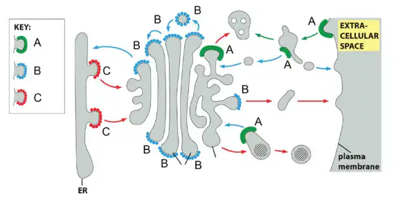
Identify the COPI-coated vesicle(s) in the diagram above.
A-only
B-only
C-only
Both A and B
Both B and C
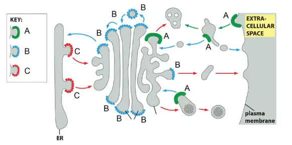
Identify which vesicle(s) in the diagram above would contain SNAREs.
A-only
B-only
C-only
Both A and C
A, B, and C
Vesicle formation, targeting, and fusion are processes facilitated by:
Rab proteins, glycolipids, and SNARE proteins, respectively.
SNARE proteins, Rab proteins, and dynamin, respectively.
Coat proteins, Ras proteins, and SNARE proteins, respectively.
Coat proteins, Rab proteins, and SNARE proteins, respectively.
Shell proteins, Erk proteins, and SNAP proteins, respectively.
A pathway NOT used to deliver material to lysosomes is:
Phagocytosis.
Autophagy.
Endocytosis.
Constitutive exocytosis.
Macropinocytosis.
Protein N-linked glycosylation…
Is the attachment of complex carbohydrates on Asn residues in only specific motifs of newly synthesised proteins
Is a very common type of protein glycosylation
Is a co-translational modification initiated in the ER
Is involved in the quality control of protein folding
All of the above
Important characteristic(s) of lysosomes include….
The high concentration of cytoskeletal proteins defining the structure of the lysosome
The presence of many hydrolases involved in the digestion of various biomolecules
An alkaline environment that differs from the pH in the extracellular environment
An abundance of nuclear pore complexes that allow rapid movement of protein from the cytosol into the lysosome
None of the above
Autophagy….
Degrades unwanted proteins and organelles
Degrades unwanted bacterial cells
Degrades phage
Is a form of programmed cell death
Is a form of apoptosis
A cell expresses a transmembrane protein that is cleaved at the plasma membrane to release an extracellular fragment. The fragment binds to receptor proteins on nearby cells and activates signaling pathways resulting in altered gene expression patterns in the cells. What form of intercellular signaling does this represent?
Contact-dependent signaling.
Paracrine signaling.
Synaptic signaling.
Endocrine signaling.
Autocrine signaling.
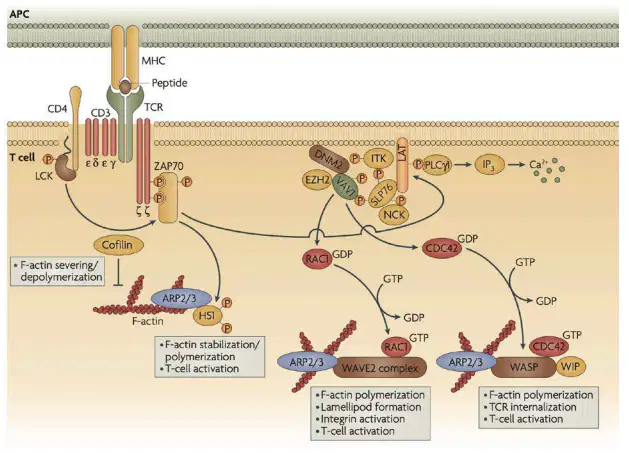
What is the expected phenotypic outcome of the activation of the pathway depicted in the figure?
Increased glucose uptake into the cell
Activation of the T-cell through changes in cytoskeletal dynamics
Suppression of the immune response
Initiation of cell apoptosis
Which of the following is NOT a common second messenger in cell signaling?
Proline
Ca²⁺
Diacylglycerol
Cyclic adenosine monophosphate
Inositol trisphosphate
Intracellular signalling occurs by ____ hydrolysis or _____ transfer and exchange.
GTP / ATP
Ca²⁺ / ATP
ATP / GTP
ATP / Ca²⁺
cAMP / IP₃
Whereas the cholera toxin ADP-ribosylates the α subunit of stimulatory G protein (Gs), thereby blocking GTP hydrolysis, pertussis toxin ADP-ribosylates the α subunit of inhibitory G protein (Gi) and prevents interaction with the receptor. What is the expected effect of these toxins on the concentration of intracellular cAMP?
Cholera toxin tends to increase cAMP concentration, whereas pertussis toxin tends to decrease cAMP concentration
Cholera toxin tends to decrease cAMP concentration, whereas pertussis toxin tends to increase cAMP concentration
They both tend to increase cAMP concentration
They both tend to decrease cAMP concentration
None of the above
Consider a signaling protein that is only made up of one SH2 domain and two SH3 domains. This protein is most likely …
a monomeric G protein.
a guanine nucleotide exchange factor.
a kinase associated with receptor tyrosine kinase signaling.
an adaptor protein.
a negative regulator of receptor tyrosine kinase signaling.
G-Protein coupled receptors (GPCRs) are 7 transmembrane domain receptors that upon ligand binding …
Open their ion channel to enable transport across the membrane.
Activate a trimeric G protein α-subunit.
Autophosphorylate, thereby becoming activated.
Cause release of Ca²⁺ stores.
Activate dimeric GTPases.
Cytochalasin B is toxic to our cells through which mechanism?
It caps microtubule ends and leads to their depolymerization.
It binds to tubulin subunits and prevents microtubule polymerization.
It binds along actin filaments and stabilizes them.
It caps the plus end of actin filaments and prevents actin polymerization.
All of the above.
During actin filament elongation phase, actin subunits add …
Faster to filament (+) ends.
Faster to filament (−) ends.
Equally to both filament ends.
Along the length of filaments.
None of the above.
Mutations that cause defective keratin filament networks are in genes encoding components of which cytoskeleton class?
Actin.
Microtubules.
Intermediate filaments.
Myosin.
Dynamin.
Which important cell structure is formed from microtubules?
Contractile bundles.
Stress fibres.
Nuclear lamina.
Basal lamina.
Centrosome.
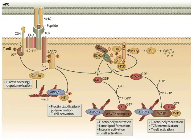
How does the ARP2/3 complex contribute to the overall pathway depicted in the figure?
It phosphorylates ZAP70.
It mediates F-actin depolymerization.
It stabilizes and polymerizes F-actin to support T-cell activation.
It recruits integrins to the plasma membrane.
The actin-nucleating protein formin has flexible “whiskers” containing binding sites that help recruit actin subunits in order to enhance polymerization by this protein. What protein would you expect to bind to these sites?
Thymosin.
Profilin.
Cofilin.
Gelsolin.
Tropomodulin.
How is microtubule stabilization and destabilization regulated within cells?
The balance of cellular ATP levels.
Protein factors such as kinesin-13.
The balance of cellular GTP levels.
Gamma (γ)-tubulin ring complexes.
Protein factors such as formin.
The myosin motor protein …
Assembles on intermediate filaments and generates movement through GTP hydrolysis
Assembles on intermediate filaments and generates movement through ATP hydrolysis
Assembles on actin filaments and generates movement through GTP hydrolysis
Assembles on actin filaments and generates movement through ATP hydrolysis
Assembles on microtubules and generates movement through GTP hydrolysis
The motor proteins associated with microtubules are …
Dynein and kinesin
Dynein and myosin
Dynamin and myosin
Myosin and kinesin
Dynamin and kinesin
DNA damage arrests the cell in G₁ through …
Phosphorylation of p53
Production of p21 Cdk inhibitor protein
Inactivation of G₁/S-Cdk
Inactivation of S-Cdk
All of the above
Cell-cycle control depends on …
Bicyclins and Ccks
Cyclins and Cdks
The circadian rhythm
The nuclear clock
None of the above
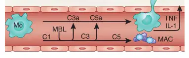
Which immune event is occurring in the figure with the formation of the membrane attack complex (MAC)?
Direct neutralization of bacteria by antibodies
Destruction of bacterial cells by punching holes in their membranes
Activation of macrophages to engulf bacteria
Recruitment of neutrophils to the site of infection
The two key events within the cell cycle are …
Replication of protein and cell merging
Replication of RNA and cell division
Replication of RNA and cell merging
Replication of DNA and cell division
Replication of DNA and cell joining
Which cellular activity is most closely associated with caspases?
Meiosis.
Exocytosis.
Cytokinesis.
Necrosis.
Apoptosis.
A tight junction requires …
Claudin and occludin
Claudin and occultin
Clonin and occludin
Clonin and occultin
None of the above
For cell-cell junctions, scaffold proteins, such as the ZO proteins, can …
Bind multiple adherens junctions together
Bind multiple tight junctions together
Bind tight junctions to other junction types
Bind desmoplakin junctions to other junction types
Bind hole junctions to other junction types
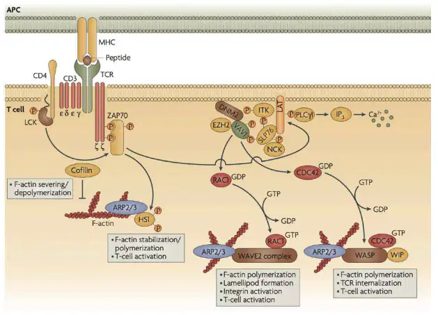
What role do RAC1 and CDC42 play in the molecular process shown?
They function as signaling molecules that regulate GTP binding and F-actin polymerization.
They function as protein kinases that directly phosphorylate TCR
They regulate the degradation of the antigen presented by the MHC.
They bind to integrins to initiate signal transduction.
In a classic experiment, H. V. Wilson studied aggregation of mechanically dissociated individual sponge cells from two different species. He found that the cells of each species would adhere to one another but not to cells of the other species. The most likely factor causing this result would be that …
The different species express different keratins.
The different species express different lamins.
The different species express different integrins.
The different species express different actins.
The different species express different cadherins.
The desmosome connects …
Actin cytoskeleton between two cells
Actin cytoskeleton between a cell and the extracellular matrix
Intermediate filaments between two cells
Intermediate filaments between a cell and the extracellular matrix
Integrin to α-catenin
A gap junction …
Enables free diffusion of proteins between neighbouring cells
Does not contribute to cell-cell adhesion
Does not allow an electrical connection between cells
Spans both cells with a total of 2 transmembrane connexin subunits
None of the above
How does hyaluronic acid differ from other glycosaminoglycans of the extracellular matrix?
It lacks sulfate groups.
It is generally not covalently linked to proteins.
It is not assembled in the Golgi apparatus.
It can be several megadaltons in mass.
All of the above.
Talin is to the actin-linked cell-matrix junction as …
E-cadherin is to the adherens junction.
E-cadherin is to the tight junction.
α-catenin is to the adherens junction.
α-catenin is to the tight junction.
Plekstrin is to the desmosome.
Cell movement over an extracellular matrix involves …
Desmosomes and intermediate filaments.
Desmosomes and microtubules.
Integrins and microtubules.
Integrins and actin.
Integrins and kinesin.
Which of the following is the term used to describe a thin, sheet-like meshwork of extracellular matrix components that can be found underlying epithelial cells?
Basal lamina
Basement layer
Gap junction
Cell wall
Collagen
Components of the innate immune system include …
A cells and B cells.
B cells and T cellls
Neutrophils and B cells.
Neutrophils and T cells
Macrophages and Neutrophils.
Components of the adaptive immune system include …
A cells and B cells.
B cells and T cells.
Neutrophils and B cells.
Neutrophils and T cells
Macrophages and Neutrophils.
The immune cell type involved in triggering apoptosis in targeted cells is a …
B cell
Cytotoxic T cell
Helper T cell
Regulatory T cell
Cardiomyocyte
Dendritic cells …
Can activate B cells in the peripheral tissues
Can activate B cells in the lymph nodes
Can activate T cells in the peripheral tissues
Can activate T cells in the lymph nodes
None of the above
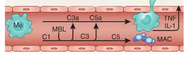
What role do C3a and C5a play in the immune response depicted in the figure?
They promote phagocytosis of bacterial cells by macrophages.
They stimulate the production of antibodies.
They act as chemoattractants, recruiting immune cells to the site of infection.
They inhibit the formation of the membrane attack complex (MAC).
Mutations in the important cancer-critical gene encoding p53 is commonly found in cancers. What type of mutations are these expected to be?
Loss-of-function mutation in both copies.
Loss-of-function mutation in one copy.
Gain-of-function mutation in both copies.
Gain-of-function mutation in one copy.
None of the above.
In intrinsic apoptosis, cytochrome c binds to which protein complex to activate caspase-9?
Apoptosome
Death-inducing signaling complex (DISC)
Death receptor
Mitochondrial membrane
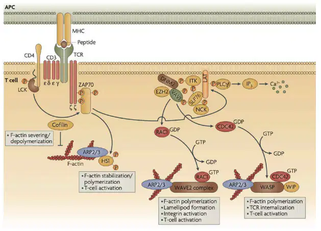
Which molecular process depicted in the figure is involved in the activation of F-actin polymerization and stabilization?
RAC1 activation and the ARP2/3 complex
CDC42 activation and WASP complex
Binding of IP3 to calcium channels
Both A and B

Identify what type of membrane protein A and B are and whether they function by passive or active transport mechanism.
Protein A is a sodium-glucose symporter (active transport), and Protein B is a glucose uniporter (passive transport).
Protein A is a glucose uniporter (passive transport), and Protein B is a sodium-glucose symporter (active transport).
Protein A is a sodium-glucose symporter (passive transport), and Protein B is a glucose uniporter (active transport).
Protein A is an enzyme (active transport), and Protein B is a glucose uniporter (passive transport).

Describe what junction(s) may be involved in the area circled in the diagram and what functional role they play in the function of the tissue.
Tight junctions - prevent leakage of extracellular fluid
Gap junctions - allow communication between cells
Desmosomes - provide mechanical strength
Adherens junctions - connect actin filaments between cells
What did Shemin eat to investigate how heme was made in animals?
A. Radioactive glycine
B. Vitamin C
C. Iron supplements
D. Protein shakes
What are two main destinations for protein after being made in the ER and processed in the Golgi apparatus?
Plasma membrane and extracellular space
Nucleus and mitochondria
Cytoplasm and ribosomes
Endoplasmic reticulum and Golgi apparatus
Proteins that are synthesised in the cytosol are trafficked to the nucleus through which of the following mechanisms?
Through the endoplasmic reticulum
Via nuclear pore complexes
By vesicular transport
Through the Golgi apparatus
Compare and contrast the smooth ER and rough ER.
Smooth ER is involved in lipid synthesis, while rough ER is involved in protein synthesis.
Smooth ER has ribosomes on its surface, while rough ER does not.
Both smooth ER and rough ER are involved in protein synthesis.
Rough ER is involved in detoxification, while smooth ER is not.
What is the purpose of mannose 6-phosphate in cells?
It acts as a signal for protein targeting to lysosomes.
It tags proteins for secretion outside the cell.
It activates proteins for use in the cytosol.
It directs proteins to the plasma membrane.
How is an extracellular signal converted into an intracellular signal?
The signal binds to a transporter protein, which directly moves the signal inside the cell.
The signal binds to a receptor on the cell membrane, triggering a signaling cascade inside the cell.
The signal enters the cell through endocytosis, where it binds to intracellular organelles.
The signal directly passes through the membrane and binds to cytoplasmic proteins.
Which of the following is NOT a way that cells can adjust their sensitivity to an extracellular signal?
By altering the number of receptors on the cell surface
Desensitization by receptor phosphorylation
Receptor endocytosis and degradation
Modifying gene expression to produce more receptors
Reducing the concentration of intracellular second messengers
Listeria monocytogenes uses the bacterially-encoded ActA protein to hijack one part of a eukaryotic cell’s cytoskeleton. Identify the cytoskeleton filament and host accessory protein to facilitate this hijacking
Microtubules, Tubulin
Actin filaments, Profilin
Intermediate filaments, Myosin
Actin filaments, Tropomyosin
Fill in the blanks below with the most appropriate word (2 marks): Cdk levels are __________________ throughout cell-cycle _______________ regulate checkpoints in cycle cell progression
Cdk levels are constant throughout cell-cycle Cyclins regulate checkpoints in cycle cell progression
Cdk levels are variable throughout cell-cycle Proteins regulate checkpoints in cycle cell progression
Cdk levels are decreasing throughout cell-cycle Enzymes regulate checkpoints in cycle cell progression
Cdk levels are increasing throughout cell-cycle Hormones regulate checkpoints in cycle cell progression
Which process describes intrinsic apoptosis?
A process involving the release of cytochrome c from mitochondria
A process triggered by external signals binding to death receptors
A process that involves phagocytosis of apoptotic cells
A process that results in necrosis
It was observed some time ago that the genomes of unicellular eukaryotic cells lack the genes necessary to make proteins associated with tight junctions. Why might this be?
Unicellular eukaryotes do not form tissues so they don't require tight junctions.
They have alternative mechanisms for cell adhesion.
Tight junctions are only used to connect cell membranes to the extracellular matrix.
Their cellular structure does not support tight junction formation.
Which type of cell-cell junction connects actin filaments of adjacent cells?
Tight junctions
Adherens junctions
Desmosomes
Gap junctions
Which of these is NOT a function of the basal lamina?
Provides structural support to epithelial cells by interacting with integrins and other adhesion molecules
Facilitates cell adhesion and acts as a barrier to macromolecules, particularly in filtration processes
Serves as a foundation for cell attachment and regulates cell behavior such as proliferation and differentiation
Directly mediates nutrient exchange between epithelial cells and connective tissues by acting as a conduit for growth factors
Describe one structural characteristic of collagens.
Collagens are composed of two polypeptide chains wound together in a double helix.
Collagens are composed of three polypeptide chains wound together in a triple helix.
Collagens are composed of multiple beta-sheets stacked in a planar configuration.
Collagens are made of a single chain of amino acids arranged in a random coil.
Describe one functional characteristic of collagen
Collagens allow tissues to stretch beyond their natural limits without damage.
Collagens provide structural support and resist stretching forces in connective tissues
Collagens primarily facilitate nutrient exchange in connective tissues.
Collagens function as enzymes that break down tissue during repair processes.
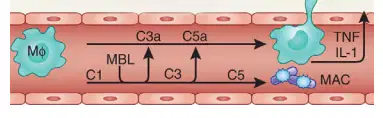
Which of the following innate immune system events likely occurred before the situation depicted in the figure?
Phagocytosis of the bacteria by macrophages
Complement activation through recognition of bacterial cells
Production of memory cells by T lymphocytes
Secretion of antibodies by B cells

What is the first molecular process that occurs after the T cell receptor (TCR) recognizes the antigen presented by the major histocompatibility complex (MHC)?
Activation of ARP2/3 complex
Phosphorylation of ZAP70
Binding of GTP to RAC1
F-actin severing and depolymerization
Which of the following events triggers extrinsic apoptosis?
DNA damage within the cell
Mitochondrial release of cytochrome c
Binding of death ligands to death receptors on the cell surface
Release of calcium ions from the endoplasmic reticulum
In the extrinsic apoptosis pathway, which caspase is directly activated by the death receptor signaling?
Caspase-3
Caspase-8
Caspase-9
Caspase-6
Which of the following events initiates the intrinsic pathway of apoptosis?
Activation of caspase-8
Binding of ligands to death receptors
Release of cytochrome c from the mitochondria
Activation of cell surface receptors

