
Font size
 Worksheets
Human Anatomy and Physiology
Total questions: 16
Worksheet time: 1hrs 3mins

Understanding the Anatomy of the Foot
The human foot is a remarkable structure that plays a crucial role in our ability to walk, run, and jump. It consists of 26 bones, which include 7 tarsal bones, 5 metatarsal bones, and 14 phalangeal bones. These bones are held together by a complex network of ligaments and fascia, and they are moved by numerous muscles. The tarsal bones, which form the ankle, include the talus and calcaneus. Additionally, the navicular, cuboid, and three cuneiform bones make up the instep of the foot.
The Structure of the Foot
The foot is designed to absorb forces and provide a stable base of support. It is also structured to form several arches that are essential for its function. These arches help support body weight, absorb shock during movement, and provide space for blood vessels, nerves, and muscles on the bottom of the foot. There are four main arches in the foot:
Metatarsal Arch: This arch is created by the distal ends of the metatarsals, stretching from the first to the fifth metatarsal.
Transverse Arch: This arch runs across the tarsal bones, forming a half dome shape.
Medial Longitudinal Arch: Originating from the medial side of the calcaneus, this arch extends to the first metatarsal. The main supporting ligament of this arch is the plantar calcaneonavicular ligament, which acts like a spring, returning the arch to its normal shape after being stretched.
Lateral Longitudinal Arch: This arch is located on the lateral side of the foot and follows a similar structure to the medial longitudinal arch, formed by the calcaneus, cuboid, and the fifth metatarsal.
Plantar Fascia and Foot Joints
The plantar fascia, also known as the plantar aponeurosis, is a thick band of fibrous tissue that begins at the calcaneus and extends to the distal ends of the metatarsals. It helps support the foot against downward forces. The joints of the foot are categorized into three regions:
Forefoot: This includes the interphalangeal, metatarsophalangeal, and intermetatarsal joints.
Midfoot: This region consists of the tarsometatarsal and midtarsal joints.
Rearfoot: The subtalar joint is part of this region.

Muscle Movements of the Foot
The movements of the foot are produced by both extrinsic and intrinsic muscles. Extrinsic muscles originate in the lower leg, while intrinsic muscles originate within the foot. Key movements include dorsiflexion, plantarflexion, inversion, eversion, abduction, adduction, pronation, and supination, which are primarily controlled by the extrinsic muscles. The phalanges' movements are managed by the intrinsic muscles.

Nerve and Blood Supply
The nerve supply to the foot comes from the medial and lateral plantar nerves, which are branches of the tibial nerve. These nerves supply all intrinsic muscles on the plantar surface of the foot. The deep peroneal nerve supplies the extensor digitorum brevis on the dorsal surface.
For blood supply, the anterior and posterior tibial arteries primarily supply the foot. The dorsum of the foot receives blood from the dorsal pedal artery and the dorsal metatarsal arteries, which branch from the anterior tibial artery. The plantar aspect is supplied by the lateral plantar artery, medial plantar artery, and the plantar arterial arch, all branching from the posterior tibial artery.
The primary venous structure in the foot is the dorsal venous arch, which drains into the medial and lateral plantar veins, eventually combining to form the posterior tibial vein and the fibular vein.
Conclusion
The anatomy of the foot is intricate, with its bones, muscles, ligaments, and blood supply working together to provide essential functions. Understanding the anatomy of the foot not only helps in appreciating its complexity but also highlights the importance of foot health in our daily activities. Whether we are walking, running, or jumping, our feet play a key role in our mobility and overall well-being.
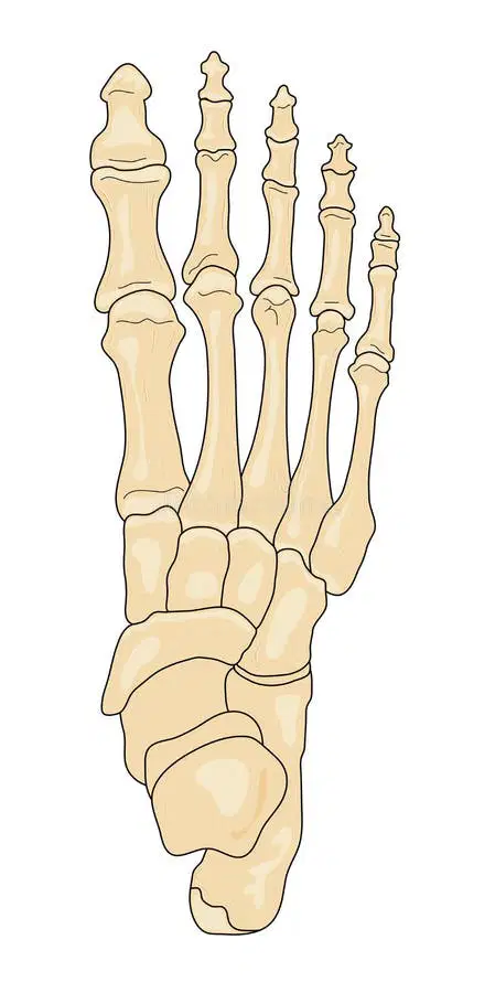
The foot consists of (a) bones, which include (b) tarsal bones, (c) metatarsal bones, and (d) phalangeal bones.
Match the arch with the image and description.

Transverse Arch

Medial Longitudinal Arch

Lateral Longitudinal Arch

Metatarsal Arch
Match the movement with the image

Toe abduction

Toe adduction

Foot pronation

Foot supination

Toe flexion

Understanding the Anatomy of the Ankle and Lower Leg Bones
The anatomy of the human body is intricate and fascinating, particularly when it comes to the lower leg and ankle. This region, which extends from just below the knee to above the ankle, is essential for movement and balance. It consists of two primary bones: the tibia and the fibula. The tibia, which is thicker and located more towards the middle of the body, is often referred to as the shinbone. In contrast, the fibula is thinner and positioned laterally, or on the outer side of the leg.
The Ankle Joint
The ankle joint, also known as the talocrural joint, plays a crucial role in our mobility. It is formed by three main components: the distal end of the tibia, referred to as the medial malleolus; the distal end of the fibula, known as the lateral malleolus; and the talus, a cube-shaped tarsal bone that fits snugly between these two malleoli.
This joint allows for two primary movements: plantarflexion, which is the action of pointing the toes downward, and dorsiflexion, which is bringing the toes upwards towards the shin. The subtalar joint, located below the talus, facilitates additional movements of inversion (turning the sole of the foot inward) and eversion (turning it outward).

Ligaments and Articulations
The stability of the lower leg and ankle is maintained by several ligaments. The tibia and fibula are connected by a fibrous tissue called the interosseous membrane, which runs the entire length of these bones. Additionally, the anterior and posterior tibiofibular ligaments help to secure the connection between the two bones at the ankle.
One of the most important ligaments in this area is the deltoid ligament, which provides stability to the medial side of the ankle. This strong ligament, along with the anatomical structure of the lateral malleolus extending further down than the medial malleolus, limits the ankle's ability to evert. As a result, eversion ankle sprains are less common compared to inversion sprains.
The three key lateral ligaments involved in ankle stability are the anterior talofibular ligament, the posterior talofibular ligament, and the calcaneofibular ligament. While these ligaments help to limit inversion, they are more vulnerable to injury, particularly during activities that involve sudden changes in direction.

Muscles and Movement
The muscles in the lower leg are responsible for facilitating movement at the ankle joint. The main movements associated with the talocrural joint are dorsiflexion and plantarflexion. Dorsiflexion is achieved by the contraction of muscles located anteriorly, while the posterior muscles contribute to plantarflexion.
Inversion and eversion movements occur at the subtalar joint. Muscles that cross the ankle joint laterally cause eversion, and those crossing medially facilitate inversion. This coordinated action of muscles and tendons allows for a wide range of movements necessary for walking, running, and jumping.


Nerve and Blood Supply
The lower leg is innervated primarily by the common peroneal nerve, which branches into two smaller nerves: the superficial peroneal nerve and the deep peroneal nerve. These nerves are crucial for muscle control and sensation in the lower leg and foot. Additionally, the tibial nerve, which runs along the back of the leg, supplies the ankle and foot with nerve signals.
Blood supply to the ankle and lower leg is provided by the anterior tibial artery and the posterior tibial artery. Blood drainage occurs through the peroneal vein, posterior tibial vein, and anterior tibial vein, ensuring that oxygen-rich blood reaches the muscles and tissues, while waste products are efficiently removed.
Surface Anatomy
To better understand the physical layout of the ankle and lower leg, it’s essential to recognize key landmarks. The ankle can be visually assessed from both lateral and medial views, allowing for the identification of the malleoli and other anatomical features. These landmarks are crucial for medical professionals when diagnosing conditions or injuries related to the ankle and lower leg.
In summary, the anatomy of the ankle and lower leg is complex yet vital for everyday movements and activities. Understanding this anatomy helps us appreciate the body's design and the importance of maintaining the health of these structures.
What are the two primary bones in the lower leg?
Tibia and Fibula
Femur and Patella
Radius and Ulna
Humerus and Scapula
Which ligament provides stability to the medial side of the ankle?
What movement is achieved by the contraction of anterior muscles in the lower leg?
Dorsiflexion
Plantarflexion
Inversion
Eversion

Understanding the Anatomy of the Knee
The knee is one of the most complex joints in the human body, playing a critical role in our ability to move and perform daily activities. Its intricate structure allows for a wide range of motion, but it also makes the knee susceptible to injuries, particularly in sports that place extreme stress on this joint. Understanding the anatomy of the knee can help us appreciate its importance and the functions it performs.
Structure of the Knee Joint
The knee joint is primarily a hinge joint, which means its main movements are flexion (bending) and extension (straightening). However, the knee also allows for a small degree of rotation, making it more complex than a true hinge joint. The stability of the knee depends on several key components, including ligaments, the joint capsule, and the muscles surrounding it.
Bones of the Knee
There are four primary bones that make up the knee joint:
Femur - the thigh bone, which is the upper part of the knee.
Tibia - the shin bone, which supports the majority of the body’s weight.
Fibula - a smaller bone located next to the tibia.
Patella - also known as the kneecap, it is located in front of the knee joint.
These bones connect to form several important articulations, including where the femur meets the tibia (the tibiofemoral joint), the femur meets the patella, and the femur meets the fibula.
The joint capsule, which is the largest joint capsule in the body, surrounds the knee. This capsule is lined with a synovial membrane that produces synovial fluid, helping to lubricate the joint.
Menisci
The knee also contains two important fibrocartilage structures called menisci:
Medial Meniscus
Lateral Meniscus
These C-shaped discs sit on top of the tibial plateau (the flat part of the tibia) and enhance the stability of the knee by helping the rounded femoral condyles fit better onto the flat tibial surface. The menisci also help absorb shock and reduce stress on the joint.


Ligaments of the Knee
Ligaments are strong bands of connective tissue that help stabilize the knee joint. The major ligaments include:
Anterior Cruciate Ligament (ACL): This ligament prevents the tibia from sliding forward relative to the femur and helps stabilize the knee during activities such as running and jumping.
Posterior Cruciate Ligament (PCL): This ligament works in opposition to the ACL, preventing the tibia from sliding backward relative to the femur.
Medial Collateral Ligament (MCL): This ligament stabilizes the knee against forces that push the knee inward.
Lateral Collateral Ligament (LCL): This ligament stabilizes the knee against forces that push the knee outward.
Together, these ligaments work to provide stability and support to the knee, allowing us to perform various movements while reducing the risk of injury.
Muscles of the Knee
Several muscle groups are essential for the proper functioning of the knee:
Quadriceps: This group of muscles, located at the front of the thigh, is responsible for knee extension. The quadriceps include the rectus femoris, vastus medialis, vastus intermedius, and vastus lateralis.
Hamstrings: Located at the back of the thigh, the hamstrings facilitate knee flexion. This group includes the biceps femoris, semitendinosus, and semimembranosus.
Other muscles, such as the gastrocnemius and popliteus, also play a role in knee movement, particularly in flexion and rotation.


Nerve and Blood Supply
The proper function of the knee is supported by an intricate network of nerves and blood vessels. The primary nerves that innervate the knee include:
Tibial Nerve: Supplies the hamstrings and gastrocnemius muscles.
Common Peroneal Nerve: Innervates part of the biceps femoris muscle.
Femoral Nerve: Supplies the quadriceps and sartorius muscles.
Blood supply to the knee is primarily provided by branches of the popliteal artery, which emerges from the femoral artery. This ensures that the knee receives the necessary nutrients and oxygen to function effectively.
In conclusion, the knee is a remarkable joint that combines complex structures to allow for movement and stability. Understanding its anatomy can help us better appreciate its role in our daily activities and the importance of taking care of this vital joint.
What is the primary function of the quadriceps muscle group in the knee?
Knee extension
Knee flexion
Knee rotation
Knee stabilization
Which ligament in the knee prevents the tibia from sliding forward relative to the femur?
Anterior Cruciate Ligament (ACL)
Posterior Cruciate Ligament (PCL)
Medial Collateral Ligament (MCL)
Lateral Collateral Ligament (LCL)
What is the role of the menisci in the knee joint?
Enhance stability and absorb shock
Facilitate knee extension
Prevent tibia from sliding forward
Innervate the knee muscles
Match the following
Tibia
Shin bone
Fibula
Lateral ankle bone
Patella
Kneecap
Femur
Thigh bone
Match the following.
Anterior Cruciate Ligament
prevents the tibia from sliding forward
Posterior Cruciate Ligament
prevents the tibia from sliding backward
Medical Collateral Ligament
prevent knee going inward
Lateral Collateral Ligament
prevents knee going outward

Understanding the Anatomy of the Thigh, Hip, Groin, and Pelvic Region
The human body is a complex structure, and understanding its anatomy is crucial for various fields such as medicine, sports science, and physical education. This text will explore the anatomy of the thigh, hip, groin, and pelvic region, focusing on the bones, muscles, nerves, and blood supply involved.
The Thigh
The thigh is the part of the leg that lies between the hip and the knee. It is primarily composed of the femur, which is the longest and strongest bone in the body. The femur is designed to provide maximum support and mobility during weight-bearing activities, such as walking or running. At the upper end of the femur, the proximal head articulates with the acetabulum of the pelvis to form the hip joint. Meanwhile, the distal femoral condyles connect with the tibia at the knee joint.
The Pelvis
The pelvis is a bony ring formed by two innominate bones, the sacrum, and the coccyx. Each innominate bone consists of three parts: the ilium, ischium, and pubis. The pelvis supports the spine and trunk, transferring their weight to the lower limbs. Additionally, it serves as an attachment point for muscles of the trunk and thigh and protects the pelvic organs. The hip and pelvis together form the core for full-body movement, with the body’s center of gravity located just in front of the upper part of the sacrum. Injuries to the hip or pelvis can significantly impact an athlete's mobility and functioning.


Ligaments and Muscles
The hip joint is formed by the articulation of the femur with the innominate bone. The head of the femur fits into a deep socket called the acetabulum, stabilized by strong ligaments and a joint capsule. The sacrum is connected to other parts of the pelvis by strong ligaments, forming the sacroiliac joint, which allows for slight backward and forward movements.
Thigh Muscles
The muscles located on the anterior part of the thigh are known as the quadriceps, which function to extend the knee. One of the quadriceps, the rectus femoris, also assists in flexing the hip. The sartorius muscle, also located on the anterior thigh, flexes the hip and rotates the thigh outward. On the posterior side of the thigh, three hamstring muscles act as flexors of the knee and extend the hip.
The medial muscles, known as the adductor group, include the gracilis, pectineus, and adductor magnus, longus, and brevis. These muscles collectively act as adductors of the hip, bringing the thigh closer to the body's midline.
Hip Muscles
Muscles of the hip can be categorized into anterior and posterior groups. The anterior group includes the iliacus and psoas muscles, which flex the thigh on the trunk. The posterior group comprises the tensor fasciae latae and gluteus medius, which abduct the hip; the gluteus maximus, which extends the hip; and the gluteus minimus, which internally rotates the hip. Additionally, six deep lateral rotators play crucial roles in hip movement.


Nerve Supply and Blood Supply
The nerves that emerge from the sacral plexus include the tibial and common peroneal nerves, which form the largest nerve in the body, the greater sciatic nerve. This nerve supplies the muscles of the thigh and lower leg. The lumbar plexus, consisting of fibers from the first four lumbar nerves, contains the femoral nerve, which branches to supply the thigh and lower leg.
Blood Supply
The main arteries that supply the thigh include the medial circumflex femoral, deep femoral, and femoral arteries. At the level of the fourth lumbar vertebra, the aorta divides into two common iliac arteries, which further divide into internal and external iliac arteries. The external iliac artery is the primary artery supplying blood to the lower limb.
The major veins in this region are the superficial great saphenous and femoral veins. The common iliac vein drains blood from the lower body, while the internal iliac vein ascends to join the external iliac vein, forming the common iliac vein, which drains the pelvis and adjoining areas.
Surface Anatomy
The surface anatomy of the thigh can be observed from both anterior and posterior views. The anterior view displays the quadriceps muscle group, which is vital for knee extension, while the posterior view reveals the hamstring group, crucial for knee flexion. Understanding these surface structures is essential for recognizing muscle function and identifying potential injury sites.
In summary, the anatomy of the thigh, hip, groin, and pelvic region is integral to understanding human movement and function. This knowledge is vital for athletes, medical professionals, and anyone interested in the mechanics of the human body.
What is the primary bone that composes the thigh?
Femur
Tibia
Fibula
Humerus
Which muscle group is responsible for extending the knee?
Quadriceps
Hamstrings
Adductors
Gluteus Maximus
What is the function of the gluteus maximus muscle?
Extends the hip
Flexes the knee
Adducts the thigh
Rotates the thigh outward

Understanding the Anatomy of the Shoulder
The shoulder complex is a vital part of the human body, allowing for a wide range of movements and activities. It consists of three main bones: the clavicle, scapula, and humerus. These bones come together to form four major articulations, or joints: the sternoclavicular joint, the acromioclavicular joint, the glenohumeral joint, and the scapulothoracic joint.
The Bones of the Shoulder Complex
Clavicle: Commonly known as the collarbone, the clavicle serves as a strut between the shoulder blade and the sternum (breastbone). It is the only bone that directly connects the upper limb to the trunk, forming the sternoclavicular joint. Although this joint is relatively weak due to its bony structure, it is supported by strong ligaments that stabilize the clavicle in place.
Scapula: Also known as the shoulder blade, the scapula is flat and triangular in shape. It has several important features, including the acromion process, which is the highest point of the shoulder. The scapula plays a significant role in shoulder motion by providing attachments for various muscles and ligaments.
Humerus: This is the long bone of the upper arm. The head of the humerus fits into the shallow socket of the scapula known as the glenoid cavity, forming the glenohumeral joint, which is commonly referred to as the shoulder joint. This joint is known for its great mobility, allowing the arm to move in multiple directions.
Stabilizing Ligaments
Ligaments are tough bands of tissue that connect bones and provide stability to the joints. In the shoulder complex, several ligaments surround the four joints:
Sternoclavicular Ligament: This ligament supports the sternoclavicular joint, anchoring the clavicle to the sternum.
Acromioclavicular Ligament: This helps maintain the position of the clavicle relative to the acromion process of the scapula.
Coracoacromial Ligament: This ligament connects the coracoid process of the scapula to the acromion, forming an arch that protects the shoulder joint.
Glenohumeral Ligaments
The glenohumeral joint is supported by the glenohumeral ligaments, which form a capsule around the joint. These ligaments, along with the rotator cuff muscles, help to keep the humerus in place within the shallow glenoid cavity. The rotator cuff is a group of muscles that play a crucial role in shoulder stability and movement.
The Muscles of the Shoulder
The muscles that cross the glenohumeral joint enable dynamic motion and provide stability. Movements at this joint include:
Flexion: Raising the arm forward.
Extension: Lowering the arm back down or moving it backward.
Abduction: Lifting the arm away from the body.
Adduction: Bringing the arm back toward the body.
Rotation: Twisting the arm inward or outward.

Muscles acting on the glenohumeral joint can be grouped into three categories:
Muscles from the Axial Skeleton: These include the latissimus dorsi and the pectoralis major, which attach to the humerus and help with powerful movements.
Muscles from the Scapula: This group includes the deltoid, teres major, coracobrachialis, and the rotator cuff muscles (subscapularis, supraspinatus, infraspinatus, teres minor). These muscles are essential for stability and motion at the shoulder joint.
Muscles Connecting the Scapula to the Axial Skeleton: This group includes the levator scapulae, trapezius, rhomboids, and serratus anterior and posterior. These muscles stabilize the scapula, allowing for efficient shoulder movement.


Nerve and Blood Supply
The nerve supply to the shoulder complex comes from a network called the brachial plexus, which is formed by the spinal nerves from the fifth cervical vertebra to the first thoracic vertebra. This network provides peripheral nerves that innervate the muscles of the upper extremity, enabling movement and sensation.
Blood is supplied to the shoulder area through the subclavian artery, which branches out into the axillary artery and then into the brachial artery. This blood supply is crucial for delivering oxygen and nutrients to the muscles and tissues in the shoulder region.
Conclusion
The shoulder complex is an intricate system of bones, ligaments, muscles, and nerves that work together to allow for a wide range of movements. Understanding the anatomy of the shoulder is essential for recognizing its function and importance in daily activities, sports, and rehabilitation. By studying the shoulder's structure and components, we can appreciate how this remarkable joint supports our ability to interact with the world around us.
What are the three main bones that make up the shoulder complex?
Clavicle, Scapula, Humerus
Femur, Tibia, Fibula
Radius, Ulna, Humerus
Sternum, Clavicle, Scapula
Which joint is known for its great mobility, allowing the arm to move in multiple directions?
Glenohumeral joint
Acromioclavicular joint
Sternoclavicular joint
Scapulothoracic joint
What is the role of the rotator cuff muscles in the shoulder complex?
They help to keep the humerus in place within the glenoid cavity.
They connect the clavicle to the sternum.
They form an arch that protects the shoulder joint.
They stabilize the clavicle in place.

Understanding the Anatomy of the Elbow
The elbow is a complex joint that plays a crucial role in the movement of the arm. It allows for a wide range of motions, enabling us to perform everyday activities such as lifting, throwing, and pushing. This text will explore the anatomy of the elbow, including its bones, ligaments, muscles, nerve supply, and blood supply.
Bones of the Elbow
The elbow complex is composed of three primary bones: the humerus, the radius, and the ulna.
Humerus: This is the long bone of the upper arm. The distal end of the humerus forms two important structures known as the medial and lateral epicondyles. These protrusions serve as attachment points for ligaments and muscles that stabilize and move the elbow.
Ulna: The ulna is one of the two bones in the forearm. Its prominent part, known as the olecranon process, articulates with the trochlea and the olecranon fossa on the back of the humerus. This connection is essential for the bending and straightening of the elbow.
Radius: The radius is the other forearm bone, located on the thumb side of the hand. The radial head articulates with the capitulum of the humerus, allowing for smooth movements at the elbow.
These three bones form three separate joints in the elbow complex: the humeroulnar joint, the humeroradial joint, and the radioulnar joint. Flexion (bending) and extension (straightening) primarily occur at the humeroulnar and humeroradial joints. In contrast, the radioulnar joint enables pronation (turning the forearm inward) and supination (turning the forearm outward).
Ligaments of the Elbow
Ligaments are strong connective tissues that stabilize joints. The elbow has several important ligaments:
Ulnar (Medial) Collateral Ligament: This ligament is crucial for providing stability against forces that push the elbow laterally (valgus forces). It extends from the medial epicondyle of the humerus to the proximal ulna.
Annular Ligament: This ligament wraps around the radial head, allowing it to rotate freely while maintaining stability in the elbow. It prevents dislocation of the radius.
Radial (Lateral) Collateral Ligament: This ligament helps stabilize the elbow against forces that push it medially (varus forces). It extends from the lateral epicondyle of the humerus and attaches to the annular ligament.
These ligaments work together to maintain the stability of the elbow during various activities, protecting it from injury.

Muscles of the Elbow
The elbow is also surrounded by several important muscles that facilitate its movement. The main muscles involved include:
Biceps Brachii: This muscle is primarily responsible for flexing the elbow and is located at the front of the upper arm.
Brachialis: Situated underneath the biceps, the brachialis is the primary flexor of the elbow, playing a key role in bending the arm.
Brachioradialis: This muscle assists in flexing the elbow and is located on the outer side of the forearm.
Triceps Brachii: Located at the back of the upper arm, the triceps are responsible for extending the elbow.
Supinator and Pronator Muscles: The biceps brachii and supinator muscles allow for supination of the forearm, while the pronator teres and pronator quadratus act as pronators.
These muscles work in coordination to allow for smooth and controlled movements of the elbow.

Nerve Supply
The movement of the elbow is controlled by several nerves that branch from the brachial plexus (C5-T1). The primary nerves involved include:
Musculocutaneous Nerve: This nerve innervates the biceps brachii and brachialis muscles, enabling elbow flexion.
Radial Nerve: Running along the outer side of the elbow, this nerve controls the muscles that extend the elbow.
Ulnar Nerve: This nerve runs behind the medial epicondyle and is responsible for some flexor muscles in the forearm.
Median Nerve: This nerve innervates several muscles in the forearm and hand, contributing to the overall function of the arm.
Understanding the function of these nerves is essential for recognizing how they contribute to arm movement and coordination.
Blood Supply
The blood supply to the elbow is essential for maintaining healthy tissue and facilitating movement. The subclavian artery branches into the axillary artery and then into the brachial artery. The brachial artery further divides into several branches that supply the elbow:
Deep Brachial Artery
Ulnar Recurrent Artery
Radial Recurrent Artery
These arteries anastomose (connect) with the radial and ulnar arteries of the forearm, ensuring that the elbow receives adequate blood flow.
The veins in the elbow region, including the medial cubital, basilic, cephalic, and brachial veins, work together to drain blood from the elbow area into the axillary vein, completing the circulatory loop.
Conclusion
The elbow is an intricate joint that relies on a combination of bones, ligaments, muscles, nerves, and blood vessels to function effectively. Understanding its anatomy is essential for recognizing how we use our arms in daily life and how we can protect this vital joint from injury. By appreciating the complexity of the elbow, we can better understand the importance of maintaining its health through proper care and exercise.
Which of the following bones is not part of the elbow complex?
Humerus
Radius
Ulna
Femur
What is the primary function of the Ulnar (Medial) Collateral Ligament?
To stabilize the elbow against valgus forces
To allow rotation of the radial head
To stabilize the elbow against varus forces
To flex the elbow
Which muscle is primarily responsible for extending the elbow?
Biceps Brachii
Brachialis
Triceps Brachii
Brachioradialis

Understanding the Anatomy of the Forearm
The forearm is a complex part of the human body consisting of two main bones: the ulna and the radius. These bones are essential for the movement and functionality of the arm, especially when it comes to daily activities like lifting, pushing, and gripping.
The Bones of the Forearm
The ulna is the longer of the two forearm bones and serves as a direct extension of the humerus, which is the bone of the upper arm. It is straight and larger at its upper end, where it connects to the humerus, than at its lower end near the wrist. This shape is important because it helps to stabilize the arm during movement.
In contrast, the radius is located on the lateral side of the forearm and is considered an extension of the hand. The radius is thicker at its lower end, where it connects to the wrist, than at its upper end. This unique structure allows for the rotation of the wrist and the ability to turn the palm up or down—a movement known as supination and pronation.
Articulations of the Forearm
The forearm has three key articulations, which are the connections between the ulna and radius at different points:
Superior Radioulnar Joint: This joint is located near the elbow and allows for the rotation of the radius over the ulna.
Middle Radioulnar Joint: This joint is a fibrous connection that doesn't allow for much movement but helps to maintain the position of the bones.
Distal Radioulnar Joint: Located near the wrist, this joint also allows the radius to rotate over the ulna.
These joints work together to provide flexibility and strength to the forearm, enabling a wide range of motion.
Muscles of the Forearm
The muscles in the forearm can be categorized into two main groups: flexors and extensors.
Flexors and Pronators: These muscles are located on the anterior (front) side of the forearm and are attached to the medial epicondyle of the humerus. Their primary function is to bend the wrist and fingers, allowing for gripping movements. The flexors can be further divided into superficial and deep muscles, with each group having specific functions.
Extensors and Supinators: Positioned on the posterior (back) side, these muscles are attached to the lateral epicondyle of the humerus. They are responsible for straightening the wrist and fingers and for turning the palm upward.
Understanding the roles of these muscles is crucial for activities such as typing, playing sports, and performing artistic tasks.

Nerve and Blood Supply
The nerve supply to the forearm muscles is mainly provided by two nerves:
The median nerve supplies most of the flexor muscles, which are responsible for bending movements.
The radial nerve controls the majority of the extensor muscles, which are involved in straightening movements.
The blood supply to the forearm comes from the brachial artery, which branches into the radial and ulnar arteries within the forearm. This network of arteries ensures that the muscles and bones receive the necessary nutrients and oxygen to function effectively.
Surface Anatomy of the Forearm
To understand the forearm better, it is essential to recognize its surface anatomy. Key landmarks include:
The medial and lateral epicondyles at the elbow, which serve as attachment points for muscles.
The styloid processes at the ends of the radius and ulna, which can be felt at the wrist.
These landmarks are important for medical professionals when assessing injuries or conditions related to the forearm.
Conclusion
The anatomy of the forearm is an intricate system that plays a vital role in daily activities and overall arm functionality. By understanding the bones, articulations, muscles, nerves, and blood supply of the forearm, we gain insight into how our bodies move and function. This knowledge not only enriches our appreciation of human anatomy but also helps in recognizing the importance of maintaining a healthy and active lifestyle.
What are the two main bones of the forearm?
Ulna and Radius
Humerus and Femur
Tibia and Fibula
Scapula and Clavicle
Which joint in the forearm allows for the rotation of the radius over the ulna near the wrist?
Superior Radioulnar Joint
Middle Radioulnar Joint
Distal Radioulnar Joint
Elbow Joint
What is the primary function of the flexor muscles in the forearm?
Bend the wrist and fingers
Straighten the wrist and fingers
Rotate the forearm
Provide blood supply

The Anatomy of the Wrist, Hand, and Fingers
The human wrist, hand, and fingers are intricate structures that play vital roles in daily activities, ranging from simple tasks like writing to complex movements in various sports. Understanding the anatomy of these body parts is essential for recognizing their functions and the potential for injury.
Bones
The wrist is formed by the distal ends of the radius and ulna, two long bones in the forearm. It consists of two rows of carpal bones: a proximal row of four carpal bones and a distal row of four carpal bones, which articulate with five metacarpals. The metacarpal bones connect the carpal bones above and the phalanges below, forming the metacarpophalangeal (MCP) articulations.
Each of the four fingers has three phalanges: proximal, middle, and distal. In contrast, the thumb has only two phalanges. This distinction allows for the unique grip and dexterity that the thumb provides, making it essential for various hand functions.

Ligaments
The wrist is supported by a complex series of ligaments that connect the carpal bones to each other, as well as to the radius and ulna. Notably, two ligaments are of major interest in wrist injuries: the ulnar collateral ligament and the radial collateral ligament. These ligaments help stabilize the wrist during movement.
Additionally, the flexor retinaculum crosses the volar (anterior) aspect of the carpal bones. This ligament forms the roof of the carpal tunnel, a narrow passageway that houses the median nerve. Compression of the median nerve in this tunnel can lead to conditions such as carpal tunnel syndrome, which can cause pain and weakness in the hand.
The interphalangeal joints, which connect the phalanges, also have medial and lateral collateral ligaments, along with a thickened joint capsule on the palmar surface known as the volar plate. This structure helps ensure stability and flexibility in finger movements.
Muscles
The movements of the wrist and hand are controlled by a combination of extrinsic and intrinsic muscles. Extrinsic muscles originate outside the hand but have tendons that travel into the hand, allowing for powerful movements. These muscles are primarily responsible for flexing and extending the wrist and fingers.
On the other hand, intrinsic muscles originate within the hand itself. These muscles allow for finer movements, such as abduction (moving away from the midline), adduction (moving toward the midline), and opposition (the ability to touch the thumb to the fingers). The intrinsic muscles of the hand also support the gripping actions necessary for holding objects.
Understanding these muscle groups is important, as they work together to perform complex actions. For example, while the extrinsic muscles enable a strong grip, the intrinsic muscles provide the dexterity needed for tasks like typing or playing a musical instrument.


Nerve Supply
The hand is primarily supplied by two major nerves: the ulnar and median nerves. The ulnar nerve reaches the hand by passing between the pisiform bone and the hook of the hamate bone. This nerve is responsible for controlling the movements of some of the intrinsic muscles in the hand, particularly those that affect the little finger.
The median nerve enters the palm through the carpal tunnel. It innervates three of the four thenar muscles (the group of muscles at the base of the thumb) and also supplies two of the lumbrical muscles. These nerves play critical roles in both motor control and sensory feedback, allowing for precise hand movements and the ability to feel sensations such as touch and temperature.
Blood Supply
The wrist and hand receive blood from two main arteries: the radial and ulnar arteries. The radial artery supplies blood to the muscles on the radial side (the thumb side) of the forearm and wrist, while the ulnar artery supplies the muscles on the ulnar side (the little finger side).
These arteries form two palmar arches: the superficial and deep palmar arches. These arches branch out into the digital arteries, which supply blood to the fingers. Additionally, the dorsal venous arch located on the back of the hand connects with deeper veins, such as the basilic and cephalic veins, helping to drain blood from the hand and wrist.
Surface Anatomy
Understanding the surface anatomy of the wrist and hand is crucial for both anatomical study and clinical practice. Key landmarks include the bony prominences of the wrist and the contours of the palm and fingers, which can be observed from both dorsal (back) and palmar (front) views.
By studying these anatomical features, students can gain a deeper understanding of how the wrist, hand, and fingers work together to facilitate movement and dexterity in everyday life.
What is the primary function of the intrinsic muscles of the hand?
To provide powerful movements by flexing and extending the wrist and fingers
To allow for finer movements such as abduction, adduction, and opposition
To stabilize the wrist during movement
To supply blood to the muscles on the radial side of the forearm
Which nerve is responsible for controlling the movements of some of the intrinsic muscles in the hand, particularly those affecting the little finger?
Radial nerve
Median nerve
Ulnar nerve
Sciatic nerve
What forms the roof of the carpal tunnel in the wrist?
Ulnar collateral ligament
Radial collateral ligament
Flexor retinaculum
Volar plate

Understanding the Anatomy of the Spine
The spine, also known as the vertebral column, plays a crucial role in the human body. It is one of the most complex regions, composed of numerous bones, joints, ligaments, and muscles that work together to enable movement and support. This intricate structure not only protects the spinal cord and nerves but also allows for flexibility and stability. Understanding the anatomy of the spine is essential for recognizing its functions and the potential implications of injuries, particularly in the cervical region, which can lead to serious consequences.
Bones of the Spine
The spine comprises thirty-three individual bones called vertebrae. These vertebrae are categorized into two main types: movable (or true) and immovable (or false). There are twenty-four movable vertebrae, which include the cervical, thoracic, and lumbar vertebrae. The nine immovable vertebrae are fused together to form the sacrum and coccyx. The design of the spine permits a high degree of flexibility in forward and lateral movements, while limiting backward mobility. Additionally, rotation around a central axis is possible in the neck and lower back areas.


The Cervical Spine
The cervical spine is composed of seven vertebrae, labeled C1 to C7. The first two vertebrae, known as the atlas and axis, have unique structures that allow for the support of the head and enable cervical rotation. This region is particularly vulnerable to injuries, especially in sports, making it essential for coaches and fitness professionals to understand its anatomy and risks.
The Thoracic Spine
The thoracic spine consists of twelve vertebrae, labeled T1 to T12. These vertebrae feature long transverse processes and prominent spinous processes. The first ten thoracic vertebrae articulate with the ribs, which contributes to the stability of the rib cage. Movement in this region is limited compared to other parts of the spine, providing a stable support for the upper body.
The Lumbar Spine
The lumbar spine is made up of five vertebrae, labeled L1 to L5, and is responsible for supporting the lower back. These vertebrae are the largest and thickest in the spinal column, featuring large spinous and transverse processes. Unlike the thoracic region, the lumbar spine allows for a greater range of motion, including flexion and extension, although rotation is somewhat limited due to the structure of the vertebrae.
The Sacrum and Coccyx
The sacrum is formed by the fusion of five vertebrae and connects with the two hip bones to create the pelvis. It plays a vital role in weight transmission during both sitting and standing. Ligaments around the sacrum provide stability to the pelvis. The coccyx, or tailbone, is the most inferior part of the vertebral column, made up of four or more fused vertebrae. It serves as an attachment point for muscles, including the gluteus maximus.
Intervertebral Discs and Articulations
Intervertebral discs are found between each of the cervical, thoracic, and lumbar vertebrae. These discs consist of two main components: the annulus fibrosus, which forms the outer layer, and the nucleus pulposus, a gel-like center that acts as a shock absorber. Together, they help to maintain spinal flexibility and reduce the risk of injury.
Ligaments of the Spine
Several key ligaments are essential in maintaining the stability and integrity of the spinal column. The anterior and posterior longitudinal ligaments run along the length of the spine, while the supraspinous ligament connects the tips of the spinous processes. The ligamentum flavum connects adjacent vertebrae, contributing to the overall stability of the spine.
Muscles Supporting the Spine
The muscles that extend and rotate the spine can be classified into superficial and deep groups. The superficial muscles, known as the erector spinae, consist of three columns: longissimus, iliocostalis, and spinalis. Each column is further divided into regions corresponding to the cervical, thoracic, and lumbar areas. These muscles primarily function to extend the spine.
Deep muscles, which include the interspinalis, multifidus, and rotatores, play a crucial role in spinal movement and stability. Additionally, muscles such as the sternocleidomastoid in the neck and the abdominal muscles assist in flexing the spine.


Spinal Cord and Nerves
The spinal cord is a vital part of the central nervous system, extending from the base of the brain to the first or second lumbar vertebra. It is protected by the vertebral column and serves as the main pathway for transmitting nerve signals between the brain and the rest of the body. Thirty-one pairs of spinal nerves emerge from the spinal cord, categorized into cervical, thoracic, lumbar, sacral, and coccygeal nerves.
Conclusion
Understanding the anatomy of the spine is fundamental for recognizing its importance in overall health and function. With its complex structure and multifaceted roles, the spine not only supports the body but also protects the nervous system. Awareness of the spine's anatomy can help prevent injuries and promote better health practices. As we learn more about its functions and vulnerabilities, we can better appreciate the remarkable design of this crucial body part.
What is the primary function of the spine?
To protect the spinal cord and nerves
To support the body's weight
To facilitate movement and flexibility
All of the above
How many movable vertebrae are in the human spine?
Twenty-four
Thirty-three
Seven
Twelve
Which vertebrae are responsible for supporting the head and enabling cervical rotation?
Atlas and axis
Thoracic vertebrae
Lumbar vertebrae
Sacrum and coccyx

Understanding the Anatomy of the Thorax
The thoracic cavity, commonly known as the chest, is a vital part of the human body. It is located between the base of the neck and the diaphragm, playing a crucial role in housing and protecting essential organs. The thoracic cavity consists of several components, including the thoracic vertebrae, twelve pairs of ribs, their associated costal cartilages, and the sternum. The primary functions of this cavity are to protect vital respiratory and circulatory organs and assist the lungs during breathing.

Structure of the Thorax
Within the thoracic cavity lie the lungs, heart, and thymus. The ribs, which are flat bones, are attached at the back to the thoracic vertebrae and at the front to the sternum. The upper seven ribs are known as sternal or true ribs. Each true rib connects directly to the sternum via a separate piece of cartilage called costal cartilage. The 8th, 9th, and 10th ribs are known as false ribs because they do not attach directly to the sternum; instead, they connect to the 7th rib through common cartilage before reaching the sternum. The 11th and 12th ribs are referred to as floating ribs, as they do not connect to the sternum at all but do have muscle attachments.
Thoracic Muscles
The thorax is also home to important muscles that aid in respiration. There are eleven pairs of both external and internal intercostal muscles located between the ribs. The external intercostal muscles are responsible for elevating the diaphragm during inhalation (inspiration), while the internal intercostal muscles help lower the rib cage during exhalation (expiration).
The Respiratory System
The respiratory system begins with the trachea, or windpipe, which branches into right and left primary bronchi. These bronchi further divide into smaller passages known as bronchioles, which control air resistance and ultimately lead to clusters of air sacs called alveoli. Alveoli are essential for the exchange of oxygen and carbon dioxide with the blood in the capillaries surrounding them. The lungs are elastic organs that expand and contract in response to the diaphragm's movement.
The Diaphragm
The diaphragm is a large, dome-shaped muscle that separates the thoracic cavity from the abdominal cavity. When the diaphragm contracts, it flattens, increasing the volume of the thorax and allowing air to rush into the lungs. Conversely, when the diaphragm relaxes, the elastic components of the lungs and thoracic cage decrease the thoracic volume, facilitating expiration.
Blood Supply to the Lungs
Blood flow to the lungs occurs through the pulmonary arteries, where blood is oxygenated before returning to the heart via pulmonary veins. Deoxygenated blood returns from the bronchi through both bronchial and pulmonary veins.

The Circulatory System and the Heart
The heart is the primary organ responsible for pumping oxygenated blood throughout the body. The coordinated function of the heart, blood vessels, blood, and lungs ensures that oxygen reaches the body’s tissues. The adult heart is located under the sternum, slightly to the left, positioned between the lungs and in front of the vertebral column. It is roughly the size of a clenched fist, extending from the first rib to the space between the fifth and sixth ribs.
The heart consists of four chambers: the right and left atria and the right and left ventricles. Deoxygenated blood returns from the body through the venous system to the right atrium. From there, it passes through the tricuspid valve into the right ventricle, which pumps the blood through the pulmonary valve to the pulmonary artery and into the lungs for oxygenation. The oxygen-rich blood returns to the heart via the pulmonary vein to the left atrium, passes through the mitral valve into the left ventricle, and is then ejected past the aortic valve into the aorta, supplying the entire body.
Heartbeat Cycle
A single heartbeat consists of a contraction of both atria, followed quickly by a contraction of both ventricles. This cycle requires a brief pause between the contractions of the atria and ventricles to allow the ventricles to fill with blood. The contraction of the chambers is known as systole, while relaxation is termed diastole.
Blood Supply to the Heart
The heart itself is supplied by the right and left coronary arteries, which branch off from the aorta. Cardiac veins drain into the right atrium.

The Thymus
The thymus is another important structure located in the thorax, situated just above the heart. Its primary function is to mature lymphocytes into T cells, which are crucial for the immune response against foreign substances. The thymus is relatively large in infants but gradually decreases in size after puberty.
Understanding the anatomy of the thorax is essential for recognizing how our body functions during respiration and circulation. This knowledge highlights the intricate systems that work together to sustain life and maintain health.

What is the primary function of the thoracic cavity?
To protect vital respiratory and circulatory organs and assist the lungs during breathing
To support the abdominal cavity
To aid in digestion
To store energy
Which ribs are known as 'floating ribs'?
11th and 12th ribs
1st and 2nd ribs
8th and 9th ribs
5th and 6th ribs
What is the role of the diaphragm during inhalation?
It contracts and flattens, increasing the volume of the thorax
It relaxes and expands, decreasing the volume of the thorax
It contracts and expands, decreasing the volume of the thorax
It relaxes and flattens, increasing the volume of the thorax

The Anatomy of the Abdomen
The human body is a complex structure made up of various systems that work together to maintain overall health and function. One of the most vital areas of the body is the abdominopelvic cavity, which houses many essential organs and plays a significant role in several bodily functions. This text will explore the anatomy of the abdomen, focusing on its structure, the abdominal muscles, and their functions.
Understanding the Abdominopelvic Cavity
The abdominopelvic cavity is situated between the diaphragm and the bones of the pelvis. It is bounded by the lower ribs, the abdominal muscles, and the vertebral column. This cavity contains vital organs, including those of the digestive system, reproductive system, and urinary system. The diaphragm serves as a critical boundary at the top, separating the thoracic cavity from the abdominal cavity. This separation is essential for proper respiratory function, allowing the lungs to expand and contract effectively.
In addition to these boundaries, the abdominopelvic cavity is home to organs such as the stomach, liver, spleen, pancreas, kidneys, and intestines. Each of these organs plays a unique role in the body, contributing to processes such as digestion, nutrient absorption, and waste elimination. The proper function of these organs is crucial for maintaining overall health, and any disruption can lead to significant health issues.
The Abdominal Muscles
The abdominal muscles are an integral part of the body's core and are essential for various movements and functions. There are four main abdominal muscles: the rectus abdominis, external oblique, internal oblique, and transversus abdominis. Each muscle has specific functions that contribute to the overall strength and stability of the torso.
Rectus Abdominis: This muscle runs vertically along the front of the abdomen and is commonly known as the "abs." It is attached to the rib cage above and the pubic bone below. The rectus abdominis is responsible for trunk flexion, which involves bending the torso forward, as well as rotation and lateral flexion. This muscle also plays a vital role in compressing the abdominal cavity, which is important during activities such as lifting heavy objects.
External Oblique: The external oblique muscles are located on each side of the rectus abdominis. They assist with trunk flexion, rotation, lateral flexion, and compression of the abdominal cavity. The external obliques are particularly important for movements involving twisting and bending, such as when reaching for an object or performing sports activities.
Internal Oblique: Located just beneath the external obliques, the internal oblique muscles also contribute to trunk flexion, lateral flexion, and rotation. These muscles work in conjunction with the external obliques to provide stability and strength to the trunk during movement.
Transversus Abdominis: This is the deepest layer of abdominal muscles. The transversus abdominis plays a critical role in holding the abdominal contents in place and aiding in forced expiration, such as during intense physical activity or coughing. It acts like a natural weight belt, providing support to the spine and pelvis.
All these abdominal muscles work together to perform various essential functions, including defecation, urination, and forced expiration. They provide support for the torso, maintain posture, and allow for a wide range of movements essential to daily life.

Surface Anatomy of the Abdomen
Understanding the surface anatomy of the abdomen is crucial for identifying landmarks and assessing the health of the abdominal organs. The abdomen can be divided into regions, making it easier to locate specific organs and structures. The major surface landmarks include the xiphoid process at the lower end of the sternum, the iliac crests at the top of the pelvic bones, and the pubic symphysis at the front of the pelvis.
In clinical settings, healthcare professionals often use these landmarks to examine the abdomen for signs of illness, tenderness, or abnormalities. For example, pain in the upper right quadrant may indicate issues with the liver or gallbladder, while pain in the lower left quadrant may suggest problems with the intestines.
Overall, the anatomy of the abdomen is vital for understanding how our bodies function. The interplay between the various muscles and organs within this cavity is essential for movement, digestion, and overall health. By studying the anatomy of the abdomen, students can gain valuable insights into the human body and its remarkable capabilities.
What is the primary function of the diaphragm in the abdominopelvic cavity?
To separate the thoracic cavity from the abdominal cavity
To aid in digestion
To support the abdominal muscles
To protect the pelvic bones
Which of the following muscles is responsible for trunk flexion and is commonly known as the 'abs'?
Rectus Abdominis
External Oblique
Internal Oblique
Transversus Abdominis
What is the role of the transversus abdominis muscle?
To hold the abdominal contents in place and aid in forced expiration
To assist with trunk flexion and rotation
To provide stability and strength to the trunk
To compress the abdominal cavity during lifting

Understanding the Abdominal Viscera: The Organs of the Body
The abdominal viscera refer to the collection of organs located within the abdominal cavity. These organs can be divided into two main categories: solid organs and hollow organs. Solid organs include the kidneys, spleen, liver, pancreas, and adrenal glands. Hollow organs consist of vessels, tubes, and receptacles such as the stomach, intestines, gallbladder, and urinary bladder. Understanding these organs is essential for grasping how our bodies function and maintain health.

The Urinary System
The urinary system plays a critical role in filtering waste from the bloodstream and expelling it from the body. The main organs of the urinary system include the kidneys, ureters, and urinary bladder.
Kidneys
The kidneys are two bean-shaped organs located on either side of the spine, at the back of the abdominal cavity. Their main function is to filter metabolic wastes, ions, and drugs from the blood. This filtration process helps to maintain the body’s balance of fluids and electrolytes. Once the kidneys have filtered these substances, they expel them from the body through a process known as urination.
Ureters and Urinary Bladder
The ureters are thin tubes that connect the kidneys to the urinary bladder. They transport urine from the kidneys to the bladder, where urine is stored until it is ready to be expelled. The urinary bladder is a hollow container that lies behind the pubic bone and can expand to hold urine.
The Digestive System
The digestive system is responsible for breaking down food, absorbing nutrients, and eliminating waste. Key organs in this system include the liver, gallbladder, pancreas, stomach, small intestine, and large intestine.
Liver
The liver is the largest internal organ and is located in the right upper quadrant of the body. It performs numerous functions, including detoxifying harmful substances, processing nutrients, and storing excess glucose. The liver also produces bile, a substance that helps digest fats in the small intestine.
Gallbladder
The gallbladder is a small, pear-shaped organ located beneath the liver. It stores bile produced by the liver and releases it into the small intestine after meals to aid in digestion.
Pancreas
Situated between the small intestine and the spleen, the pancreas produces digestive enzymes that break down fats, carbohydrates, and proteins. Additionally, it secretes hormones such as insulin and glucagon, which regulate blood sugar levels.
Stomach
The stomach is located between the esophagus and the small intestine. It serves as a storage and mixing chamber for food, where initial digestion occurs. The stomach secretes acids and enzymes to break down food before it moves to the small intestine.
Small and Large Intestine
The small intestine consists of three sections: the duodenum, jejunum, and ileum. Most digestion and nutrient absorption take place here. The large intestine, which includes the cecum, colon, and rectum, absorbs water and converts the remaining waste into feces.
The Lymphatic System
The lymphatic system is crucial for immune function and fluid balance. Two primary organs in this system are the spleen and thymus.
Spleen
The spleen is the largest lymphatic organ, located on the left side of the body, just beneath the diaphragm. It serves several functions: it acts as a reservoir for red blood cells, regulates their circulation, and produces antibodies to fight infections.
The Reproductive System
The reproductive system differs significantly between males and females. Understanding this system is important for recognizing how human reproduction occurs.
Female Reproductive Organs
In females, the reproductive organs include the ovaries, uterus, uterine tubes (fallopian tubes), and vagina. The ovaries produce and store eggs, releasing one egg each month into the uterine tubes. If fertilized, the egg can implant in the uterus, leading to pregnancy. If not fertilized, the body undergoes menstruation.
Male Reproductive Organs
In males, the reproductive organs consist of the testes, prostate gland, seminal vesicles, and penis. The testes produce sperm and testosterone. The prostate gland contributes fluid to the semen, which is ejected through the urethra during ejaculation.


Conclusion
The abdominal viscera consist of vital organs that perform essential functions in the body. From filtering waste in the urinary system to digesting food in the digestive system and supporting reproduction, these organs work together to maintain health and balance. Understanding these systems is crucial for appreciating the complexity of the human body and the importance of each organ's role in overall well-being.
Which of the following is a solid organ located in the abdominal cavity?
Kidneys
Stomach
Intestines
Gallbladder
What is the primary function of the liver in the digestive system?
Detoxifying harmful substances
Storing urine
Producing insulin
Absorbing nutrients
Which organ stores bile produced by the liver?
Gallbladder
Pancreas
Stomach
Spleen

Anatomy of the Head: Understanding the Bones and Structures
The human skull is a complex structure composed of 22 bones. Except for the mandible (the lower jaw), all skull bones are tightly joined together by immovable joints known as sutures. The cranium, or the protective case for the brain, consists of several key bones: the frontal, ethmoid, sphenoid, two parietal, two temporal, two zygomatic, and the occipital bones. The thickness of the skull varies in different areas, being thinner in the temporal regions, providing insight into its protective roles and structural integrity.
The Brain
The brain, also called the encephalon, is the central component of the nervous system housed within the bony cavity of the cranium. It can be divided into four main sections. The cerebrum, the largest part of the brain, is split into two hemispheres and is responsible for coordinating voluntary muscle activities, interpreting sensory impulses, and managing higher mental functions, including memory, reasoning, intelligence, learning, judgment, and emotions.
The cerebellum, located at the back of the brain, controls the coordination of skeletal muscle movements and is crucial for balance and posture. The pons, which is positioned above the medulla oblongata, regulates sleep, posture, respiration, swallowing, and bladder control. Finally, the medulla oblongata, the lowest part of the brainstem, is vital for controlling essential bodily functions such as heart rate, breathing, and blood pressure. It also manages reflex actions like coughing, sneezing, and vomiting, which are necessary for protecting the respiratory system.

Meninges and Cerebrospinal Fluid
Surrounding the brain and spinal cord are three protective membranes known as meninges. The outermost layer is called the dura mater, which is separated from the bony wall of the skull by a layer of fat that houses important arteries and veins, forming the epidural space. Beneath the dura mater lies the arachnoid layer. The subarachnoid space, located between the arachnoid and the pia mater (the innermost layer), contains cerebrospinal fluid. This fluid plays a critical role in cushioning the brain, protecting it from shocks and impacts.

Anatomy of the Teeth
Teeth are essential for various functions including chewing and speaking. Each tooth is made up of mineral salts, primarily calcium and phosphorus. The visible part of the tooth above the gumline is known as the crown, which is covered by enamel, the hardest substance in the human body. The part of the tooth that extends into the jawbone is called the root, and it is covered by a thin, bony substance known as cementum. Beneath the enamel and cementum lies dentin, a hard material that forms the bulk of the tooth. Inside the dentin is a central canal and chamber that contains the pulp, which is composed of nerves, lymphatics, and blood vessels supplying the tooth.

Anatomy of the Nose
The nose serves several important functions, including cleaning, warming, and humidifying the air we inhale. The external part of the nose is formed by a combination of bone and fibrocartilage. The nasal cavity extends from the nostrils to the choanae, openings that lead into the pharynx. The hard palate separates the nasal cavity from the oral cavity below. A nasal septum divides the nasal cavity into right and left chambers, facilitating airflow and filtration.
Anatomy of the Ear
The ear is critical for hearing and maintaining balance. It consists of three main parts: the external ear, the middle ear (or tympanic membrane), and the internal ear (or labyrinth). The middle and internal ear are specifically structured to transport auditory impulses to the brain. The eustachian tube connects the middle ear to the nose, helping to equalize pressure. The external ear is divided into the auricle (or pinna) and the external auditory canal. The auricle collects and directs sound waves into the canal, where they are processed.

Anatomy of the Eye
The eye is equipped with various protective features. It is securely placed within an oval socket formed by the bones of the head, surrounded by a cushion of soft fatty tissues. Eyelids protect the eye and prevent debris from entering. Eyelashes and eyebrows act as filters to block foreign particles. The inner lining of the eye, the conjunctiva, spreads tears that are secreted by accessory lacrimal glands. A larger gland above the eye produces additional fluid to wash away foreign particles. The eye itself is protected by the sclera, a tough outer layer, and the cornea, a transparent center that allows light to enter. Light passes through the cornea, then through the lens and vitreous body, collectively focusing images on the retina, where they are detected by the optic nerve.
Understanding the anatomy of the head, including the skull, brain, teeth, nose, ear, and eye, reveals not only the complexity of human physiology but also the intricate relationships between these structures and their essential functions for survival and interaction with the environment.
Which bone is not part of the cranium?
Mandible
Frontal
Parietal
Occipital
What is the primary function of the cerebellum?
Coordination of skeletal muscle movements
Interpreting sensory impulses
Regulating sleep and respiration
Controlling heart rate and blood pressure
What is the hardest substance in the human body?
Enamel
Dentin
Cementum
Bone

The Anatomy of the Skin: A Closer Look
The skin is the largest organ of the human body, playing a vital role in protecting us from the environment and helping to regulate various bodily functions. It is composed of three main layers: the epidermis, dermis, and subcutis. Each layer has its own unique structure and function, which contribute to the overall health and protection of the body.
The Epidermis
The epidermis is the outermost layer of the skin and serves as a critical barrier against external threats. It protects against invading microorganisms, foreign particles, chemicals, and harmful ultraviolet (UV) rays. One of the key functions of the epidermis is to prevent the loss of water and electrolytes, which is essential for maintaining hydration and overall health.
Within the epidermis, the stratum corneum is a sub-layer that acts as a permeability barrier. It allows only small molecules to diffuse into the lower regions of the epidermis, thus protecting against environmental irritants, toxins, and pathogens. Another important component of the epidermis is the melanin layer, which determines skin pigmentation and provides protection from the damaging effects of UV radiation.
The Dermis
Beneath the epidermis lies the dermis, a thicker layer composed mainly of connective tissue. The dermis provides mechanical support to the epidermis and contains a rich supply of blood vessels, nerves, sweat glands, hair follicles, and sebaceous glands.
One of the notable features of the dermis is the presence of dermal papillae, which are small, finger-like projections that extend into the epidermis. This interlocking arrangement increases the surface area of contact between the dermis and epidermis, helping to anchor the two layers together and prevent the epidermis from slipping away.
Adnexal Structures: Hair, Sebaceous Glands, and Sweat Glands
Hair follicles, which are located within the dermis, play an important role in the structure of the skin. Hair grows from these follicles and extends into the dermis, where it receives nourishment from blood capillaries. Small muscles called arrectores pilorum are attached to each hair follicle. When these muscles contract—often in response to cold or fear—they pull the hair upright, causing the familiar “goosebumps” effect. This reaction helps to increase the insulating effect of hair, aiding in the body’s temperature regulation.
Surrounding the hair follicles are sebaceous glands, which secrete an oily substance known as sebum. This oil helps to keep the hair and skin moisturized and protects against bacterial infections.
Sweat glands are another crucial component of the skin. They help cool the body by producing sweat, which evaporates from the skin surface. There are two types of sweat glands: eccrine and apocrine. Eccrine glands are present at birth and are found throughout the skin, while apocrine glands develop during adolescence and are larger, typically located in areas with hair, such as the armpits and groin. The sweat produced by these glands contains antibacterial agents that help control infections.
Nails and Sensory Nerve Endings
Nails are another important feature of the skin. They are composed of hard, keratinized cells that protect the tips of our fingers and toes. The nail matrix, located beneath the proximal nail fold, is responsible for the growth of the nail plate.
The dermis also contains sensory nerve endings that provide our sense of touch. These peripheral nerves allow us to detect sensations such as pressure, temperature changes, and pain, enabling us to interact with our environment safely.
The Subcutis
The innermost layer of the skin is the subcutis, which is primarily composed of fat. Subcutaneous fat serves several essential functions: it provides insulation to help regulate body temperature, stores energy, and increases the mobility of the skin over the underlying tissues. This layer plays a crucial role in protecting internal organs and maintaining overall body health.
In conclusion, the anatomy of the skin is complex and multifaceted. Each layer—the epidermis, dermis, and subcutis—works in harmony to protect the body, regulate temperature, and provide sensory information. Understanding the structure and function of the skin is essential for appreciating how it contributes to our overall health and well-being.
What is the primary function of the epidermis?
To act as a barrier against external threats
To provide mechanical support to the skin
To store energy and provide insulation
To produce sweat and regulate temperature
Which layer of the skin contains hair follicles and sebaceous glands?
Epidermis
Dermis
Subcutis
Stratum corneum
What is the role of the subcutis layer in the skin?
To protect against UV radiation
To provide insulation and store energy
To act as a permeability barrier
To produce melanin
Understanding the Immune System: Our Body's
Defense Mechanism
The immune system is a complex network of cells and molecules that work together to protect the body from harmful invaders, known as pathogens. Pathogens can include bacteria, viruses, fungi, and parasites. They can enter our bodies through various means, such as cuts, inhalation, or ingestion. The immune system is equipped with defenses that are both immediate and long-lasting, ensuring that we can fight off infections effectively.
The Two Branches of the Immune System
The immune system consists of two main branches: the innate immune response and the adaptive immune response.
Innate Immune Response
The innate immune response is the body's first line of defense against pathogens. This response is non-specific, meaning it does not target specific pathogens but rather acts against all types of invaders. When a pathogen breaches the body’s physical barriers—like the skin and mucous membranes—the innate immune cells quickly recognize the threat.
Key players in the innate immune response include:
Neutrophils: These cells are among the first to arrive at the site of infection. They can engulf and destroy pathogens using antimicrobial molecules.
Macrophages: These are larger immune cells that also engulf and digest pathogens. They play a crucial role in alerting the adaptive immune system by presenting pieces of the invader, known as antigens, on their surface.
Dendritic Cells (DCs): Similar to macrophages, dendritic cells capture pathogens and present antigens to the adaptive immune cells, activating them.
Adaptive Immune Response
The adaptive immune response is more specialized and takes longer to activate, usually days to weeks. It is tailored to recognize specific pathogens and remember them for future encounters. The key players in this response are B cells and T cells.
B Cells: When B cells recognize a foreign antigen, they differentiate into plasma cells. Plasma cells are responsible for producing antibodies. Antibodies are proteins that bind to specific regions on pathogens, neutralizing them and preventing them from entering host cells.
T Cells: There are different types of T cells, including helper T cells and cytotoxic T cells. Helper T cells assist B cells in recognizing antigens and can also activate other immune cells. Cytotoxic T cells directly kill infected cells.
Immunological Memory
One of the most remarkable features of the adaptive immune response is its ability to create immunological memory. After an initial exposure to a pathogen, some B cells and T cells become memory cells. These memory cells remain in the body for long periods and enable a faster and more effective response if the same pathogen invades again. This is why vaccinations are effective; they prepare the immune system by mimicking a real infection without causing disease.
Types of Immunity
Immunity can be classified into two main categories: natural immunity and acquired immunity.
Natural Immunity
Natural immunity develops as a result of natural exposure to pathogens. When the immune system successfully eliminates an invading pathogen, it becomes primed to respond quickly and effectively if that pathogen reappears. This is often referred to as innate immunity.
Acquired Immunity
Acquired immunity can develop in two ways:
Artificially: This occurs through vaccination, where a person is exposed to weakened or inactive parts of a pathogen. The immune system responds by creating memory cells, preparing the body for future infections.
Passively: In some cases, antibodies can be injected into a person to provide immediate protection. This is often used in emergency situations or to protect individuals who cannot produce their own immune response.
Conclusion
The immune system is a vital part of our health, working tirelessly to defend against invading pathogens. Understanding how it operates—from the immediate response of the innate system to the specialized actions of the adaptive system—helps us appreciate the complexity and efficiency of our body's defense mechanisms. Through vaccinations and natural exposure, we can enhance our immunity and better protect ourselves from infectious diseases. As science continues to advance, we gain more insights into how to strengthen our immune responses and improve public health.
What are the two main branches of the immune system?
Innate immune response and adaptive immune response
Innate immune response and acquired immune response
Adaptive immune response and natural immune response
Natural immune response and acquired immune response
Which cells are responsible for producing antibodies in the adaptive immune response?
B Cells
T Cells
Neutrophils
Macrophages
What is the role of cytotoxic T cells in the immune system?
Directly kill infected cells
Assist B cells in recognizing antigens
Engulf and destroy pathogens
Present antigens to adaptive immune cells
Understanding the Endocrine System
The endocrine system is a complex network of glands that plays a crucial role in regulating various functions in the human body. It is responsible for producing and secreting hormones directly into the bloodstream, which then transport these chemical messengers to different target organs and cells. Hormones are vital for regulating numerous body functions, including growth and development, tissue repair, metabolism, reproduction, sexual function, sleep, and even mood.
Key components of the endocrine system include several primary glands: the pituitary gland, pancreas, hypothalamus, thyroid gland, parathyroid glands, testes, ovaries, adrenal glands, and parts of the gastrointestinal tract. Each of these glands serves a specific purpose, selecting and extracting chemicals from the blood, processing them, and ultimately secreting transformed chemical products. These products coordinate the functions of one or more organs throughout the body.
One of the most important glands in the endocrine system is the hypothalamus, located in the brain. It acts as the neural control center, regulating the function of other endocrine glands. The hypothalamus sends signals in the form of releasing hormones, which stimulate specific glands to secrete stimulating hormones into the bloodstream. This process is essential for maintaining homeostasis, or balance, within the body.
The endocrine system operates through a feedback mechanism. When the concentration of a hormone increases in the blood, the hypothalamus and the respective gland will reduce the secretion of the stimulating hormone. This feedback loop helps to maintain stable blood concentrations of hormones, ensuring that the body functions efficiently.
In summary, the endocrine system is vital for the proper functioning of the human body. By producing and regulating hormones, it influences a wide range of processes, from growth to mood regulation. Understanding how this system works is essential for appreciating the complexity of human biology and health.
What is the primary function of the endocrine system?
To produce and secrete hormones into the bloodstream
To digest food and absorb nutrients
To transport oxygen throughout the body
To protect the body from infections
Which gland acts as the neural control center in the endocrine system?
Pituitary gland
Thyroid gland
Hypothalamus
Adrenal gland
How does the endocrine system maintain stable blood concentrations of hormones?
Through a feedback mechanism
By continuously increasing hormone production
By storing excess hormones in glands
By rapidly breaking down hormones

