
 Worksheets
Test 2 - DS 3361 Ch9 The Liver SP25
Total questions: 50
Worksheet time: 25mins
You are scanning a patient with a known mass in the medial segment of the left lobe of the liver. What landmark can you use to identify the left medial segment separate from the anterior segment of the right lobe of the liver?
Lt portal vein
Ligamentum teres
Ligamentum venosum
Middle hepatic vein
You are asked to rule out the presence of a recanalized paraumbilical. Which anatomic structure is a useful landmark in location of this structure?
Ligamentum teres
Ligamentum venosum
Coronary ligament
Hepatoduodenal ligament
You have detected a mass anterior and to the left of the ligamentum venosum. This mass is located in what lobe of the liver?
Left lobe
Caudate lobe
Right lobe
Quadrate lobe
All of the following are sonographic appearance of cavernous hemangioma except
Hyperechoic with posterior enhancement
Well define borders
May project with calcifications
Elevated serum bilirubin
A patient is referred with RUQ tenderness and a history of oral contraceptive use. A solid hypoechoic mass is identified in the Rt lobe of the liver. Color Doppler reveals hypervascularity of the mass. Which of the following scenarios is most likely?
Hydatid liver disease
Hepatic lipoma
Hepatic abscess
Hepatic adenoma
A liver ultrasound on a 49 yr old obese male demonstrates diffuse increased echogenicity with a focal hypoechoic area anterior to the portal vein. This most likely represents
Liver cirrhosis with hepatocellular carcinoma
Hydatid disease
Fatty metamorphosis of the liver with focal sparing
Metastatic disease most likely due to a colon primary
You are scanning a patient with suspected liver cirrhosis. All of the following are sonographic features of cirrhosis except
surface nodularity
increased vascular markings
altered echo texture
ascites
An ultrasound evaluation of liver cirrhosis should include a search for which associated complication?
portal hypertension
mesenteric ischemia
splenic infarction
cholelithiasis
Ultrasound findings of an abdominal study on a 51 yr old female include enlargement of the hepatic veins and IVC in an otherwise normal appearing liver. These findings are most consistent with which of the following?
budd chiari syndrome
rt sided heart failure
lt side heart failure
portal hypertension
Focal fatty liver is most commonly found in which location?
Medial to the ascending branch of the Lt portal vein
Anterior to the right hepatic vein
Lateral, inferior tip of the Rt lobe
Anterior to the portal vein at the porta hepatis
A 52 yr old male with known liver cirrhosis presents for an abdominal ultrasound. You will carefully evaluate the liver to rule out the presence of any focal mass because of which statement below?
Patients with liver cirrhosis are at increased risk for hepatocellular carcinoma
Patients with liver cirrhosis tend to develop multiple cysts in their liver and pancreas
Metastatic disease occurs commonly with cirrhosis
The presence of regenerative nodules rules out cirrhosis.
You have examined a patient with a diagnosis of acute hepatitis. What are the ultrasound features correlates to this diagnosis?
Liver parenchyma is more echogenic
Hepatosplenomegaly
Gallbladder wall markedly thickened
All of the above
A patient is referred to rule out hepatomegaly. All of the following are useful indicators of hepatomegaly except
Rounding of the inferior border of the liver
Longitudinal measurement of the right lobe exceeding 15.5 cm
Extension of the right lobe inferior to the lower pole of the right kidney
Increased diameter of the main portal vein greater than 1 cm
You have identified a single homogeneous hyperechoic lesion measuring 2.4cm in the posterior aspect of the right lobe of the liver. What is the most common etiology of a mass fitting this description?
Hepatic adenoma
Cavernous hemangioma
Hepatocellular carcinoma
Focal fatty sparing
A patient is referred for a sonogram of the liver to rule out metastatic disease. Which of the following describes the sonographic appearance of liver metastasis?
Single hypoechoic mass
Multiple hyperechoic masses
Masses of mixed echogenicity
All of the above appearances of liver metastasis may be encountered
A single large, well defined mass with smooth walls and homogeneous low level echoes are seen within the anterior right lobe of the liver in a 48 yr old female. No Doppler signals could be obtained within the mass. Which of the following conditions is the most likely etiology of this mass?
Kaposi's sarcoma
Focal nodular hyperplasia
Hepatocellular carcinoma
Hemorrhagic cyst
You are scanning a patient with a history of fever, abnormal liver function tests, and RUQ tenderness. The liver is enlarged with increased echogenicity, the gallbladder wall is thickened and thick echogenic bands are noted surrounding the portal veins. Which of the following conditions is most likely?
Fatty liver
Cirrhosis
Budd-Chiari syndrome
Hepatitis
You are evaluating a suspicious lesion to look for gas bubbles to confirm the presence of liver abscess in a patient with fever and increased WBC count. What is the sonographic appearance of the gas bubbles.
Brightly echogenic echoes with clean distal acoustic shadow
Brightly echogenic foci associated with echogenic ringdown artifact
Hypoechoic areas within the mass associated with increased through transmission
Anechoic foci with distal acoustic enhancement
Which of the following is associated with infestation by a parasite and is most prevalent in sheep and cattle-raising countries?
Budd-Chiari syndrome
Hydatid disease
Candidiasis
Hepatitis A
You are scanning the liver and notice irregularity of the surface. A nodular liver surface is associated with which of the following abnormalities?
Cirrhosis
Acute hepatitis
Fatty liver
Polycystic liver disease
The most common benign tumor in the liver is
Focal nodular hyperplasia
Hepatic adenoma
Hepatic lipoma
Cavernous hemangioma
Which of the following is most commonly associated with invasion of the portal vein?
Hepatocellular carcinoma
Cavernous hemangioma
Liver metastases
Hepatic adenoma
You have been asked to perform a liver sonogram on a patient with AIDS. Which of the following tumors is most commonly associated with this history?
Hepatocellular carcinoma
Kaposi's sarcoma
Hemangiosarcoma
Hepatic adenoma
You are scanning a 53 yr old female with a history of recent weight loss and vague abdominal pain. The liver is markedly heterogeneous and contains numerous calcified lesions. This most likely represents metastatic disease from which primary?
Non Hodgkin lymphoma
Cystadenocarcinoma of the ovary
Lung
Adenocarcinoma of the colon
During an ultrasound evaluation of the liver, a bull's eye or target lesion is identified in the anterior right lobe. The most likely etiology of this mass is
Liver abscess
Hepatic adenoma
Hepatocellular carcinoma
Liver metastasis from lung cancer
You are performing an ultrasound exam on a young female and notice a well defined solitary mass with a central scar measuring 4cm in diameter. Color Doppler reveals prominent blood vessels coursing within the scar. This most likely represents
Liver abscess
Hepatic adenoma
Focal nodular hyperplasia
Liver metastasis from lung cancer
You are performing a liver sonogram on a young female with right upper quadrant pain, sudden onset ascites and hepatomegaly. You have obtained transverse and sagittal images of the liver, common bile duct, and gallbladder according to your protocol. What else should you do?
Nothing the study is complete
Expand the study to include the kidneys to rule out associated hydronephrosis
Use color and spectral Doppler to determine patency of the portal and hepatic venous systems
Call the referring physician to get an order to perform a pelvic study to see if the patients pain is referred from an ovarian mass.
Which of the following is not true regarding cavernous hemangiomas?
Small, well defined, hyperechoic masses
Consists of a vascular network
More common in women than men
Show prominent 'water lily' sign.
Hepatofugal flow in the portal vein is a sign of
Normalcy
Hepatic artery thrombosis
Portal hypertension
Acute cholecystitis
A patient is referred for abdominal ultrasound with a high fever and RUQ pain. You document the presence of a large, rounded, homogeneous mass with low level internal echoes and poorly defined borders. The mass is located in the right lobe of the liver, adjacent to the capsule and shows increased through transmission. This most likely represents
Hemorrhagic cyst
Abscess
Hematoma
Choledochal cyst
You are scanning a patient with known liver cirrhosis and notice a focal mass within the posterior right lobe. What laboratory test would be most helpful in determining if this mass is hepatocellular carcinoma?
Serum alpha-fetoprotein
Alk phos
Serum bilirubin
Serum creatinine
You have been asked to perform an ultrasound to rule out the presence of Budd-Chiari syndrome. You will tailor your exam to include which of the following?
Volume measurement of the spleen
Doppler analysis of the hepatic venous system
Both supine and upright views of the porta hepatitis
Careful search for periaortic lymphadenopahty
You are scanning a patient with liver cirrhosis and suspected portal hypertension. In this study, assessment of the size of which of the following is most important?
Spleen
CBD
Rt hepatic vein
IVC
A recanalized paraumbilical vein may be seen as a result of which of the following
Hepatic adenoma
Portal hypertension
Hepatitis
Hepatic trauma
Which of the following would not be a sonographic finding of Budd-Chiari syndrome?
Liver appears hypoechoic in the early stages of acute thrombosis
Patent hepatic veins
In chronic cases the hepatic veins are easily studied with color Doppler
Liver appears hyperechoic and inhomogeneous in later stages
Which measurement below is the diagnostic cutoff for portal vein enlargement?
13 mm
5 mm
10 cm
1.1 cm
You are performing an ultrasound exam in a patient with a history of alcoholic liver cirrhosis. You have documented the presence of splenomegaly and dilated veins at the splenic hilum. Considering the patient's history and findings, what else should you look for?
Search for signs of acute cholecystitis
Carefully scan the spleen for the presence of infarcts
Search for the presence of portosystemic collaterals
Rule out the presence of an aortic aneurysm
You are scanning a patient with an enlarged caudate lobe and shrunken right lobe. What diffuse liver process should you suspect?
Cirrhosis
Acute hepatitis
Fatty infiltration
Hepatocellular carcinoma
You are scanning an obese patient to rule out fatty liver. Which of the following describes a common sonographic appearance of this condition?
Increased through transmission throughout the hypoechoic liver
Increased echogenicity of the liver compared to the normal kidney
Focal hypoechoic masses throughout both lobes of the liver surrounded by normal liver echotexture
Shrunken liver with surface nodularity
'Water lily sign' is a sonographic feature of
Liver adenoma
Echinococcal cyst
Hemangioma
Budd-Chiari syndrome
Passive hepatic congestion is developed due to
Hepatic cyst
Hepatic adenoma
Systemic hypertension
Congestive heart failure
Sonographic findings of the 'Hepatic candidiasis' are
Bull eye or target lesion
wheel within a wheel
small hypoechoic masses with echogenic center cores
All of the above
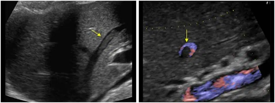
The image above represents
Thrombus in the main portal vein
Thrombus in a hepatic vein
Recannalized paraumbilical with thrombus
Thrombus in the hepatic artery
A 51 yr old man is seen at the radiology department for an abdominal ultrasound. He has elevated liver enzymes levels and increasing abdominal girth. No previous films are available for comparison. History included cholecystectomy complicated by biliary obstruction 5 years previously. The abdominal ultrasound reveals a homogeneous liver with increased echogenicity. No distinct solid or cystic masses are noted, but free fluid is noted within the abdomen. The hepatic vasculature is difficult to evaluate and the hepatic parenchyma is difficult to penetrate. Which of the following would be the best diagnosis for this patient
Hepatocellular carcinoma
Acute hepatitis
Cirrhosis
Fatty liver disease
A 41 yr old woman with long standing cirrhosis undergoes an abdominal ultrasound scan. serum bilirubin and alkaline phosphatase levels are elevated. The liver is small and difficult to penetrate. A sonolucent area is noted between the lateral and medial segment of left lobe and shows flow with color Doppler. Based on these findings what is the most likely diagnosis.
Hepatofugal flow in the hepatic artery
Hepatoma
Recanalized umbilical vein
Normal Rt hepatic vein flow
A 38 yr old woman with a history of Glycogen storage disease is seen for an abdominal ultrasound because of RUQ pain. Two hyperechoic lesions are identified in the liver. Doppler reveals flow around the periphery of each of the lesions. This would be most consistent with which of the following?
Hemangioma
Hepatocellular carcinoma
Metastasis
Liver cell adenoma
A solitary anechoic lesion is identified in the Rt lobe of a 50 yr old woman. The lesion is 2.5cm with smooth walls and acoustic enhancement. The patient is asymptomatic. The remainder of the ultrasound is unremarkable. The anechoic lesion most likely represents
An abscess
A liver cyst
A hematoma
A hepatoma
A Splenorenal shunt
Attaches the mid distal SMV to the IVC
Attaches the MPV to the SMV-Splenic vein confluence to the anterior aspect of the IVC
Attaches the splenic vein to the lt renal vein
Attaches the lt renal vein to the portal vein
A 27 year old man undergoes a RUQ ultrasound in the emergency room. The patient has RUQ pain, elevated liver enzymes, and a history of drug abuse. Ultrasound reveals a hypoechoic liver with no distinct masses. Which of the following would be the best diagnosis for this patient?
Hepatocellular carcinoma
Acute hepatitis
Cirrhosis
Fatty liver disease
The most common benign liver tumor associated with glycogen storage disease is
Cavernous hemangioma
Metastasis
Hepatic adenoma
Lipoma

