
 Worksheets
CVS Review
Total questions: 38
Worksheet time: 19mins
What are arrhythmias?
Normal heart rates produced through the SA node
Any abnormalities in pattern of cardiac activities
Flow of blood through the ventricles.
Normal patterns of cardiac activity
What is the purpose of the heart valves?
To minimize heart arrhythmias
To protect the chordae tendiae
To allow blood to move from the ventricles to the atria
To prevent any backflow of blood between atrium and ventricles
What blood vessel branches from pulmonary trunk?
Pulmonary arteries
Pulmonary valve
Pulmonary veins
Superior vena cava
What happens in ventricular systole
atria contracts
ventricles relax
ventricles contract
both ventricles and atria relax
What happens during ventricular diastole
atria relax
ventricles relax
ventricles contract
both contract
What happens in atrial systole
atria relax
ventricles contract
atria contract
atria and ventricles contract
What happens during atrial diastole
atria relax
ventricles relax
atria contract
neither
What are intercalated discs
neighboring cells that each cardiac muscle are bound to help transmit action potentials to networks of cells
support cells for conduction transmission
aid in the transmission of the SA node conduction pathway
provide cardiac muscle with structure
What is the role of myocardium during contraction?
contains the purkinje fibers for the signal transduction
contains the connective tissue for the heart to expand
muscle that regulates the heart rate pulse
the muscle that produces a twisting and squeezing action during a heart contraction
What is known as the "pacemaker"
AV node
Purkinje fibers
SA node
Bundle of His
Which blood vessel delivers oxygenated blood to the left atrium?
Pulmonary arteries
Pulmonary veins
Superior vena cava
Inferior vena cava
What are the 4 chambers of the heart?
bicuspid, mitral, pulmonary, aortic
right atrium, left atrium, right ventricle, left ventricle
left atrium, bicuspid, left ventricle, aortic
right atrium, tricuspid, right ventricle, pulmonary
Which blood vessel returns systemic blood to the heart?
pulmonary veins
coronary arteries
superior/inferior vena cava
common carotid artery
What is the superior portion of the heart called?
apex
head
base
cardio head
What is the distal end of the heart called?
apex
inferior head
base
cardiac notch
The heart consists of what type of muscle?
skeletal
connective
cardiac
smooth
What is the role of the interventricular septum
separate the left and right atrium
separate the left and right ventricles
to connect the left and right ventricles
to prevent back flow of blow
Where is the heart located within the body?
chest
thorax
pleural cavity
mediastinum
What is a fast HR called?
bradycardia
tachycardia
hypercardia
hypocardia
What is a slow HR called?
hypocardia
tachycardia
hypercardia
bradycardia
What best describes capillaries?
transports blood away from the hear
functions at the blood reservoir
vessels that permit exchange of gases, nutrients, and waste between blood and tissue
they are located between the pulmonary trunk and pulmonary veins
During an ECG, what happens at the P wave
depolarization of the atria
repolarization of the atria
ventricular repolarization
depolarization of the ventricle
During an ECG, what happens at the QRS complex?
relaxation of ventricles
contraction of atria
depolarization of ventricles
depolarization of atria
During an ECG, what occurs at the T wave?
ventricular depolarization
contraction of ventricles
atrial repolarization
ventricular repolarization
When is blood pressure the strongest in arteries?
atrial systole
ventricular diastole
ventricular systole (contraction)
atrial contraction
What are the branches of the aorta?
brachiocephalic trunk, left common carotid, left subclavian
left and right brachiocephalic
left common carotid, left subclavian, right common carotid, right subclavian
left and right common carotid
Where does the external jugular veins drain blood from?
drain the brain, and deep parts of the face and neck
drain the face, scalp, and superficial neck
drain the superior neck
drain the superficial posterior aspect of the scalp
What is a normal BP reading?
80/120
140/95
95/140
120/80
How does vasodilation affect BP?
steady BP
increases BP
decreases BP
accelerate BP
How does vasoconstriction affect BP?
increases BP
decreases BP
slows BP
steady the BP
What organ helps veins return blood to the heart?
cardiac muscle
smooth muscle
skeletal muscle
Explain Starling's Law
the greater the length of the stretched cardiac muscle fiber, the stronger the ventricular contraction that will follow
the greater the length of the stretched cardiac muscle fiber, the weaker the ventricular contraction that will follow
the short the length of the stretched cardiac muscle fiber, the stronger the stronger the ventricular contraction that will follow
the length has no relationship with the strength of the ventricular contraction
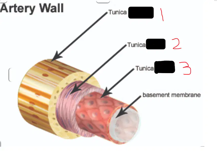
Identify the layers of the blood vessel
1: tunica interna 2: tunica media 3: tunica externa
1: tunica externa 2: tunica media 3: tunica interna
1: tunica media 2: tunica interna 3: tunica externa
1: tunica externa 2: tunica interna 3: tunica media
What is the difference in structure between veins and arteries
arteries have thin muscle layers and large lumen, veins have thick muscle layer and small lumen
arteries have thicker muscle layer and smaller lumen, veins have thin muscle layer and large lumen
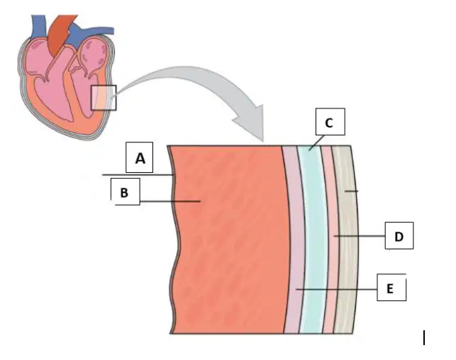
Identify letter A
parietal pericardium
endocardium
myocardium
visceral pericardium
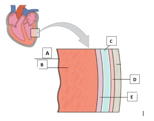
Identify letter B
myocardium
endocardium
parietal pericardium
visceral pericardium
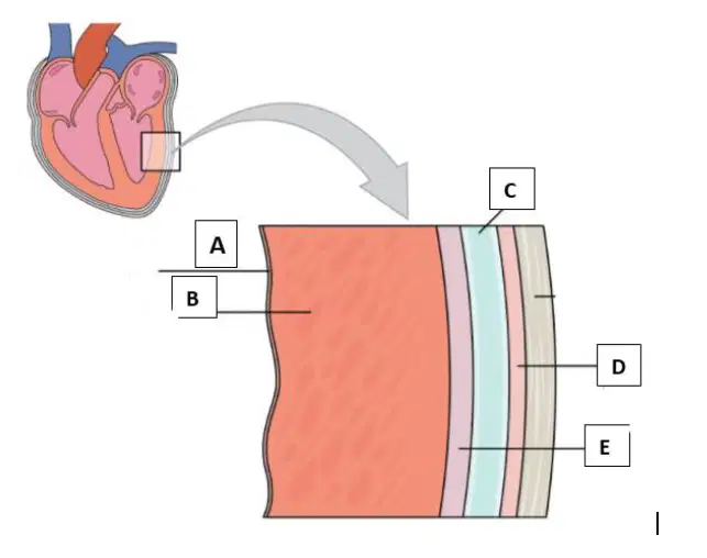
Identify letter E
endocardium
myocardium
visceral pericardium
parietal pericardium
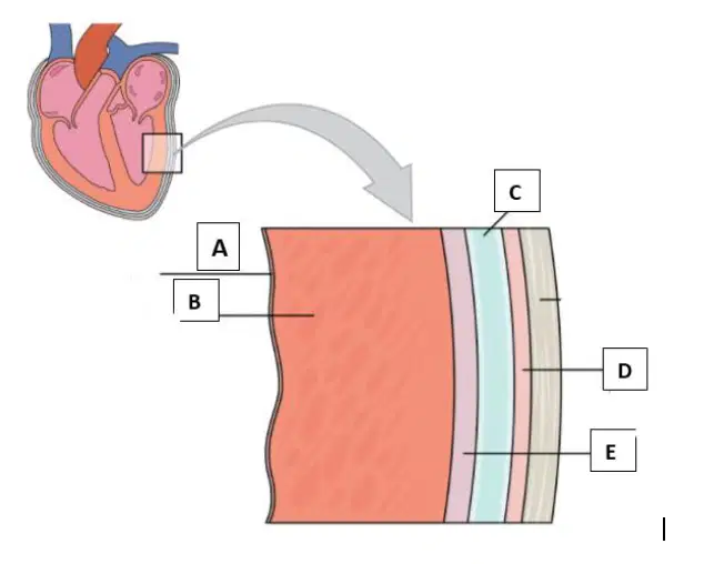
Identify letter D
myocardium
Parietal pericardium
visceral pericardium
endocardium

