
 Worksheets
Basics of the Heart
Total questions: 93
Worksheet time: 59mins
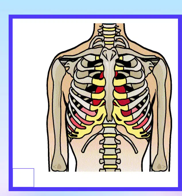
Where is the heart located?
Center of chest
Left leg
Right arm
Back of the head
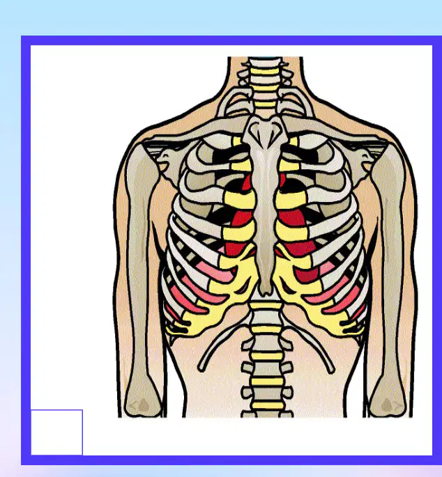
The heart is located under the ______ and between the lungs.
sternum
clavicle
rib
scapula
The heart is about the size of your _____
fist
foot
head
knee
What is the average number of heart beats per minute?
72
50
90
120
What is the total output of the heart per minute?
5 liters per minute
2 liters per minute
10 liters per minute
1 liter per minute
How much does the heart weigh?
10.6 oz or 300 grams
2 lbs or 900 grams
1 oz or 28 grams
5 lbs or 2000 grams
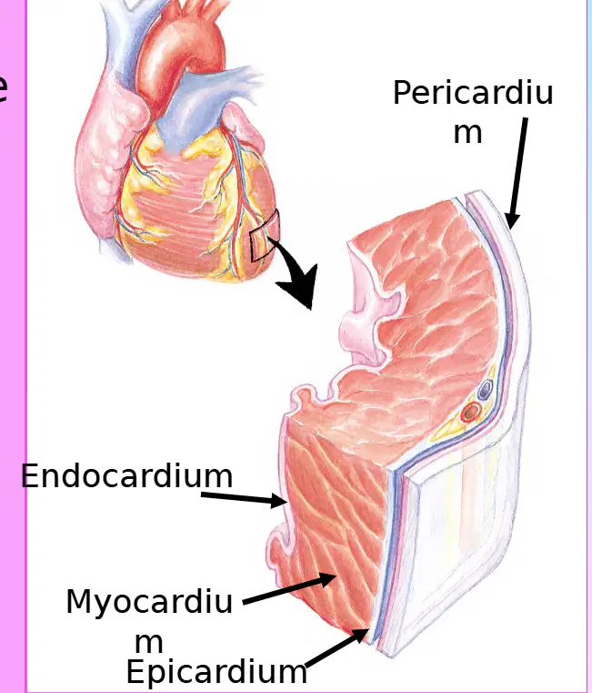
The inner layer of the heart that lines the chambers and valves, and contains the Purkinje fibers, is called the ________.
Endocardium
Myocardium
Epicardium
Pericardium
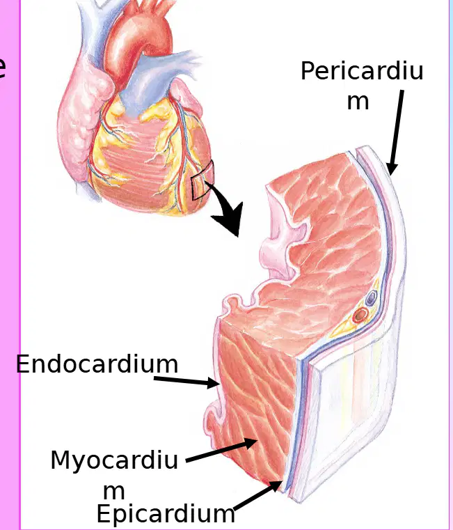
The middle, thickest muscular layer of the heart, responsible for heart contraction, is called the ________.
Myocardium
Endocardium
Pericardium
Epicardium
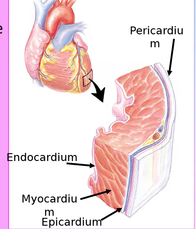
The outside, thin layer of the heart that contains the coronary arteries is called the ________.
Epicardium
Endocardium
Myocardium
Pericardium
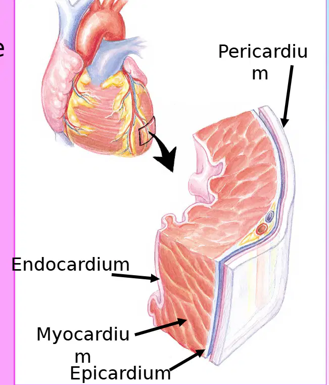
The double-layer sac that encloses the heart is called the ________.
Pericardium
Myocardium
Endocardium
Epicardium
What is the space called that is located between the parietal and visceral (epicardium) layers of the heart?
Pericardial Space
Pleural Cavity
Peritoneal Cavity
Mediastinum
How much fluid does the pericardial space normally contain?
10 - 20 ml
50 - 100 ml
100 - 200 ml
1 - 2 ml
Which of the following best describes the pericardium?
A muscle layer of the heart
A sack of tissue surrounding the heart
The innermost layer of the heart
The outermost layer of the blood vessels
The pericardium has two layers. The tough outer layer is called the ______ layer, and the inner layer that adheres closely to the heart is called the ______ or epicardium layer.
parietal; visceral
visceral; parietal
fibrous; serous
serous; fibrous
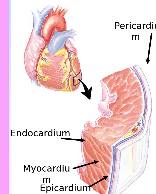
Which layer of the heart is labeled as the innermost layer in the diagram?
Endocardium
Epicardium
Myocardium
Pericardium
The visceral layer of the pericardium is also known as the epicardium.
True
False

Which part of the heart receives oxygen-poor blood from the body?
Left Atrium
Right Atrium
Left Ventricle
Aorta

The valve between the left atrium and the left ventricle is called the _________.
Mitral Valve
Tricuspid Valve
Pulmonary Valve
Aortic Valve

Which color in the circulation diagram represents oxygen-rich blood?
Blue
Red

The ________ is the lower pointed end of the heart.
Apex
Base
Septum
Atrium

The pulmonary vein carries oxygen-rich blood from the lungs to the heart.
True
False
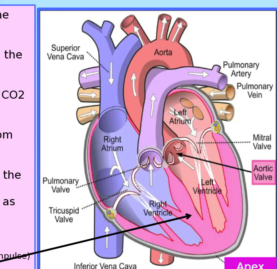
What is the function of the Right Atrium?
Receives deoxygenated blood from the body.
Pumps oxygenated blood to the lungs.
Receives oxygenated blood from the lungs.
Pumps deoxygenated blood to the body.

What does the Right Ventricle do?
What happens to blood inside the lungs?
Deoxygenated blood releases the CO2 and takes O2. Now the blood is oxygenated.
Blood becomes deoxygenated by absorbing CO2 from the air.
Blood cells are destroyed and replaced with new ones.
Blood is filtered to remove waste products.

What is the function of the Left Atrium?
Receives the now oxygenated blood from the lungs.
Pumps deoxygenated blood to the lungs.
Receives deoxygenated blood from the body.
Pumps oxygenated blood to the body.

What does the Left Ventricle do?

What is the Apex of the heart?
Base of the heart (also known as the point of maximal impulse).
Right atrium
Left ventricle
Interventricular septum
What is the interatrial septum?
What is the main function of the aorta?
Returns blood from upper body to the heart
Returns blood from lower body to the heart
Carries blood from the left ventricle to the body
Carries blood from the right ventricle to the lungs
Which vessel returns blood from the upper body to the heart?
Aorta
Superior Vena Cava
Inferior Vena Cava
Pulmonary Artery
The Inferior Vena Cava returns blood from the ______ body to the heart.
lower
upper
left
right
Refer to the diagram of the heart. Which vessel is labeled as carrying blood from the left ventricle to the body?
Aorta
Pulmonary artery
Superior vena cava
Pulmonary vein
In the body, what do arteries carry to the tissues?
Oxygenated blood
Carbon dioxide
Waste products
Hormones
In the body, what do veins carry back to the heart?
Deoxygenated blood
Oxygenated blood
Nutrients
Hormones
In the heart, which artery carries deoxygenated blood to the lungs?
Pulmonary artery
Aorta
Coronary artery
Carotid artery
In the heart, which vein carries oxygenated blood back into the heart in the left atrium?
Pulmonary vein
Superior vena cava
Inferior vena cava
Coronary sinus
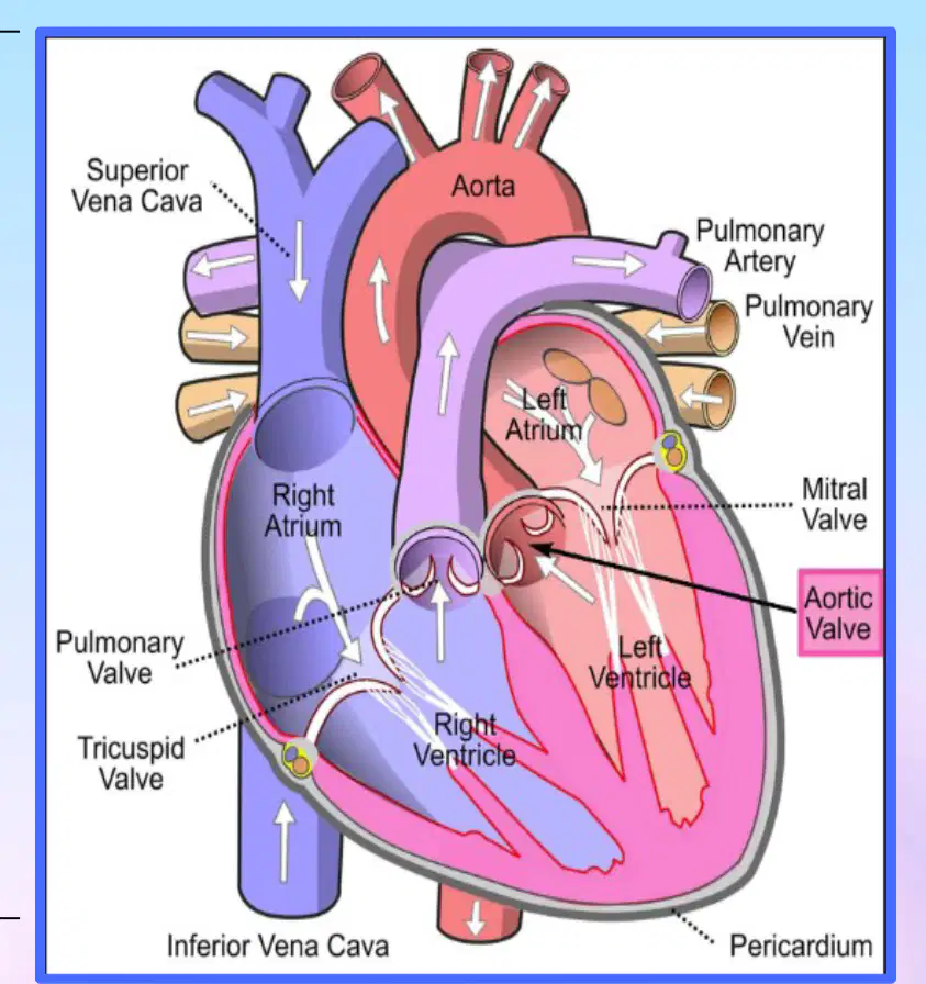
Which part of the heart receives blood from the body?
Right Atrium
Left Atrium
Right Ventricle
Left Ventricle

Which valve is located between the left atrium and the left ventricle?
Mitral Valve
Tricuspid Valve
Pulmonary Valve
Aortic Valve

Which sound is associated with the closing of the tricuspid valve?
"Lub"
"Dub"
"Ventricular gallop"
"Atrial gallop"
Which sound is associated with the closing of the pulmonary valve?
S2: "dub"
S1: "lub"
S3: "ventricular gallop"
S4: "atrial gallop"

Which sound is associated with the closing of the mitral (bicuspid) valve?
"lub"
"dub"
"ventricular gallop"
"atrial gallop"

Which sound is associated with the closing of the aortic valve?
"dub"
"lub"
"ventricular gallop"
"atrial gallop"

Which heart sound is associated with the closing of the tricuspid valve?
S1: 'Lub'
S2: 'Dub'
S3: 'Ta'
S4: 'La'

Which valve produces the S1 'lub' sound when it closes?
Mitral valve (bicuspid valve)
Pulmonary valve
Aortic valve
Tricuspid valve

The S2 'dub' sound is produced by the closing of which valve?
Tricuspid valve
Mitral valve
Pulmonary valve
None of the above
The aortic valve produces the S2 '____' sound when it closes.
dub
lub
tick
snap
What is the characteristic sound pattern of S3 early diastole?
Lub-dub-tah (KEN~TUCK~Y)
Lub-dub-dub (TEN~NES~SEE)
Lub-tah-dub (MIS~SOU~RI)
Dub-lub-tah (NEW~YORK~ER)
What are possible causes of S3 early diastole?
Fluid overload, heart failure
Mitral valve prolapse, pericarditis
Aortic stenosis, hypertrophic cardiomyopathy
Pulmonary embolism, atrial fibrillation
S3 early diastole is best heard when the patient is on which side?
Right side
Left side
Supine
Prone
What is the characteristic sound pattern of S4 late diastole?
Tah-lub-dub
Lub-dub-ta
Dub-lub-tah
Lub-tah-dub
How do you listen for heart sounds using a stethoscope?
Inch your stethoscope over the chest and do not pick it up and down
Place the stethoscope and keep moving it up and down
Only use the stethoscope's bell
Only use the stethoscope's diaphragm
Which part of the stethoscope should you use to hear S1 and S2 heart sounds?
The big bell
The little bell
The earpiece
The tubing
Which part of the stethoscope is used to hear S3, S4, and murmurs?
The big bell
The little bell
The earpiece
The tubing
Where are heart sounds best heard?
At the base
At the apex
At the sternum
At the clavicle
Why does it matter which bell of the stethoscope you use?
Big bell is for hearing big sounds (S1 & S2), little bell is for hearing low sounds (S3 & S4)
Both bells are for the same sounds
The bell does not matter
The bell is only for decoration
The stethoscope's ________ is used to listen to heart sounds.
diaphragm
bell
tube
earpiece
Refer to the diagram of the chest showing auscultation points. Which area is best for hearing the mitral valve?
Second right intercostal space
Second left intercostal space
Fifth left intercostal space (midclavicular line)
Third left intercostal space

What is the function of the Sinoatrial (SA) Node in the heart's electrical pathway?
The SA Node is the 'Natural Pacemaker' that generates electrical impulse and sends it to the left atrium and AV Node.
The SA Node acts as a valve to prevent backflow of blood into the atria.
The SA Node is responsible for pumping oxygenated blood to the body.
The SA Node regulates the opening and closing of the heart valves.

What is the role of the AV Node in the heart's electrical pathway?
The AV Node acts as the 'gatekeeper' and is the second pacemaker if the SA node fails. (40-60 bmp)
The AV Node initiates the heartbeat and sets the pace for the heart. (60-100 bmp)
The AV Node contracts the ventricles directly to pump blood out of the heart.
The AV Node supplies oxygenated blood to the heart muscle.
What does the Bundle of His do in the heart's electrical pathway?

What happens at the Right Bundle Branch (RBB) in the heart's electrical pathway?

What is the function of the Left Bundle Branch (LBB) in the heart's electrical pathway?
The electrical impulse splits, like a fork in the road, at the Left Bundle Branch.
It pumps blood from the left ventricle to the aorta.
It regulates the opening and closing of the mitral valve.
It produces red blood cells in the left atrium.

What is the role of Purkinje Fibers in the heart's electrical pathway?
Purkinje Fibers send the impulse throughout the ventricles (20-40 bpm).
Purkinje Fibers initiate the heartbeat in the right atrium.
Purkinje Fibers regulate the opening of heart valves.
Purkinje Fibers are responsible for oxygenating the blood.
What is the ability of the heart cells to initiate an electrical impulse without being stimulated by an outside source called?
Automaticity
Excitability
Conductivity
Contractility
What is the ability of the heart cells to receive and transmit electrical impulses called? Without this, the heart would not have an electrical pathway.
Conductivity
Elasticity
Contractility
Permeability
What is the term for when the heart muscle cells are stimulated by an electrical impulse, making them contract?
Contractility
Depolarization
Repolarization
Relaxation
What is the ability of the heart cells to respond to electrical impulses called? Without this, the heart would not react, resulting in no heartbeat.
Excitability
Contractility
Conductivity
Automaticity
What is the amount of blood that is pumped out of the left ventricle with each contraction called?
Stroke Volume
Cardiac Output
Heart Rate
Blood Pressure
What is the total volume of blood pumped each minute called?
Cardiac Output
Stroke Volume
Blood Pressure
Heart Rate
What is the percentage of blood pumped out of the left ventricle with each contraction during systole called?
Ejection Fraction
Stroke Volume
Cardiac Output
Preload
Diastolic - systolic = ________.
stroke volume
blood pressure
cardiac output
heart rate
Heart Rate (HR) x Stroke Volume (SV) = ________.
Cardiac output (CO)
Blood pressure (BP)
Respiratory rate (RR)
Ejection fraction (EF)
What is the normal range for Ejection Fraction (EF)?
10% - 30%
30% - 50%
50% - 70%
70% - 90%
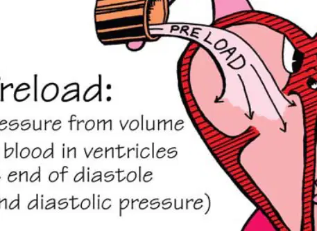
What is preload?
The resistance the ventricles must overcome to circulate blood
The amount of blood within the ventricles waiting to be pumped out
The pressure in the arteries
The amount of oxygen in the blood
What is afterload?
The amount of blood within the ventricles waiting to be pumped out
The resistance or 'pressure' that the ventricles must push against to pump blood out to the body
The amount of oxygen in the blood
The pressure in the veins
Preload is increased in which of the following conditions?
Hypertension
Hypervolemia
Vasoconstriction
Low blood pressure
Afterload is increased in ________ and vasoconstriction.
Hypertension
Hypotension
Bradycardia
Anemia
Increased afterload increases cardiac workload.
True
False
What is the isoelectric line (baseline) in an EKG/ECG waveform?
The point where the heart contracts
The line where no electrical activity is occurring in the heart
The peak of the R wave
The end of the T wave
The isoelectric line is also known as the _________.
baseline
QRS complex
P wave
T wave

What does the P wave represent in an EKG/ECG waveform?
Ventricles contracting
Atria contracting
Ventricles relaxing
Depolarization of ventricles

What is the normal duration of the QRS complex in an EKG/ECG waveform?
0.8-0.12 seconds
0.06-0.12 seconds
0.2-0.4 seconds
0.01-0.05 seconds

The T wave in an EKG/ECG waveform represents the ________ relaxing.
ventricles
atria
lungs
arteries

Which part of the EKG/ECG waveform is associated with depolarization of the ventricles?
P wave
QRS complex
T wave
J point

True or False: The P wave duration is normally between 0.8 and 0.12 seconds.
True
False
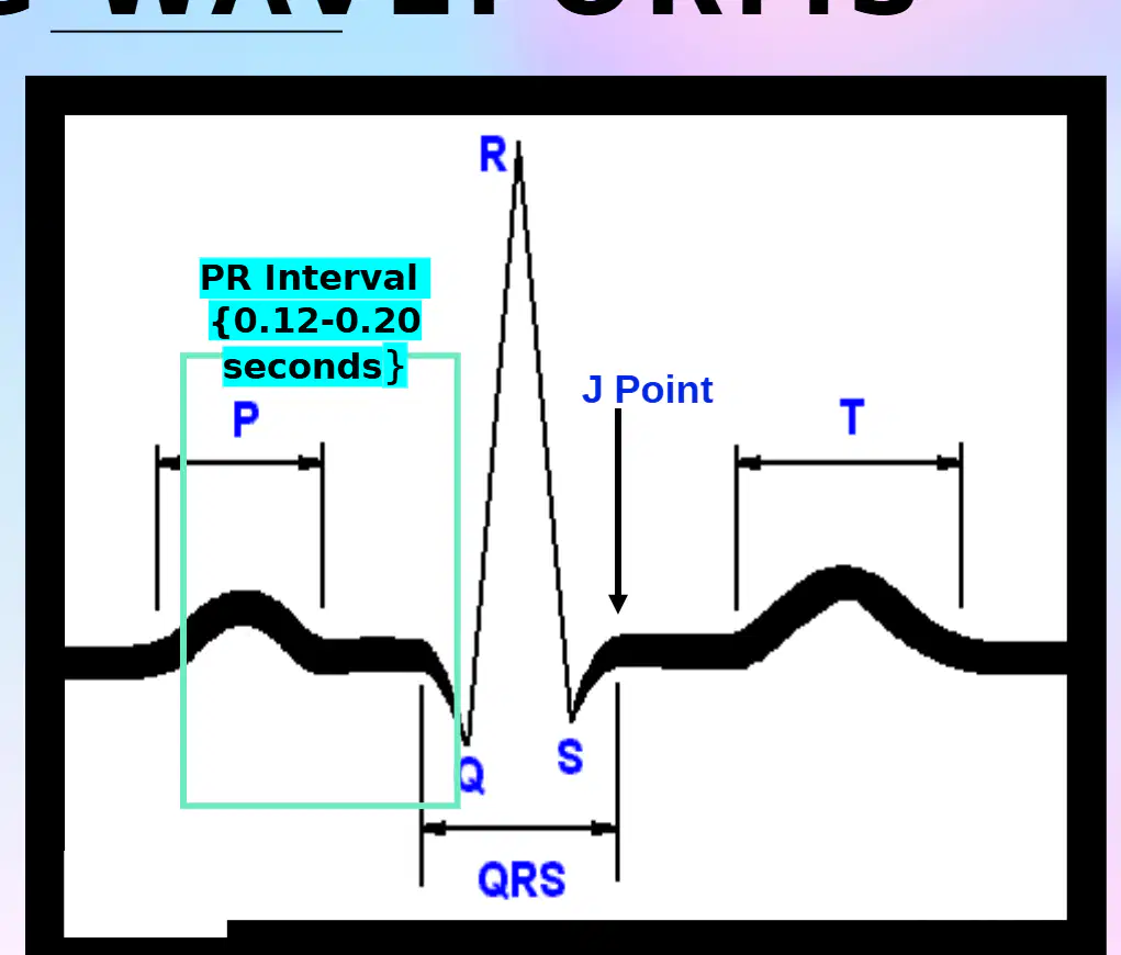
What is the normal duration of the PR Interval in an EKG/ECG waveform?
0.12-0.20 seconds
0.08-0.10 seconds
0.22-0.28 seconds
0.30-0.40 seconds
The PR Interval represents the time from the beginning of atrial depolarization to the beginning of ______ depolarization.
ventricle
atrial
nodal
bundle
Which of the following best describes the PR Interval in an EKG/ECG?
A) Measures from the beginning of the P wave to the beginning of the QRS
B) Measures from the end of the T wave to the beginning of the P wave
C) Measures from the beginning of the QRS to the end of the T wave
D) Measures from the end of the P wave to the end of the QRS
The PR Interval is the electrical impulse moving from the atria to the ventricles.
True
False
What does the ST segment represent in an EKG/ECG waveform?
The beginning of ventricular depolarization
The end of ventricular depolarization and the beginning of ventricular repolarization
The contraction of the atria
The end of the T wave
The ST segment is the end of the _______ complex to the beginning of the T wave.
QRS
P
U
PR
According to the information provided, what is happening to the ventricles during the ST segment?
They are contracting
They are starting to relax
They are depolarizing
They are repolarizing
Normally, the ST segment is on the _______ line.
isoelectric
baseline
depolarization
elevated

