
NEW
Font size
 Worksheets
Upper Limb Osteology revision
Total questions: 14
Worksheet time: 7mins
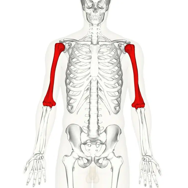
How do you tell that a humerus belongs to right or left side?
The head is directed laterally, the lesser tubercle is posterior, the greater tubercle is anterior, and the olecranon fossa is anterior.
The head is directed medially, the lesser trochanter is presented anteriorly, the greater tubercle is placed posteriorly, and olecranon fossa is placed posteriorly
The head is anterior, the greater tubercle is medial, the lesser tubercle is lateral, and the olecranon fossa is inferior.
The head is posterior, the lesser tubercle is inferior, the greater tubercle is superior, and the olecranon fossa is on the medial side.
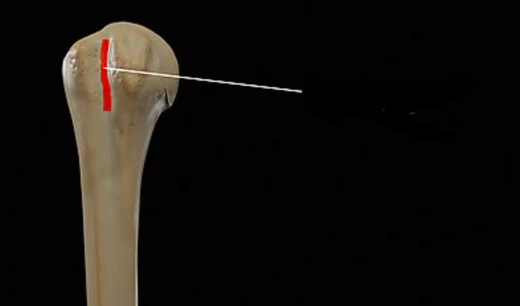
What is marked in the image on humerus?
Intertubercular sulcus/bicipital groove
Greater trochanter
Lesser trochanter
Head
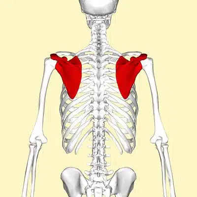
How do you know the scapula is present on body's right side or left side?
The glenoid cavity is directed medially, the spine of the scapula is anterior, and the subscapular fossa lies posteriorly.
The glenoid cavity faces inferiorly, the spine is on the inferior border, and the subscapular fossa is on the superior surface.
Glenoid cavity presents laterally, spine of scapula is present on posterior surface of bone, and subscapular fossa is present anteriorly
The glenoid cavity faces posteriorly, the spine is on the medial border, and the subscapular fossa is on the lateral side.
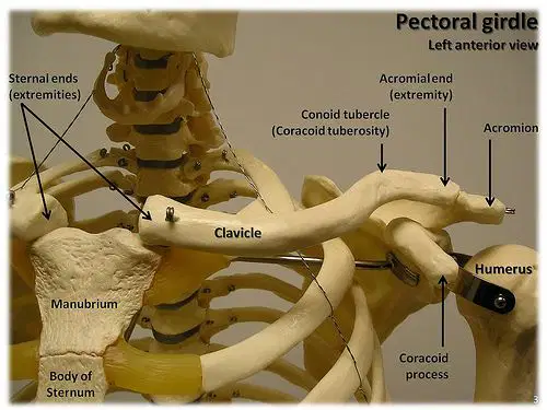
How do you know which side the clavicle belongs to?
The medial end is flattened, the lateral end is quadrangular, the shaft is convex forwards in the lateral 2/3rd and concave forwards in the medial 1/3rd.
The medial end is round, the lateral end is triangular, and the shaft is straight throughout its length.
The medial end is circular, the lateral end is cylindrical, and the shaft is convex backwards in the medial part and convex forwards in the lateral part.
Medial end is quadrangular, lateral end is flattened, convex forwards in medial 2/3rd, concave forwards in lateral 1/3rd
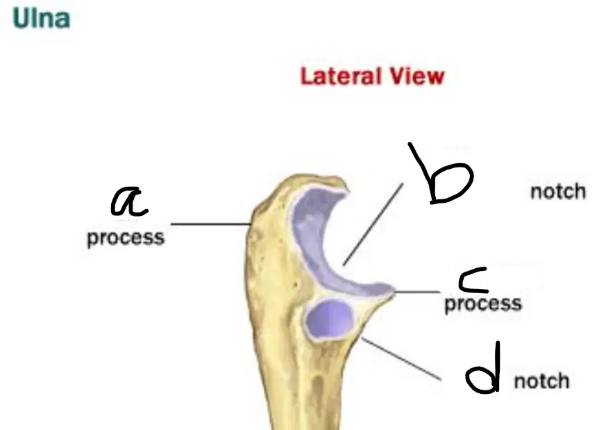
This is the Ulna. Mark the correct labelling
a) Olecranon process
b) Semilunar notch
c) Coronoid process
d) Radial notch
a) Olecranon process
b) Semilunar notch
c) Coronoid process
d) Radial notch
a) Olecranon process
b) Trochlear notch
c) Coronoid process
d) Radial notch
a) Olecranon Process
b) Semilunear, trochlear notch
c) coronoid process
d) radial notch
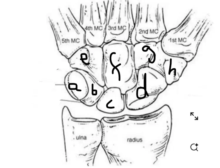
The proximal row of carpal bones is shown on image, a, b, c, d.
a) Pisiform
b) Triquetrum
c) Scaphoid
d) Lunate
a) Pisiform
b) Triquetrum
c) Scaphoid
d) Lunate
a) Pisiform
b) Triquetrum
c) Scaphoid
d) Lunate
a) Pisiform
b) Triquetrum
c) Scaphoid
d) Lunate
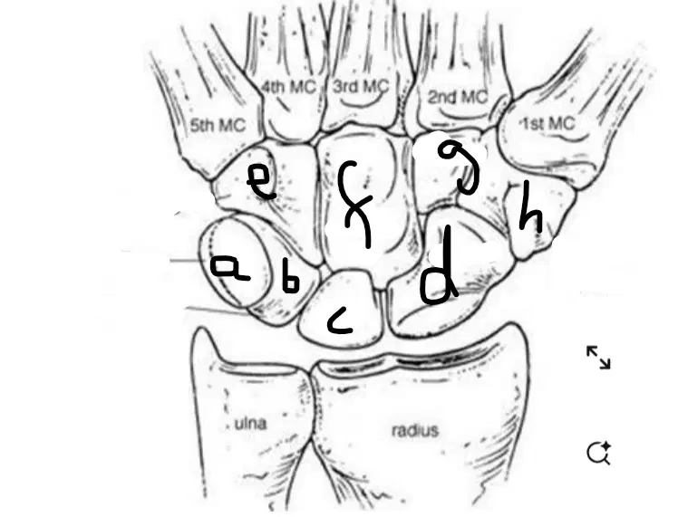
The distal row of carpal bones is marked by e,f,g,h.Label the row.
e) capitate
f) trapezium
g) hamate
h) trapezoid
e) capitate
f) hamate
g) trapezium
h) trapezoid
e) capitate
f) hamate
g) trapezoid
h) trapezium
e) hamate
f) capitate
g) trapezoid
h) trapezium
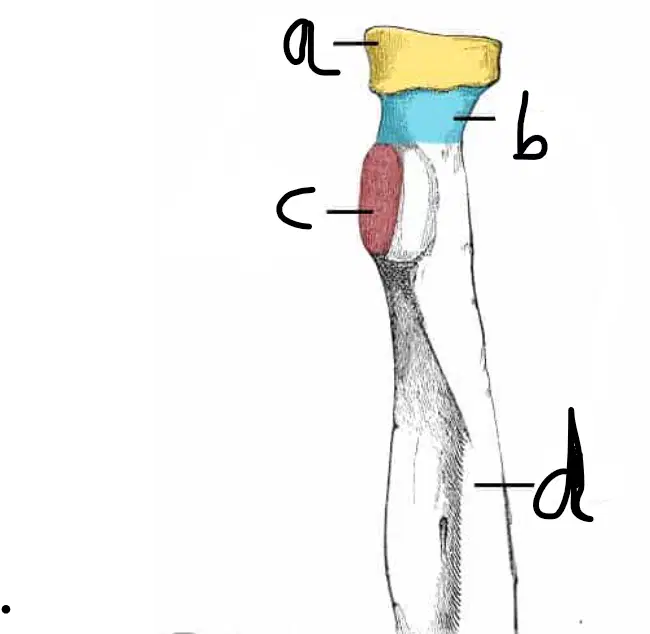
Label parts of radius.
a) head
b) neck
c) tuberosity (radial)
d) shaft
a) neck
b) head
c) neck
d) shaft
a) neck
b) head
c) tuberosity (radial)
d) shaft
a) neck
b) head
c) shaft
d) radial tuberosity
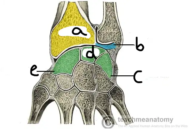
Label the wrist joint.
a) lunate
b) ulna
c) triquetrum
d) scaphoid
e) lunate
a) lunate
b) triangular disc
c) triquetrum
d) scaphoid
e) lunate
a) lunate
b) triangular disc
c) triquetrum
d) radius
e) scaphoid
a) radius
b) triangular disc
c) triquetrum
d) lunate
e) scaphoid
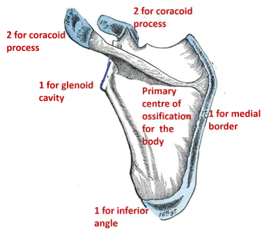
Which of the following is the primary center of ossification for the scapula?
a) Body of the scapula
b) Coracoid process
c) Glenoid cavity
d) Inferior angle
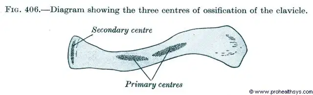
How many ossification centers are involved in the development of the clavicle?
a) 2 primary centers and 1 secondary center
b) 1 primary center and 2 secondary centers
c) 3 primary centers only
d) 2 secondary centers and no primary center
Which of the following best describes a Colles' fracture?
a) Fracture of the distal radius with dorsal displacement of the distal fragment
b) Fracture of the proximal ulna with anterior dislocation of the radial head
c) Fracture of the distal radius with volar (anterior) displacement of the distal fragment
d) Fracture of the radius and dislocation of the distal radioulnar joint
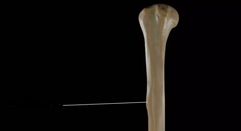
Which of the following statements is true regarding the deltoid tuberosity of the humerus?
a) It serves as the insertion point for the deltoid muscle
b) It is located on the distal end of the humerus near the epicondyles
c) It is a groove on the posterior surface for the radial nerve
d) It articulates with the scapula at the glenoid cavity
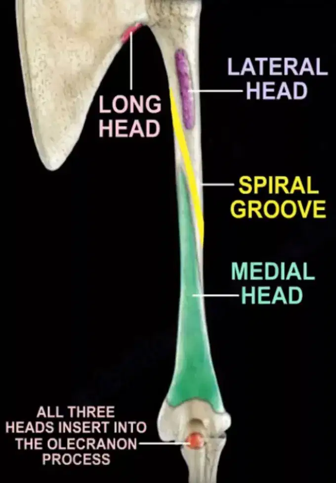
Which 2 structures pass in radial groove ?
Profunda Brachii artery and Radial Nerve
Brachial artery and Ulnar Nerve
Nutrient Artery and Axillary Nerve
Ulnar Nerve and Axillary Artery

