
NEW
Font size
 Worksheets
GLAUCOMA
Total questions: 18
Worksheet time: 9mins
A 55-year-old man was brought to the hospital’s emergency department by his family with complaints of pain in his right eye radiating to the right side of the head, accompanied by nausea and vomiting for the past 3 hours. He also experienced blurred vision, redness, and watering of the eye. No history of trauma or similar episodes.
Ophthalmological examination revealed:
VOD: 1/300
VOS: 20/40
TOD: 60 mmHg
TOS: 18 mmHg
Right anterior segment: mixed conjunctival injection, hazy cornea, shallow anterior chamber, fixed mid-dilated pupil
Left eye: within normal limits.
What is the most likely diagnosis?
Ocular Hypertension
Normal-Tension Glaucoma
Primary Open-Angle Glaucoma
Acute Primary Angle-Closure Glaucoma
Chronic Primary Angle-Closure Glaucoma
A 55-year-old man was brought to the hospital’s emergency department by his family with complaints of pain in his right eye radiating to the right side of the head, accompanied by nausea and vomiting for the past 3 hours. He also experienced blurred vision, redness, and watering of the eye. No history of trauma or similar episodes.
Ophthalmological examination revealed:
VOD: 1/300
VOS: 20/40
TOD: 60 mmHg
TOS: 18 mmHg
Right anterior segment: mixed conjunctival injection, hazy cornea, shallow anterior chamber, fixed mid-dilated pupil
Left eye: within normal limits.
What is the pathophysiology of the above condition?
Vascular insufficiency of the optic nerve
Presence of hemorrhagic particles blocking the trabecular meshwork
Degeneration of the trabecular meshwork and Schlemm’s canal, partly due to genetics
Inflammatory cells blocking aqueous humor flow in the trabecular meshwork
Pupillary block due to risk factors like shallow anterior chamber and narrow angles
A 55-year-old man was brought to the hospital’s emergency department by his family with complaints of pain in his right eye radiating to the right side of the head, accompanied by nausea and vomiting for the past 3 hours. He also experienced blurred vision, redness, and watering of the eye. No history of trauma or similar episodes.
Ophthalmological examination revealed:
VOD: 1/300
VOS: 20/40
TOD: 60 mmHg
TOS: 18 mmHg
Right anterior segment: mixed conjunctival injection, hazy cornea, shallow anterior chamber, fixed mid-dilated pupil
Left eye: within normal limits.
What is the definitive treatment for this patient?
Goniotomy
Trabeculotomy
Trabeculectomy
Laser Trabeculoplasty
Peripheral Iridectomy
A 65-year-old woman came to the primary care clinic complaining of blurry vision in both eyes, described as tunnel vision for the past 2 years, worsening over the past month. No other symptoms or systemic disease.
Examination:
VODS: 1/60
TODS: 35 mmHg
Anterior segment: appears quiet, fixed mid-dilated pupils
What is the next investigation to confirm the diagnosis?
Fundoscopy
Gonioscopy
Fundus Photography
OCT of the optic nerve
Visual field perimetry
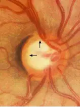
Based on fundoscopy, what is the most likely diagnosis?
Ocular Hypertension
Normal-Tension Glaucoma
Primary Open-Angle Glaucoma
Acute Primary Angle-Closure Glaucoma
Chronic Primary Angle-Closure Glaucoma
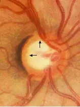
What is the pathophysiology of this condition?
Vascular insufficiency of the optic nerve
Hemorrhagic particles blocking the trabecular meshwork
Degeneration of trabecular meshwork and Schlemm’s canal, partly due to genetics
Inflammatory cells blocking aqueous humor flow
Pupillary block due to shallow anterior chamber and narrow angles
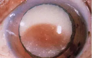
A 70-year-old man came to the primary clinic complaining of blurry vision in his right eye for the past year, worsening in the last month. Associated with redness and pain. No systemic disease.
Examination:VOD: 1/300
VOS: 2/60
TOD: 38 mmHg
TOS: 20 mmHg
Right eye image shows signs suggestive of:
Phacolytic Glaucoma
Phacomorphic Glaucoma
Primary Open-Angle Glaucoma
Acute Primary Angle-Closure Glaucoma
Chronic Primary Angle-Closure Glaucoma
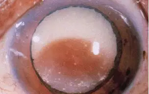
A 70-year-old man came to the primary clinic complaining of blurry vision in his right eye for the past year, worsening in the last month. Associated with redness and pain. No systemic disease.
Examination:VOD: 1/300
VOS: 2/60
TOD: 38 mmHg
TOS: 20 mmHg
What is the pathophysiology of the condition above?
Intumescent cataract blocking the pupil and narrowing the anterior chamber angle
Hypermature cataract causing lens protein leakage, leading to inflammatory reaction and trabecular meshwork blockage
Degeneration of trabecular meshwork and Schlemm’s canal
Inflammatory cells blocking aqueous humor flow
Pupillary block due to shallow anterior chamber
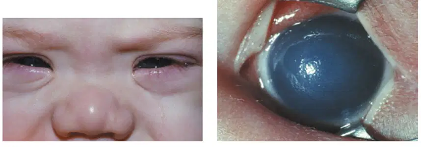
A 1-year-old boy is brought to the clinic by his mother with complaints of an enlarged left eye since birth, excessive tearing, and photophobia.
Examination shows :
Buphthalmic left eye, hazy cornea, blepharospasm, and epiphora.
What is the pathogenesis?
Hemorrhagic particles blocking the trabecular meshwork
Degeneration of trabecular meshwork and Schlemm’s canal
Inflammatory cells blocking aqueous humor flow
Isolated trabeculodysgenesis causing incomplete separation of the iridocorneal angle
Pupillary block due to shallow anterior chamber
A 55-year-old man came to a general clinic with complaints of bilateral blurry vision for the past year. Describes progressive tunnel vision in the last 6 months. No other complaints.
Ophthalmology exam:
VODS: 20/80
TODS: 30 mmHg
Anterior segment and chamber: normal
Optic disc: cupping, CDR 0.7
Visual field: narrowed
What is the diagnosis?
Glaucoma Suspect
Ocular Hypertension
Normal-Tension Glaucoma
Primary Open-Angle Glaucoma
Primary Angle-Closure Glaucoma
A 55-year-old man came to a general clinic with complaints of bilateral blurry vision for the past year. Describes progressive tunnel vision in the last 6 months. No other complaints.
Ophthalmology exam:
VODS: 20/80
TODS: 30 mmHg
Anterior segment and chamber: normal
Optic disc: cupping, CDR 0.7
Visual field: narrowed
What is the pathomechanism of blurred vision in the acute glaucoma case described previously?
High IOP irritates ciliary body due to epithelial damage
Sudden IOP increase causes pain, nausea, vomiting
Corneal edema and accompanying uveitis from increased IOP
Damage to optic nerve axons and ganglion cells
A 55-year-old man complains of bilateral blurry vision worsening over 1 year, with tunnel vision for 6 months.
Exam:
VODS: 20/80
TODS: 25 mmHg
Anterior segment: normal
Optic disc cupping (CDR 0.6), narrowed visual field
What is the initial treatment?
Timolol 0.75% 2x/day
Betaxolol 1% 2x/day
Pilocarpine 0.4% 2x/day
Acetazolamide 500 mg once daily
Prostaglandin analog 1x/day
Increased IOP without visual field or optic nerve damage is characteristic of:
Primary Open-Angle Glaucoma
PACG
Plateau Iris Syndrome
Ocular Hypertension
Which of the following is NOT a risk factor for PACG?
Shallow anterior chamber angle
Myopia
Short axial length
Small corneal diameter
Older age
Which form of open-angle glaucoma occurs without elevated IOP?
Ocular Hypertension
Normal-Tension Glaucoma
PACS
PAS
Which of the following is NOT a cause of secondary open-angle glaucoma?
Intumescent Cataract
Long-term steroid use
Traumatic Hyphema
Uveitis (inflammatory cells)
Where is aqueous humor produced?
Trabecular Pathway
Uveoscleral Pathway
Ciliary Processes
Ciliary Artery
Which of the following is NOT a physiological mechanism of aqueous humor entering the posterior chamber?
Active Secretion
Ultrafiltration
Simple Diffusion
Ultrasecretion

