
 Worksheets
Heart
Total questions: 123
Worksheet time: 2hrs 55mins
Central portion of thorax occupied by (a)
Majority of heart is positioned in __ and __chest
(a)
Apex of heart should be directed toward (a)
Base of heart lies horizontal to (a)
Which of the following correctly describes the size of the heart?
Finger
Closed fist
Upper arm
Foot
Blunt rounded point of cone
(a)
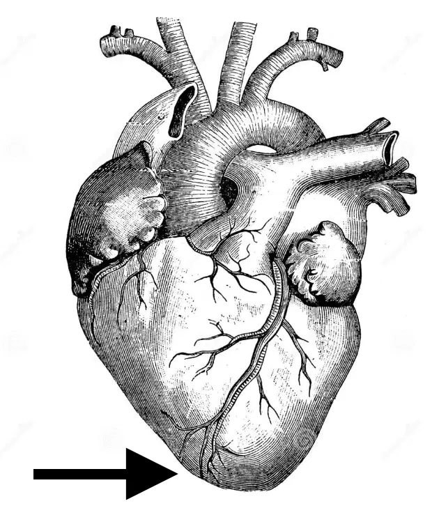
The arrow is pointing to the:
Apex
Base
Flat part at opposite of end of cone
(a)

a membrane, or sac, that surrounds your heart. It holds the heart in place and helps it work properly
(a)
Which of the following is the three layers of the heart wall?
Epicardium, Myocardium, endothelial
Epithelial, Myocardial, Echothelial
Epicardium, Myocardium, Endocardium
This serous membrane of smooth outer surface of heart
(a)
Middle layer composed of cardiac muscle cell and responsibility for heart contracting
(a)
Smooth inner surface of heart chambers
(a)
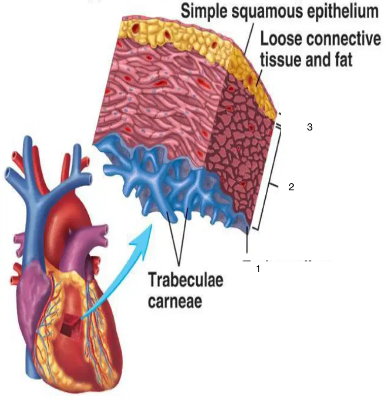
1
(a)
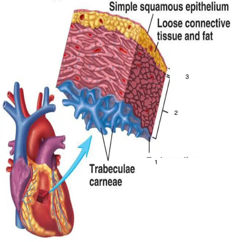
2
(a)
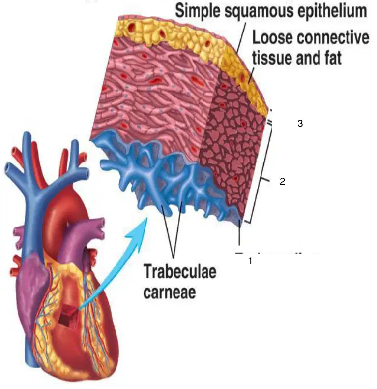
3
(a)
How many atria are in the heart?
1
2
3
4
How many ventricles are in the heart?
1
2
3
4
How many chambers of the heart are there total?
1
2
3
4
• Superior vena cava
• Pulmonary veins
• Inferior Vena cava
(a)
• Aorta
• Pulmonary trunk
(a)
Appendages
(a)
Filling chambers of the heart
(a)
Two upper chambers • Right and left
(a)
The atria are separated by a partition called (a) septum
Superior portion of each atrium has a small triangular extension called appendage or (a)
Within appendages and extending out to anterior surfaces of atria are (a) muscles
Connected to two great veins: IVC and SVC
(a)
Receives deoxygenated blood from the body via the SVC, IVC, and coronary sinus
(a)
Receives four pulmonary veins
(a)
Receives oxygenated blood from the lungs via the pulmonary veins
(a)
Receives deoxygenated blood from the right atrium via the right atrioventricular office and sends it to the lungs via the pulmonary trunk
(a)
Receives oxygenated blood from the left atrium via left atrioventricular office and sends it to body via aorta
(a)
Two lower chambers • Right and left
(a)
Pumping chambers of the heart
(a)
The ventricles are medially separated by (a) septum
Ventricles have (a) walls when compared to atria
Left ventricle is almost (a) times thicker than right
Which ventricle is connected to the pulmonary artery?
Right
Left
Which ventricle is connected to the aorta?
Right
Left
Left myocardium is relatively (a) than the right ventricle
Right ventricle is also more trabeculated than left and contains (a) prominent muscular bands
Parietal band
Crista supraventricularis,
Septal band
Moderator band
(a)
On external surface of heart, ventricles are separated by anterior and posterior (a)
Ventricles are separated from the atria by:
(a)
are grooves that contain coronary vessels, all of which are embedded in fat
(a)
(a) are located between atria and ventricles and are anchored at one end to annulosus fibrosus
Ventricles, in turn, are connected to outflow tracts with semilunar (half-moon–shaped) valves controlling exit of blood from heart
True
False
• Tricuspid
• Bicuspid or Mitral
(a)
• Aortic
• Pulmonary
(a)

Prevent blood from flowing back
(a)
Right AV valve
(a)
Contains three leaflets
• Anterior
• Posterior
• Septal
(a)
The tricuspid valve sits between the __ and __
Right atrium, right ventricle
Left atrium, left ventricle
The pulmonary valve seperates the:
Right ventricle, pulmonary artery
Left ventricle, pulmonary artery
Left atrium, pulmonary artery
Right atrium, pulmonary artery
The mitral valve seperates the:
Right ventricle, left ventricle
Right atrium, aorta
left atrium, left ventricle
Left atrium, right atrium
The aortic valve seperates the:
Left ventricle, aorta
Right ventricle, aorta
Right atrium, aorta
Left atrium, aorta
Left AV valve
(a)
Two leaflets
• Anterior • Posterior
(a)
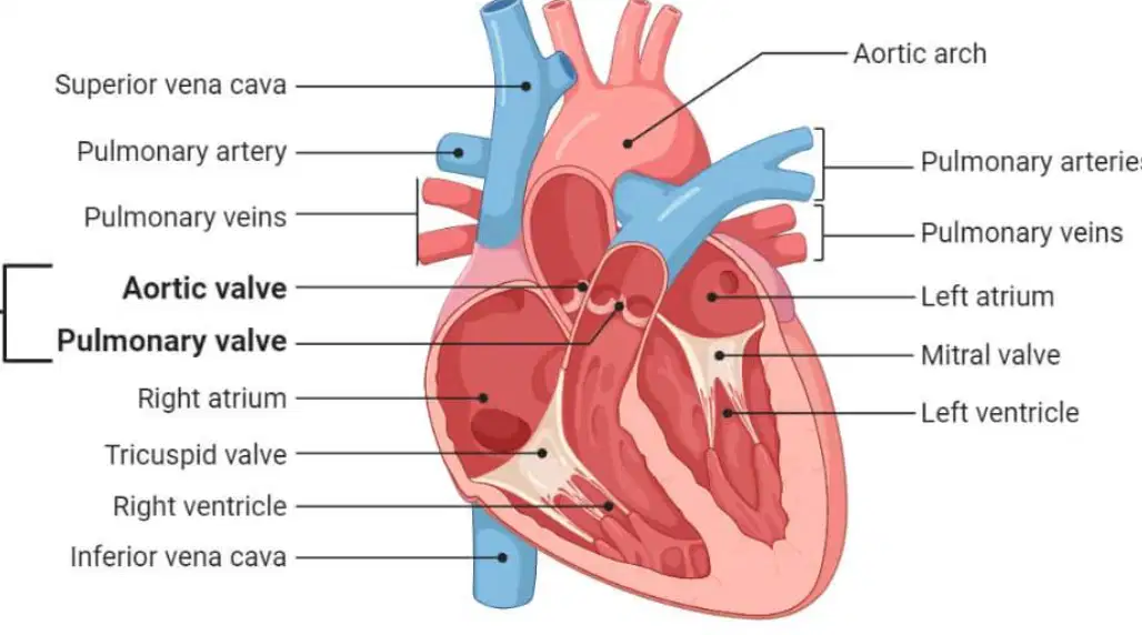
Which of the following are semilunar valves?
Tricuspid
Mitral
Pulmonary
Aortic
Located at junction of left ventricle and aorta
(a)
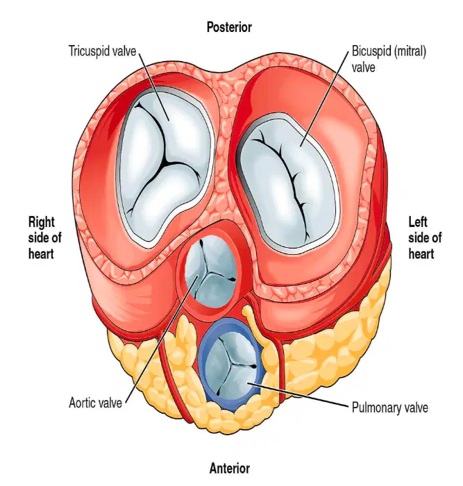
Contains three cusps
Right coronary
Left coronary
Noncoronary
(a)
Located at junction of right ventricle and pulmonary artery
(a)
Contain three cusps
• Anterior
• Right
• Left
(a)
pump deoxygenated blood to lungs and oxygenated blood to vessels and tissues of the body
(a)
Blood returning from upper portion of body enters right atrium via (a)
Deoxygenated blood from lower body enters right atrium by way of:
(a)
Deoxygenatedbloodfrom heart itself through coronary sinus enters:
(a)
From Right atrium, oxygen- depleted blood flows through Tricuspid valve and into (a)
Right ventricle then pumps blood past Pulmonic valve and into (a)
Main pulmonary artery bifurcates into __ and __ pulmonary arteries
(a)
Pulmonary: These in turn enter each of the (a) , where blood is reoxygenated in pulmonary circuit
Oxygenated blood is returned from lungs to Left atrium through four
(a)
Blood then passes from Left atrium, through Mitral valve, and into __ __
(a)
Left ventricle then pumps blood past Aortic valve and into (a)
From here, oxygenated blood is distributed to heart and rest of body through arterial system; this is start of (a)
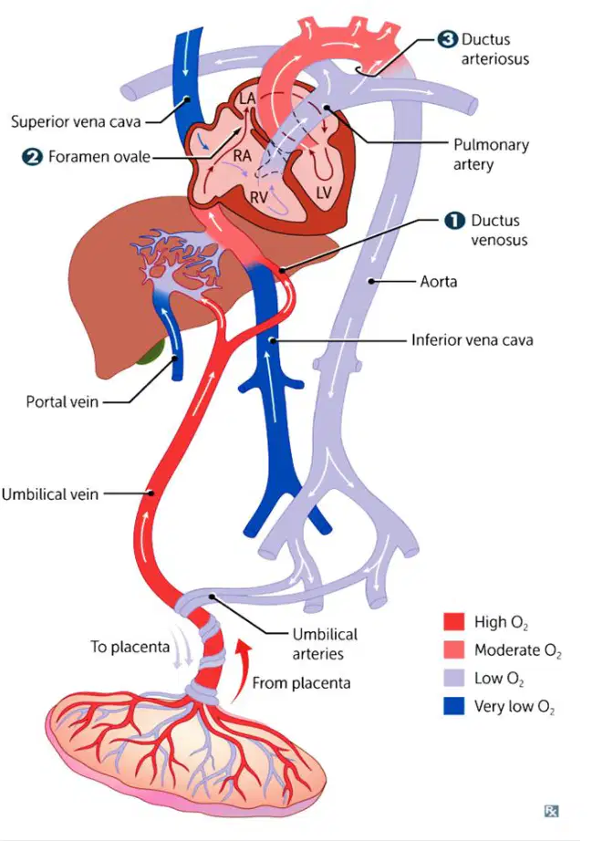
where small fetal vessels on surface of villi are bathed by maternal blood.
(a)
Umbilical veins return (a) blood from placenta
Less oxygenated blood from superior vena cava and small portion of blood from IVC empty into right atrium and into right ventricle
(a)
At the entrance of the liver, the umbilical vein branches into theductus venosus, which is the first shunt in the fetal circulation
(a)
The __ __ RT and LT are major blood vessels in your body, supplying blood to the heart
(a)
They make it possible for the heart to beat and pump blood throughout the body
(a)
These arteries and their branches supply all parts of the heart muscle with blood.
(a)
Two major coronary arteries:
Superior and inferior
Medial and lateral
Right and left
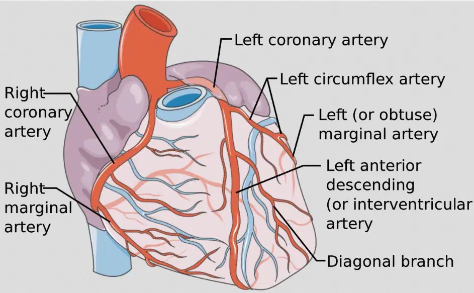
Origin of coronaries is the (a) just posterior to valve in region of right and left sinuses of Valsalva

Heart also has a venous system that courses over its surface and drains into (a)
Pressure in ventricles is greater than that in atria; AV valves close, semilunar valves open, and blood is ejected from ventricles
(a)
Atria contract, forcing AV valves to reopen and remainder of blood in atria to be propelled into ventricles
(a)
is most echogenic structure of the heart and is often seen as bright or white color
(a)
Blood or any other fluids appear (a) or black
(a) and papillary muscles are homogeneous and appear medium gray
Thin, mobile leaflets, or (a) cusps, have slightly increased echogenicity when compared with heart muscle
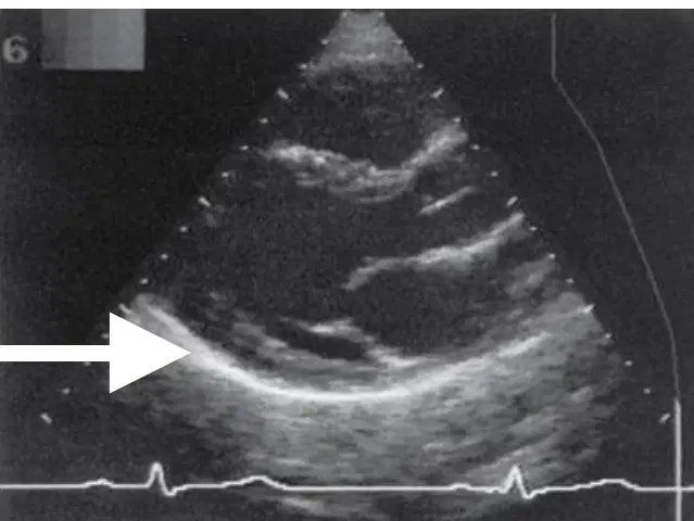
The arrow is pointing to the:
(a)
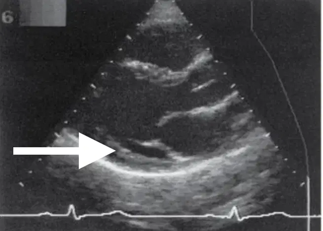
The arrow is pointing to the:
(a)
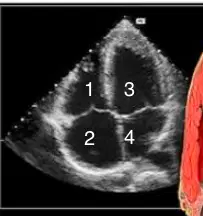
1
(a)
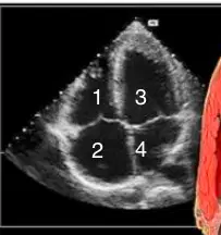
2
(a)
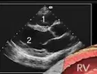
1
(a)
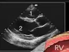
2
(a)
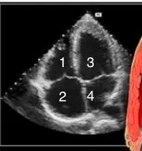
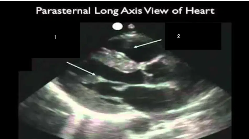
Which basic view is this:
Paraseternal long axis
Parasternal short axis
Apical 4 chamber
Suxiphoid 4 chamber
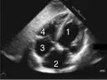
1
(a)
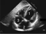
2
(a)
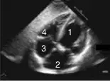
3
(a)
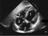
4
(a)

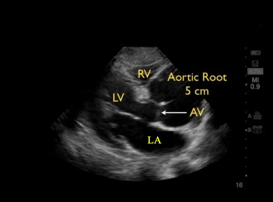
This image shows the:
Subxiphoid 4 chamber
Apical 4 chamber
Parasternal long axis
Parasternal short axis
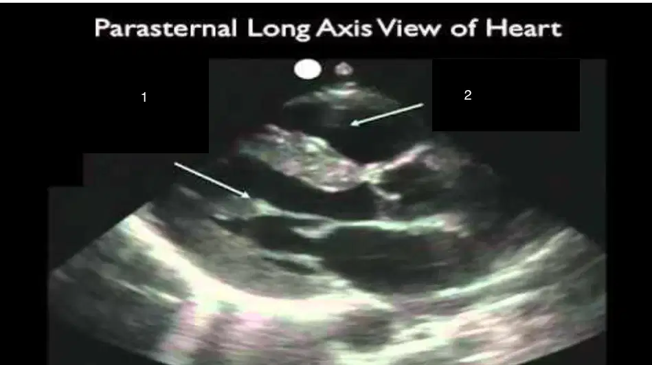
1
(a)
2
(a)
RV
(a)

AV
(a)
1
(a)
2
(a)
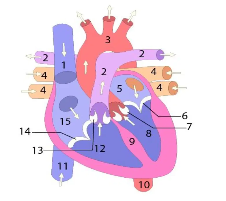
3
(a)
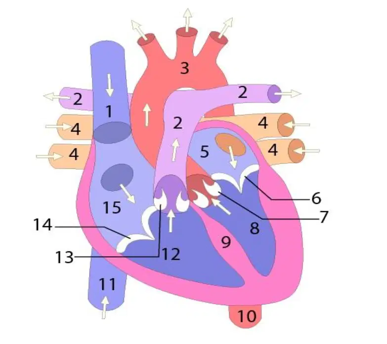
4
(a)

5
(a)
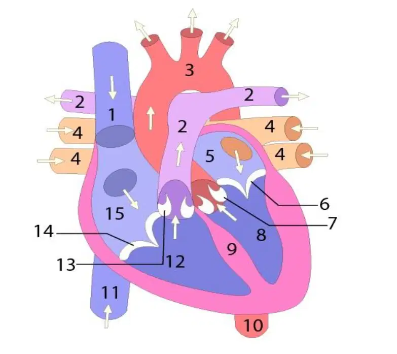
6
(a)
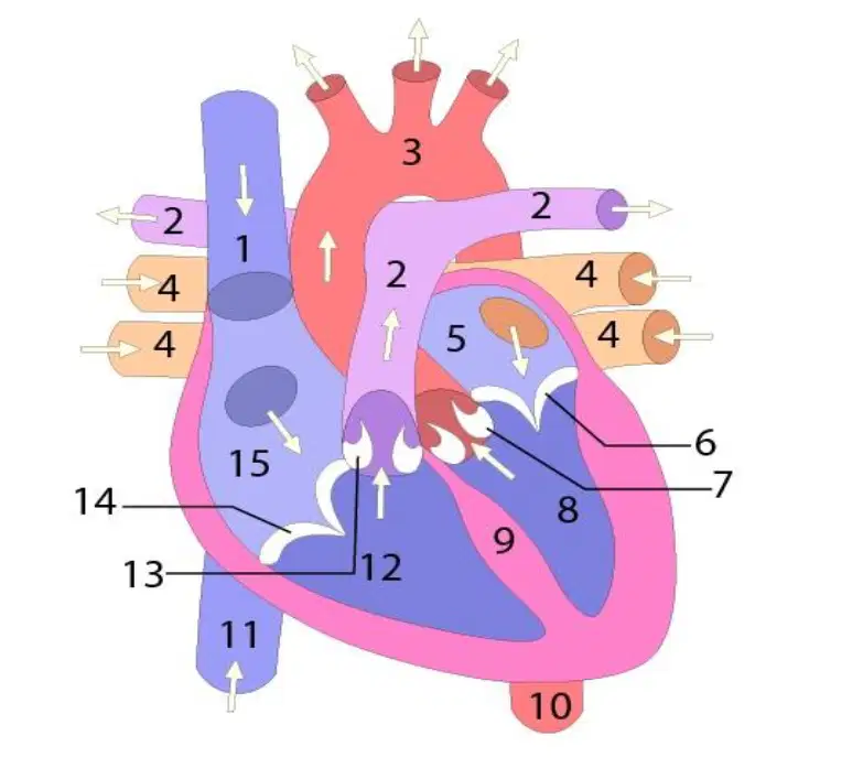
7
(a)
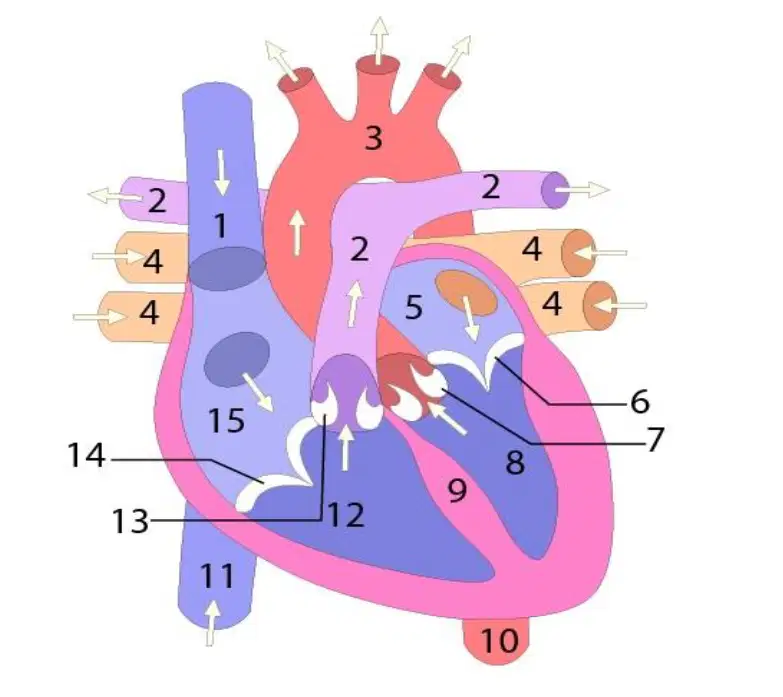
8
(a)
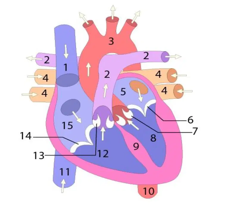
9
(a)

10
(a)
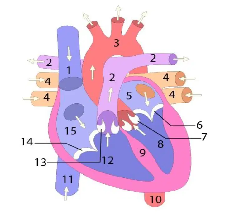
11
(a)

12
(a)

13
(a)

14
(a)

15
(a)

