
 Worksheets
OCT and Glaucoma Quiz
Total questions: 45
Worksheet time: 24mins
Which retinal nerve fiber layer (RNFL) region is most commonly affected in early glaucoma?
Central fovea
Nasal
Temporal Inferior
Temporal
Temporal Superior
During a routine eye examination, you are assessing a patient with suspected glaucoma using OCT. Which layers will you focus on during the examination?
inner plexiform layer
retinal nerve fibre layer
outer plexiform layer
retinal ganglion cell layer
Which of the following is typically a clinical sign of glaucoma, rather than a direct quantitative OCT parameter?
Alpha Zone
RNFL thinning
Disc hemorrhage
GCC thinning
When interpreting OCT results in glaucoma, which factor would most significantly limit the reliability of the scan?
Stage of glaucoma
Signal strength
Presence of cataract
Artefacts
CIRRUS normative comparison for RNFL is based on ?
Race
Age
Disc size
Age and disc size
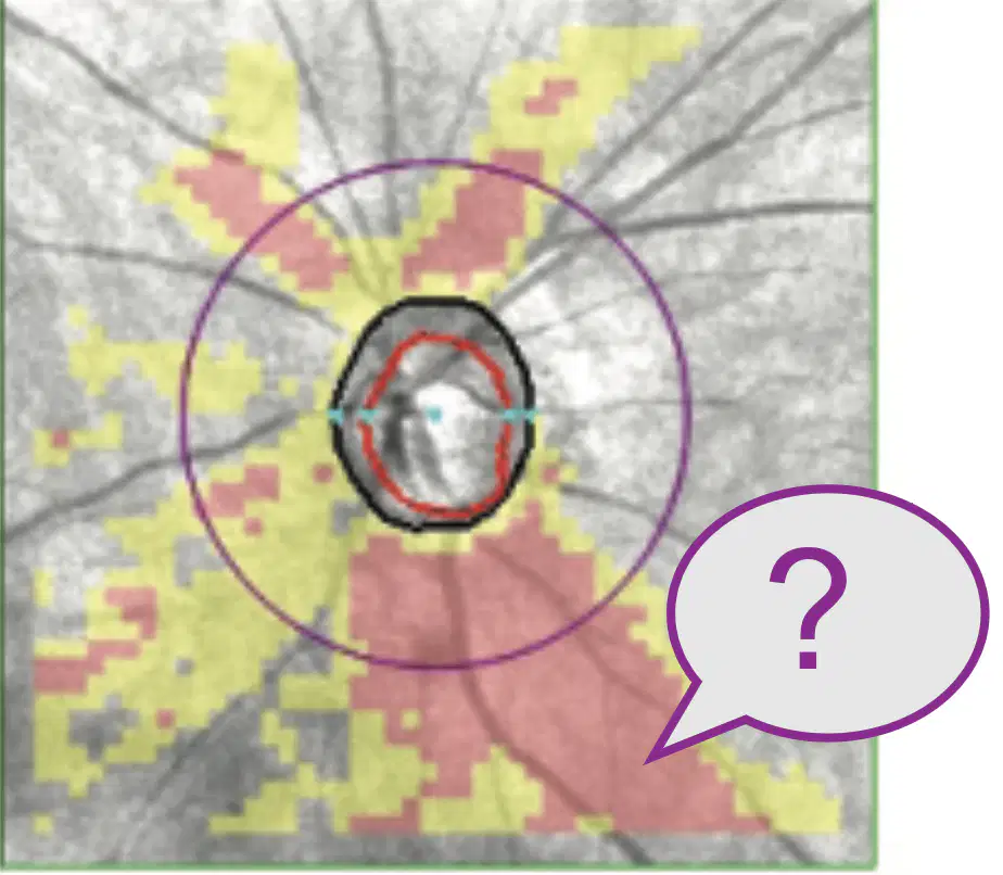
What does the region indicated by the question mark depict ?
The thinnest 5% of RNFL thickness measurements
The thinnest 1% of RNFL thickness measurements
5%-95% of RNFL measurement
Thickest 5% of
all
RNFL measurements
Wedge artefact
A patient's OCT scan shows significant thinning in the inferotemporal quadrant of the RNFL. Which part of the visual field is most likely to be affected?
(a)
What is the 'red disease' in OCT interpretation for glaucoma?
OCT data shows no sign of abnormality in the presence of thinning
significant thinning of the retinal nerve fiber layer (RNFL) associated with glaucoma
OCT mistakenly indicates significant thinning
In OCT Angiography (OCT-A), a decrease in vessel density in which specific area is most strongly correlated with glaucomatous damage?
Superficial Peripapillary capillary plexus
Choroicapillaris
Deep capillary plexus
All of the above
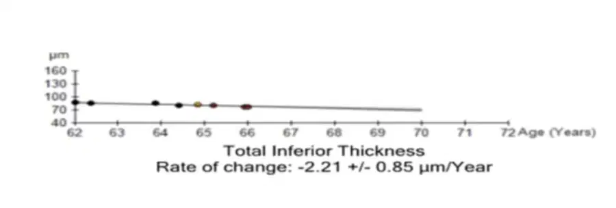
You are reviewing an OCT for a glaucoma patient and see this graph What does this indicate?
Stable
Artefact
Improvement
Likely Progression
Possible Progression
A reliable macular GCC scan is most useful for detecting early glaucoma in which scenario?
Dense cataract
Advanced cupping
Myopic disc suspect
Inferior neuroretinal rim thinning with normal 24-2 visual field
In Cirrus OCT which of the following scans will be unreliable?
Signal strength 7
Signal strength 5
Signal Strength 4
Signal strength 6
Signal strength 8
What is the primary reason that a low signal strength on an OCT scan makes the results unreliable?
Comparison with normative database becomes invalid
Artificially increased RNFL thickness
Media opacity
Artificially decreased RNFL thickness
Segmentation errors
On an RNFL deviation map, a small, isolated red patch is observed in a patient with no other signs of glaucoma. What is the most likely explanation?
The 'floor effect'
The machine has reached its limit for minimum RNFL thickness measurement
Artefacts and the patient needs to be re scanned
The lower part of the scan is cut off
What is the fundamental physical principle upon which Optical Coherence Tomography (OCT) operates?
Fluorescence detection
Absorption of radiowaves
Interference
Refraction of X rays
Polarization
The 'segmentation' process in OCT refers to:
Manual adjustment of scan during acquisition by the operator
Automated identification of retinal layer boundaries
Removal of artefacts from the scan
Maintaining patient fixation
Post processing of the scan
Which of the following best describes the 'normative database' used in OCT analysis for glaucoma?
ONH parameters of patients with advanced glaucoma
RNFL parameters of patients with early glaucoma
HVF, ONH and macular parameters of normal individuals
OCT measurements of healthy sample of participants
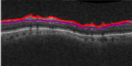
What is this image, a part of the OCT scan called?
B scan
A single cross sectional image of the retina
A scan
Z scan
One superpixel is made of -- PIXELS
(a)
What is the primary clinical significance of performing a 'macular GCL-IPL' (Ganglion Cell Layer - Inner Plexiform Layer) thickness scan in glaucoma?
vulnerable zone of damage in early glaucoma
progression in advanced glaucoma
detecting pattern of damage
immune to floor effect
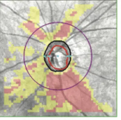
The color-coded maps (green, yellow, red) on an OCT printout are generated by comparing the patient's data to?
The values from patients last visit
Parameters of patients with glaucoma
Healthy individuals in the population
Parameters from an age matched dataset of healthy participants
Standard normal retinal thickness
A reliable OCT scan of a healthy individual shows a suspicious red area. What can cause such a finding?
Pre perimetric glaucoma
Uncooperative patient
Artifact
Anatomical variation
An OCT cube scan is built from a series of many individual slices stacked together. What is the technical term for one of these single cross-sectional 'slices'?
Z scans
Brightness scans
A scans
C scans
What is true regarding the GCL-IPL?
Immune to floor effect
Thinning in early glaucoma
no artefacts
has a larger normative database
Paths taken by the two beams in OCT imaging are:
Imaging the retina and fixation for the patient
Cornea and optic nerve
Reference path and sample path
If the OCT's segmentation algorithm incorrectly identifies the boundary of the RNFL, what is the most likely consequence on the final report?
Low Signal Strength
Thicker RNFL
Thinner RNFL
Scan is flagged as unreliable by the machine software
Inaccurate RNFL thickness
In which of the following patients would the OCT's normative database be LEAST reliable for interpretation?
65 year old with Nuclear sclerosis grade 2
55 year old with mild NPDR
28 year old with axial length 29.5mm
30 year old disc suspect
What kind of OCTA artefact is typically caused by the shadowing effect of retinal blood vessels?
(a)

The components of the scan are marked with number. Which number/numbers show localized or focal progression?
1
2
3
4
What is the primary purpose of viewing an OCT scan in the 'en face' or 'C-scan' view?
Cross sectional view of all the layers
Choroidal thickness measurement
Visualize retinal layer from a frontal aspect or perspective
To check signal strength
Why is a loss of ganglion cells in the macula a particularly concerning sign in glaucoma?
Indicates NTG
Affected only in glaucoma
Signifies early visual loss
Easy to treat
Who is widely credited with inventing Optical Coherence Tomography (OCT) ?
Eric Swanson
James Fujimoto
Joel Schumann
David Huang
All of the above
The difference between Time-Domain OCT (TD-OCT) and Spectral-Domain OCT (SD-OCT) is
SD OCT has better signal strength
SD OCT has a moving mirror and spectrometer
SD OCT has faster scan acquisition
SD OCT has more axial resolution
Why was the measurement of the Retinal Nerve Fiber Layer (RNFL) thickness around the optic disc particularly appealing for early OCT applications in glaucoma research and clinical practice?
Thickest layer of the retina
Easily imaged
Information about patients vision
Primary site of damage in glaucoma
Integration of AI with OCT:
Automate image acquisition
Replace the need for ophthalmologists
Improve diagnostic accuracy
Reduce the cost of OCT devices
What is the key difference in the data acquisition method between Time-Domain OCT and Spectral-Domain OCT?
Pupil dilation
Laser wavelength
Circular versus cube scans
Moving versus stationary reference mirror
AI in OCT:
Increases scan speed
Improves scan quality
Improves scan resolution
Identifies patterns
What is a common reason/reasons why optical coherence tomography (OCT) artefacts are frequently overlooked in clinical practice?
Machines do not flag artefacts
Clinicians do not look carefully
Patients move during scan acquisition
Clinicians do not segment the layers
Which of the following condition/conditions can be a frequent cause of artefacts leading to an artificially thickened or normal-appearing Retinal Nerve Fiber Layer (RNFL) in glaucoma patients?
Cataract
Dry eye
Erm
Retinal schisis
Clinicians need to be highly suspicious of a Retinal Nerve Fiber Layer (RNFL) thickness measurement value of:
50 microns thickness
95 micron thickness
10 micron thickness
Flagged as Yellow
Name the artefact appearing as a wavy undulating image in the scan.
(a)
What happens to RNFL thickness in posterior uveitis ?
Thinner
Thicker
Does not affect RNFL thickness/OCT measurements
Affects Scan acquisition
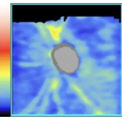
What does this image show?
(a)
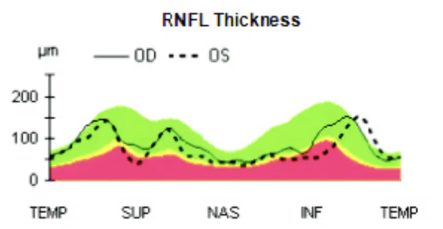
What clinical finding does this image show ?
(a)
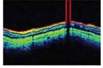
This image illustrates a
(a)

