
 Worksheets
Intraoral Radiographic Anatomy
Total questions: 46
Worksheet time: 23mins

Which radiolucent anatomic landmark is indicated by the black arrows?
Incisive foramen
Superior foramina of the incisive canal
Median palatal suture
Lateral fossa
Nasal cavity
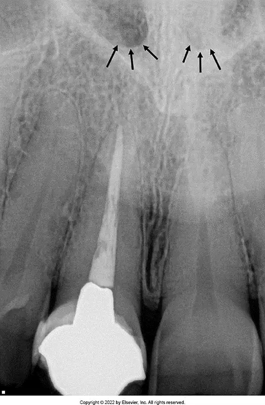
Which small, round radiolucent anatomic landmark(s) are indicated by the black arrows?
Lateral fossa
Median palatal suture
Nasal cavity
Inferior nasal conchae
Superior foramina of the incisive canal

Which radiolucent anatomic landmark is indicated by the white arrows?
Incisive foramen
Nasal cavity
Nasal septum
Median palatal suture
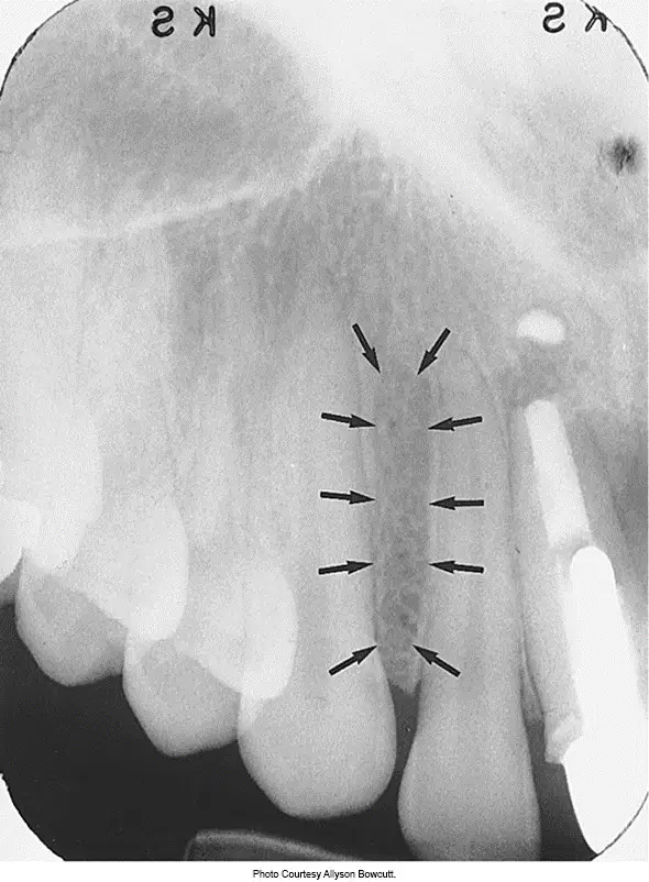
Which radiolucent anatomic landmark is indicated by the black arrows?
Nasopalatine suture
Lateral fossa
Incisive foramen
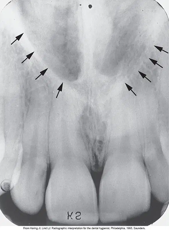
Which large radiolucent anatomic landmark is indicated by the black arrows?
Nasal septum
Median palatal suture
Nasal cavity
Maxillary sinuses

Which radiopaque anatomic landmark is indicated by the black arrows?
Nasal septum
Floor of the nasal cavity
Anterior nasal spine
Inferior nasal conchae
Maxillary sinus
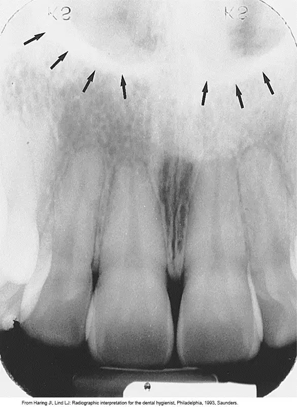
Which radiopaque anatomic landmark is indicated by the black arrows?
Soft tissue of the nose
Floor of the nasal cavity
Anterior nasal spine
Inferior nasal conchae
Maxillary sinus
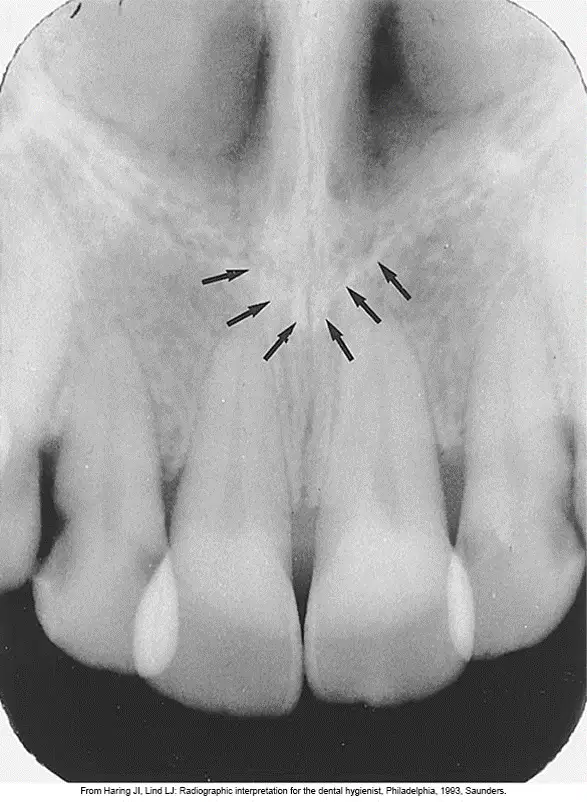
Which radiopaque anatomic landmark is indicated by the black arrows?
Nasal septum
Floor of the nasal cavity
Anterior nasal spine
Inferior nasal conchae
Maxillary sinus

Which radiopaque anatomic landmarks are indicated by the white arrows?
Nasal septum
Floor of the nasal cavity
Anterior nasal spine
Inferior nasal conchae
Maxillary sinus
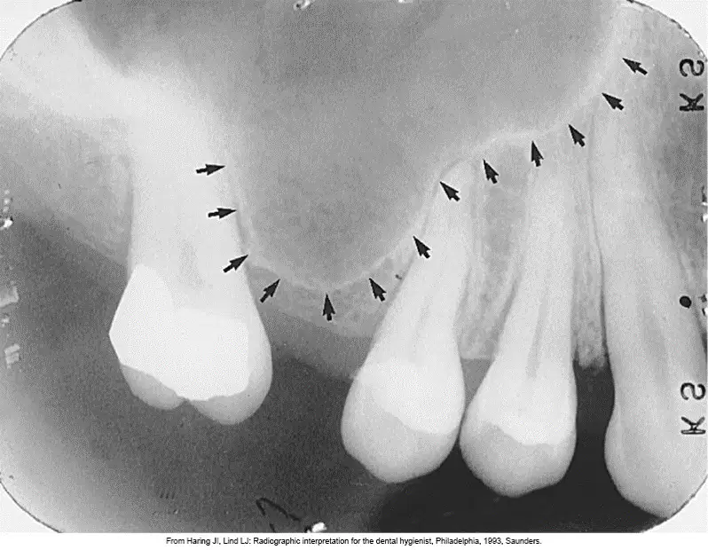
Which radiolucent anatomic landmark is seen above the apices of maxillary molars, indicated by the black arrows?
Maxillary tuberosity
Zygomatic process of the maxilla
Maxillary sinus
Nutrient canals
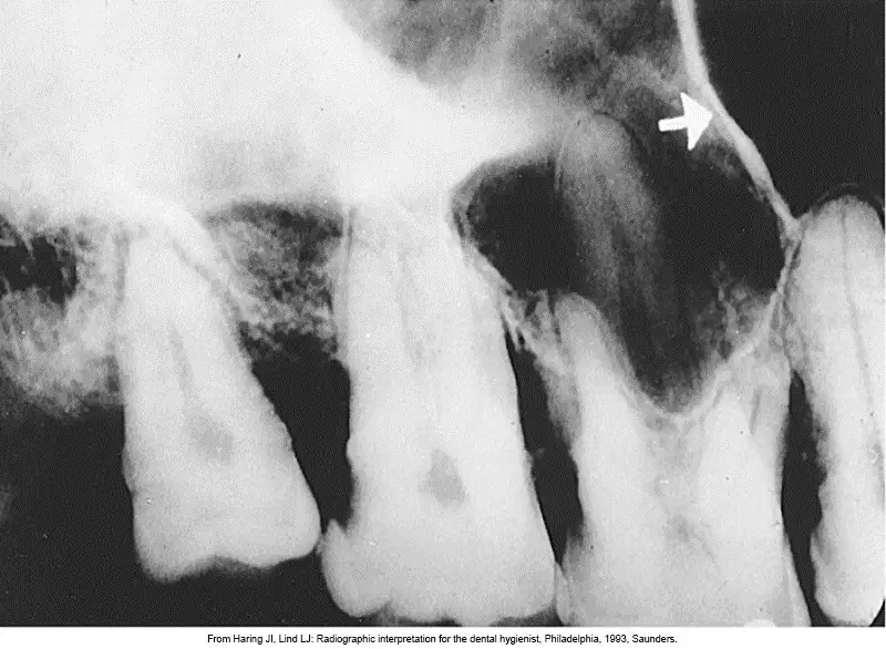
Which radiopaque anatomic landmark is seen within the maxillary sinus and indicated by the white arrow?
Septum
Nutrient canals
Inverted Y
Maxillary tuberosity
Hamulus
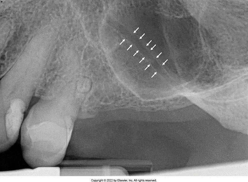
Which radiolucent anatomic landmark, bounded by radiopaque bands is seen within the maxillary sinus and indicated by the white arrows?
Septa
Nutrient canals
Inverted Y
Maxillary sinus
Zygomatic process of the maxilla
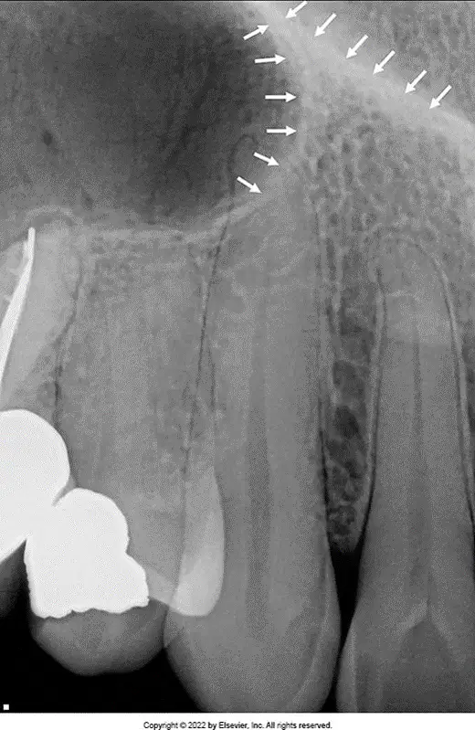
Which radiopaque anatomic landmark is indicated by the white arrows?
Septa
Nutrient canals
Inverted Y-line of Ennis
Maxillary tuberosity
Zygoma
The Inverted Y is made up of which two other anatomic landmarks?
Floor of the maxillary sinus and floor of the nasal cavity
Nasal septum and floor of the maxillary sinus
Zygomatic process and maxillary sinus

The radiopaque bulge indicated by the white arrows is which radiopaque anatomic landmark?
Maxillary tuberosity
Hamulus
Zygomatic process of the maxilla
Zygoma
Genial tubercles
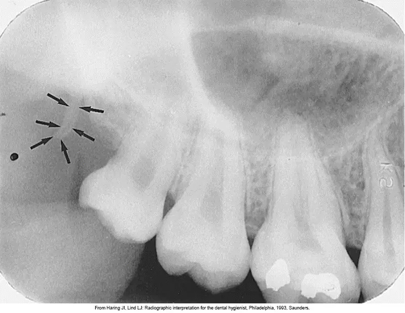
Which anatomic landmark is a radiopaque hook-like landmark located posterior to the maxillary tuberosity?
Hamulus
Zygomatic process of the maxilla
Zygoma
Genial tubercles
Lingual foramen
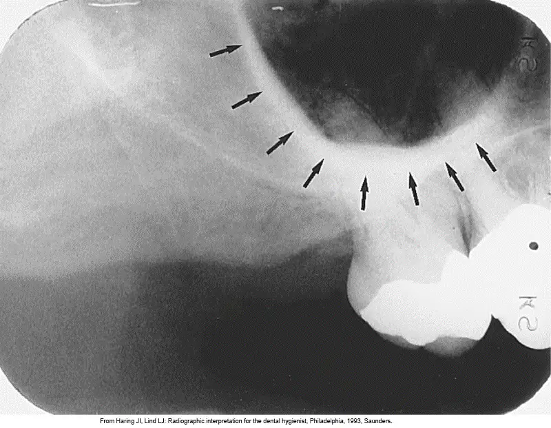
Which J-shaped or U-shaped radiopacity is seen here, indicated by the black arrows?
Zygomatic process of the maxilla
Zygoma
Genial tubercles
Lingual foramen
Mental ridge
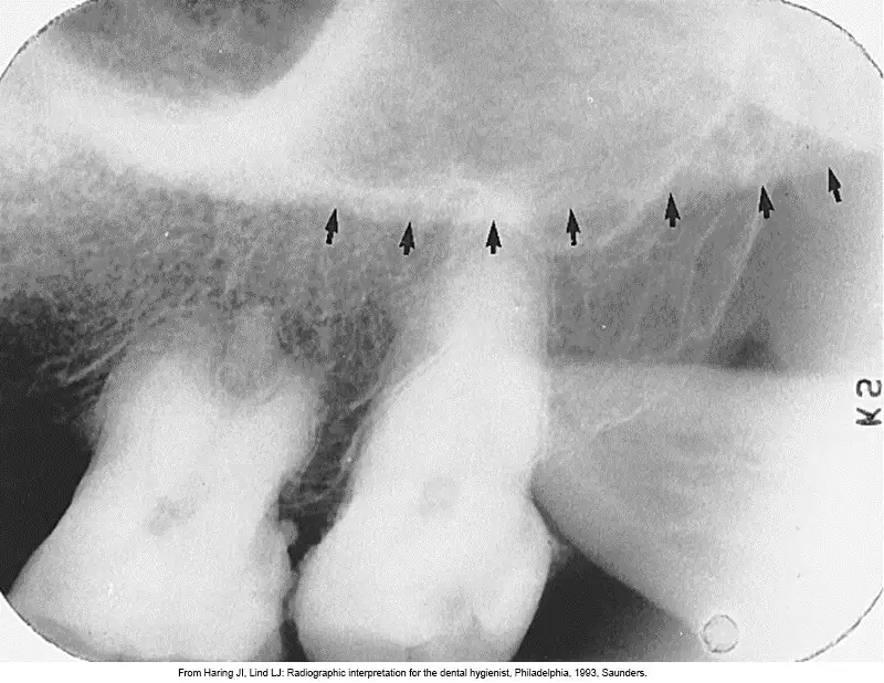
Which anatomic landmark is seen as a diffuse radiopaque band extending posteriorly from the zygomatic process?
Zygoma
Genial tubercles
Maxillary sinus
Mental ridge
Coronoid process

Which ring-shaped radiopacity is seen below the mandibular incisors?
Genial tubercles
Lingual foramen
Mental ridge
Mental fossa
Mental foramen
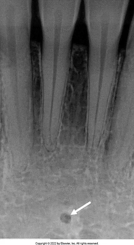
Which anatomic landmark is seen as a small radiolucent dot below the mandibular incisors, indicated by the white arrow?
Genial tubercles
Lingual foramen
Mental fossa
Mental foramen
Mental ridge
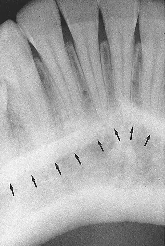
Which anatomic landmark is seen as a thick radiopaque band extending from the premolar region to the incisor region? (indicated by the black arrows)
Mental ridge
Mental fossa
Mental foramen
Mandibular canal
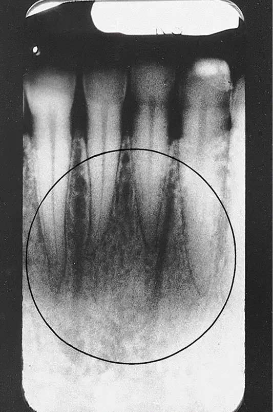
Which radiolucent anatomic landmark is indicated by the black circle?
Mental fossa
Mental foramen
Mandibular canal
Mylohyoid ridge
External oblique ridge
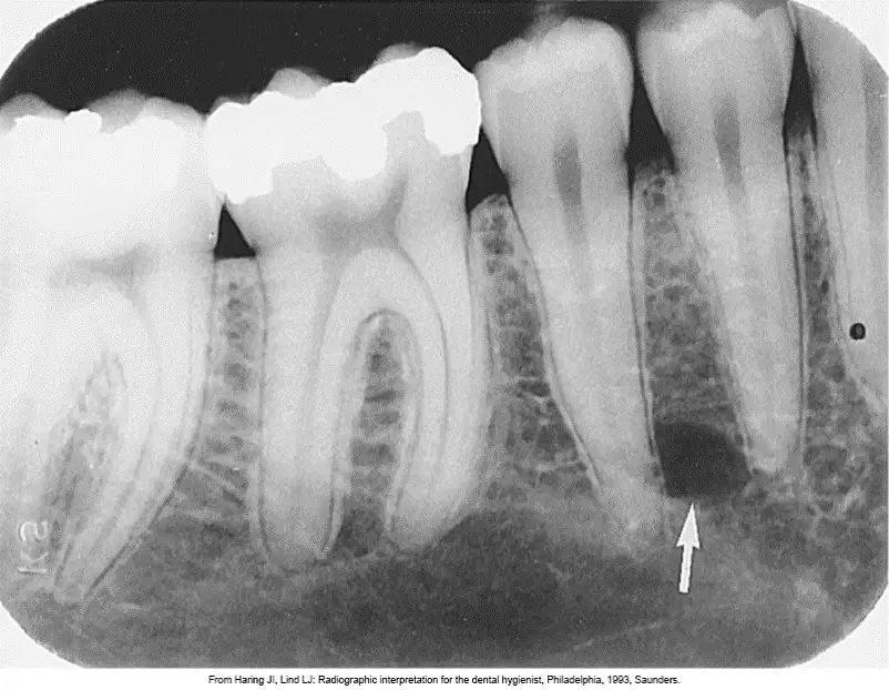
Which small, round, radiolucent anatomic landmark is frequently misdiagnosed as a periapical lesion?
Lingual foramen
Mental fossa
Mental foramen
Mandibular canal
Genial tubercles
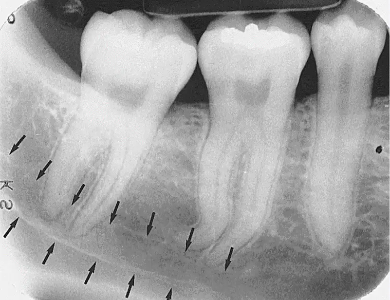
Which mandibular anatomic landmark is seen as a radiolucent band outlined by two thin radiopaque lines, indicated by the black arrows?
Mandibular canal
Mylohyoid ridge
External oblique ridge
Anterior border of the ramus
Submandibular fossa

What is the dense radiopaque band that extends downward and forward from the molar region, indicated by the black arrows?
Mylohyoid ridge
External oblique ridge
Anterior border of the ramus
Submandibular fossa
Coronoid process
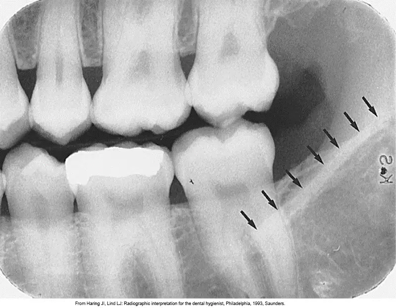
What is the dense radiopaque band that extends downward and forward from the anterior border of the ramus of the mandible, indicated by the black arrows?
Mylohyoid ridge
External oblique ridge
Mandibular canal
Submandibular fossa
Genial tubercles
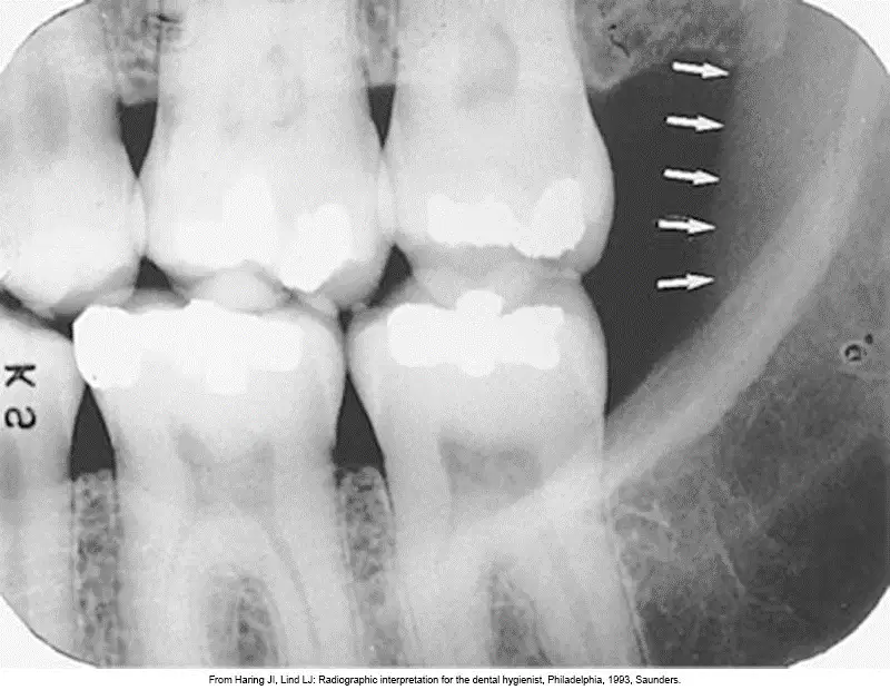
On this bite-wing image, we see a slightly radiopaque vertical band that is posterior to the molar region. What is this anatomic landmark?
Anterior border of the ramus
Submandibular fossa
Coronoid process
External oblique ridge
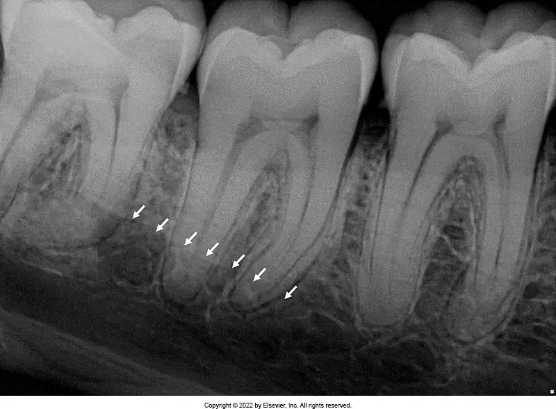
Which radiolucent anatomic landmark is seen in the mandibular molar region below the mylohyoid ridge, indicated by the white arrows?
Submandibular fossa
Lateral fossa
Mandibular canal
Periodontal ligament space
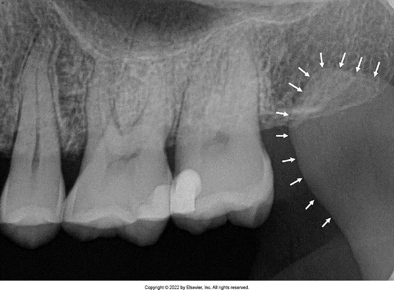
Which mandibular anatomic landmark shows up in maxillary images as a triangular radiopacity posterior to, or superimposed over, the maxillary tuberosity region?
Coronoid process
Anterior border of the ramus
Mental fossa
Mental ridge
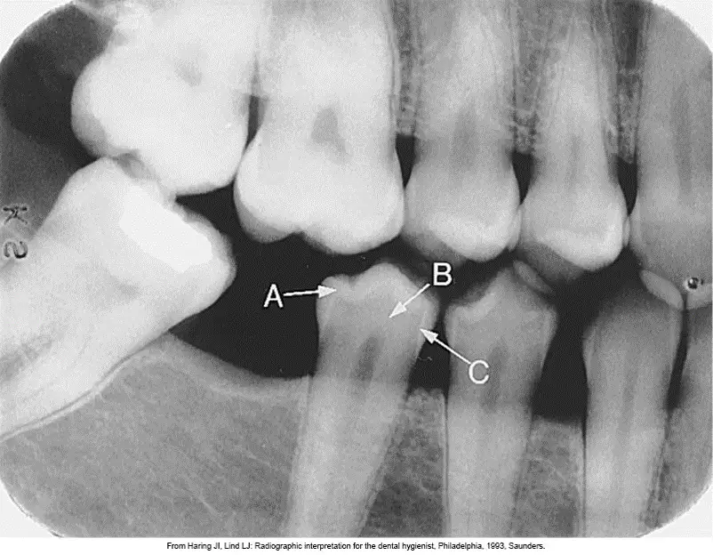
Which tooth structure is indicated by the letter B?
Enamel
Dentin
Dentino-enamel junction
Pulp chamber
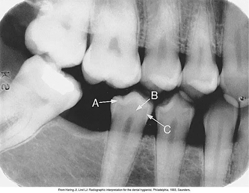
Which tooth structure is indicated by the letter C?
Enamel
Dentin
Dentino-enamel junction
Pulp chamber
Which tooth structure shows up on a radiograph as the most radiolucent tooth structure?
Enamel
Dentin
Dentino-enamel junction
Pulp chamber

What is the dense radiopaque line surrounding the root of a tooth, seen in this image, indicated by the white arrows?
Lamina dura
Periodontal ligament space
Alveolar crest
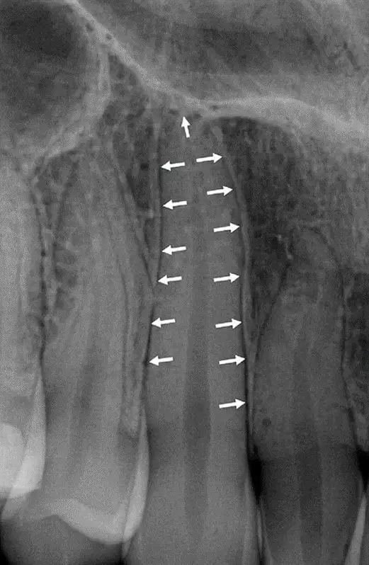
What is the radiolucent line surrounding the root of a tooth, seen in this image, indicated by the white arrows?
Lamina dura
Periodontal ligament space
Alveolar crest
Radiopaque structures appear light or white in a radiographic image.
True
False
Structures that are cavities, depressions or openings in bone such as a sinus, fossa, canal or foramen will allow x-rays to penetrate through them and expose the receptor. These areas will appear radiolucent or black on radiographic images.
True
False

What is making the radiopaque border indicated by the arrows?
Soft tissue of nose
Lip line
Gumline
Maxillary torus
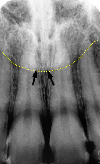
What is making the radiopaque border indicated by the yellow dotted line?
Soft tissue of nose
Lip line
Gumline
Maxillary torus
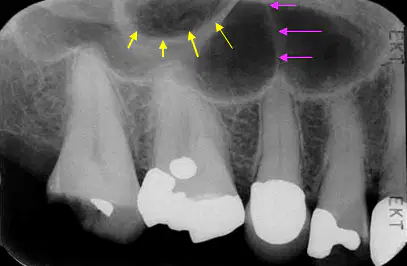
What is the curved, radiopaque anatomical structure indicated by the yellow arrows?
Sinus septation
Zygoma
Zygomatic process of the temporal bone
Zygomatic process of the maxilla
What is the radiopaque anatomical structure indicated by the pink arrows?
Sinus septation
Zygoma
Zygomatic process of the temporal bone
Zygomatic process of the maxilla
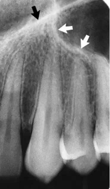
What is the straight, radiopaque line indicated by the black arrow?
Nasal cavity border
Maxillary sinus border
Nasolabial fold
Neurovascular canal
What is the curved, radiopaque line indicated by the white arrow?
Nasal cavity border
Maxillary sinus border
Nasolabial fold
Neurovascular canal
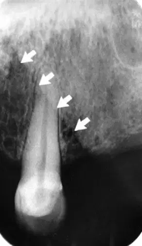
What is the straight, radiopaque border indicated by the white arrows?
Nasal cavity border
Maxillary sinus border
Nasolabial fold
Neurovascular canal
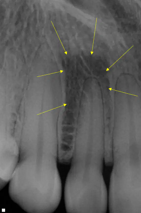
What is the radiolucent region indicated by the arrows?
Periapical cyst
Lateral/ Incisive fossa
Soft tissue of the nose
Incisive foramen
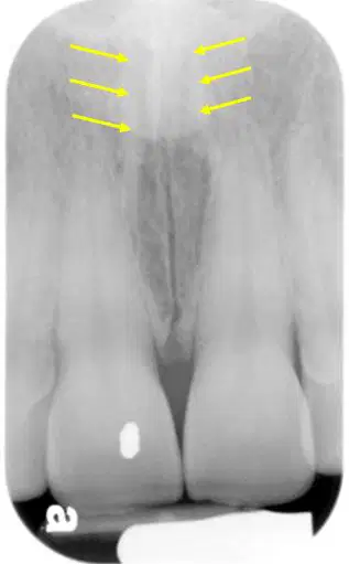
What is the vertical, columnar structure surrounded by cortical bone indicated by the arrows?
Incisive foramen
Nasopalatine canal
Nasal septum
Soft tissue of the nose
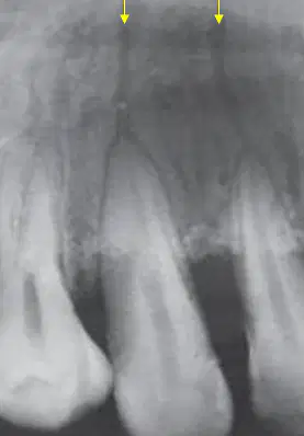
Name the two vertical, radiolucent lines extending apical from the roots.
Fracture lines
Maxillary sinus
Sinus septations
Neurovascular (nutrient) canals

