
Font size
 Worksheets
Abdomen Final Exam Study Guide
Total questions: 94
Worksheet time: 49mins
Ultrasound contrast agents are used for:
Help reduce or eliminate current limitations of ultrasound and doppler blood flow detection
Treating infections detected by ultrasound
Reducing the noise in ultrasound machines
Measuring bone density with ultrasound
The microbubbles of contrast agents are made up of:
gas enclosed in a shell
liquid
solid
plasma
Harmonic imaging works by:
Utilizing the nonlinear behavior of tissues to generate higher frequency echoes for improved image quality.
Using only the fundamental frequency of the ultrasound wave for imaging.
Applying contrast agents to enhance the visibility of blood vessels.
Relying solely on Doppler effect for tissue characterization.
How can we avoid the microbubbles being destroyed?
Low acoustic power according to MI
By increasing agitation and exposure to sunlight
By adding strong acids to the solution
By freezing the microbubbles rapidly
Contraindications for an ultrasound-guided interventional procedure include which of the following?
patient declines procedure
bleeding disorder
lack of safe path for needle
uncooperative patient
The type of equipment used for a core biopsy and an FNA is:
A core needle for core biopsy and a fine needle for FNA
A scalpel for both procedures
A vacuum device for both procedures
A laser probe for core biopsy and a syringe for FNA
The advantages of using ultrasound guidance during a procedure include:
Improved visualization of anatomical structures
various positioning / angles
comfort the patient
lack or radiation
portable and shorter time
An inconclusive result during a procedure could be caused by:
insufficient sample size
accurate measurements
necrotic tissue in sample
wrong amount of tissue
Select the correct order of the steps for an ultrasound-guided procedure.
Patient positioning, probe placement, image acquisition, needle insertion
Needle insertion, image acquisition, probe placement, patient positioning
Image acquisition, patient positioning, probe placement, needle insertion
Probe placement, needle insertion, patient positioning, image acquisition
What might cause a needle to deviate during a procedure?
Tissue the needle is trying to pass through, watch the needle for bending
The color of the needle
The temperature of the room
The type of gloves worn by the practitioner
What is a gastric bezoar?
Trichobezoars- hair balls in young women; Phytobezoars- vegetable matter; Concretions- inorganic materials (sand, gravel, etc.)
A type of stomach ulcer caused by bacteria
A benign tumor found in the stomach lining
A digestive enzyme produced in the stomach
What is the normal shape and echogenicity of the spleen?
Half moon shaped with homogeneous and slightly hyperechoic compared to the liver
Oval shaped with heterogeneous and hypoechoic compared to the liver
Round shaped with anechoic texture
Irregular shaped with mixed echogenicity
What is Crohn disease?
A recurrent granulomatous inflammatory disease that affects the terminal ileum and/or colon, regional enteritis
A viral infection of the liver
A genetic disorder causing excessive mucus production
A degenerative neurological condition
What is a leiomyosarcoma?
A rare, malignant tumor originating from smooth muscle cells within the stomach wall, unpredictable and highly resistant
A benign tumor arising from adipose tissue
A malignant tumor of the lymphatic system
A cancer originating from epithelial cells
Describe pseudomyxoma peritonei.
Caused by cancerous cells that produce excessive mucin, gelatinous ascites, with tumors that cause fibrosis of tissue. Digestion is impeded. If left untreated, tumors and mucin will fill the abdominal cavity, and can be lethal. Organs can become compressed and function is destroyed.
A rare autoimmune disorder characterized by progressive muscle weakness and skin rash, primarily affecting the limbs and face.
A congenital metabolic disorder resulting in the accumulation of copper in the liver, brain, and other vital organs.
A chronic inflammatory bowel disease that causes ulcers in the lining of the large intestine and rectum.
How accurate is contrast enhanced ultrasound when differentiating between benign and malignant masses?
can confirm characteristics of malignancy
Low accuracy
can confirm characteristics of benign
high accuracy
What is bilirubin?
Forms when blood cells breakdown (important to detox liver); Bilirubin is the main pigment in bile
A hormone produced by the pancreas that regulates blood sugar levels
A protein responsible for oxygen transport in the blood
An enzyme that breaks down carbohydrates in the mouth
Describe the ultrasound appearance of the portal veins vs the hepatic veins.
Portal veins are more echogenic
Portal veins are less echogenic
Portal veins are not visible on ultrasound
Portal veins and hepatic veins have identical echogenicity
How is the main portal vein formed?
Union of SMV and splenic vein
Union of IVC and hepatic vein
Union of renal vein and splenic vein
Union of SMV and renal vein
Describe Morrison’s pouch.
Between right lobe and right kidney; First place for fluid will accumulate
Between spleen and left kidney; Common site for abscess
Between bladder and uterus; Site for ectopic pregnancy
Between pancreas and duodenum; Site for pseudocyst formation
What is Glisson’s capsule?
Surrounds the liver
Surrounds the heart
Surrounds the kidney
Surrounds the spleen
Describe fatty infiltration.
Increased accumulation of lipids within liver cells; Acquired and reversible
Decreased lipid content in liver cells; Congenital and irreversible; Causes: malnutrition, acute infection, and vitamin deficiency
Increased protein accumulation in liver cells; Acquired and reversible; Causes: excessive protein intake, chronic infection, and autoimmune disorders
Decreased accumulation of carbohydrates in liver cells; Acquired and reversible; Causes: fasting, dehydration, and excessive exercise
What are the signs and symptoms of acute pancreatitis?
Severe epigastric pain that radiates to back, nausea and vomiting, malaise, abdominal distension, fever, jaundice, and history of gallstones and alcohol abuse
Mild right lower quadrant pain, constipation, and weight gain
Chronic cough, hemoptysis, and night sweats
Intermittent chest pain relieved by rest, palpitations, and syncope
Which structures divide the liver into right and left lobes?
Middle hepatic vein and falciform ligament
Round ligament and ligamentum venosum
Inferior vena cava and gallbladder fossa
Right hepatic vein and caudate lobe
What does amylase do?
Breaks down a complex carbohydrate, into simpler sugars like glucose, which the body can then use for energy
Transports oxygen in the blood
Breaks down proteins into amino acids
Helps in the clotting of blood
The risk factors for pancreatic carcinoma are:
Blacks, Males, Diet high in fat, smoking, Family history correlation, Chronic pancreatitis, and more common after 60
Young age, vegetarian diet, regular exercise, and low BMI
Females, low-fat diet, non-smokers, and no family history
Children, high fiber diet, absence of chronic diseases, and early diagnosis
What does glucagon do in the body?
Increase blood glucose levels, counteracts insulin
Decrease blood glucose levels, mimics insulin
Promotes fat storage, lowers metabolism
Suppresses appetite, increases insulin secretion
Which lab values are associated with HCC?
LFT normal or elevated and AFP may be elevated
LFT always decreased and AFP always normal
LFT normal and AFP always decreased
LFT always elevated and AFP always decreased
What landmark can be used to identify the caudate lobe of the liver?
Located posterior superior surface of the right lobe; Landmarks include: IVC, ligamentum venosum, and left lobe
Located anterior to the gallbladder; Landmarks include: portal vein and hepatic artery
Located inferior to the left lobe; Landmarks include: falciform ligament and round ligament
Located adjacent to the spleen; Landmarks include: splenic vein and pancreatic tail
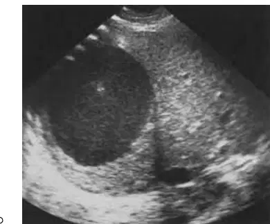
Describe an amoebic abscess and what you might see on ultrasound.
Contracted by ingestion of ameba in food or water; Subcapsular, low amplitude cyst fill in, thickened irregular walls, and good through transmission
Caused by viral infection; Appears as a solid mass with calcifications and shadowing
Results from bacterial infection; Presents as multiple small hyperechoic nodules with no through transmission
Due to fungal infection; Shows a well-defined anechoic lesion with posterior shadowing
31. An intercostal image of the main portal vein is taken to:
Avoid bowel gas interference
Enhance color Doppler signals
Reduce patient discomfort
Increase image magnification
The ligamentum teres used to be UMBILICAL VEIN in fetal life, and it can be found in the ___________ in the adult.
liver
ligamentum venousm
falciform ligament
body
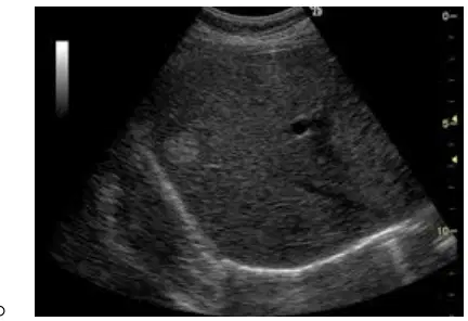
The ultrasound appearance of a cavernous hemangioma is:
A well-defined, hyperechoic lesion with possible posterior acoustic enhancement
A poorly defined, hypoechoic mass with irregular borders
classically round, highly echoic, well defined, solid mass, posterior enhancement, homogenous, and avascular
A solid, hypoechoic mass with calcifications
The _____________ is an echogenic line that connects the GB and the PV on ultrasound.
main lobar fissure
ligamentum teres
porta hepatis
hepatic vein
The vessels that are part of the portal triad include:
hepatic artery, portal vein, bile duct
hepatic vein, portal vein, bile duct
hepatic artery, hepatic vein, bile duct
hepatic artery, portal vein, lymphatic vessel
What does the ligamentum terms look like on US
It appears as a hyperechoic linear structure and is found by identifying the falciform ligament.
It appears as a hypoechoic round mass and is found near the gallbladder.
Triangle in LL inferior to LPV
It appears as a hyperechoic dot and is found in the spleen.
The direction in which blood should be flowing in the portal vein is:
From the gastrointestinal tract to the liver
Towards the liver (hepatopetal)
From the heart to the portal vein
From the kidneys to the liver
What is the normal measurement for the liver?
13-15 cm
8-10 cm
18-20 cm
20-25 cm
What is the normal measurement for the portal vein?
No greater than 13 mm
No greater than 20 mm
No greater than 8 mm
No greater than 18 mm
What is the normal measurement for the splenic vein?
No greater than 10 mm
No greater than 20 mm
No greater than 5 mm
No greater than 15 mm
What is the normal measurement for the SMA?
No greater than 10 mm
No greater than 20 mm
No greater than 5 mm
No greater than 15 mm
What is the normal measurement for the GB wall?
Less than 3 mm
Less than 5 mm
More than 3 mm
More than 5 mm
What is the normal measurement for the pancreas head?
3.5 cm or less AP
5.0 cm or less AP
2.0 cm or less AP
4.5 cm or less AP
What is the normal measurement for the pancreas neck?
1.0-2.0 cm or less AP
2.5-3.5 cm AP
0.2-0.5 cm AP
3.0-4.0 cm AP
What is the normal measurement for the pancreas body?
2.5 cm or less AP
4.0 cm or less AP
1.0 cm or less AP
3.5 cm or less AP
What is the normal measurement for the pancreas tail?
2.5 cm or less AP
4.0 cm or less AP
1.0 cm or less AP
3.5 cm or less AP
What is the normal measurement for the pancreatic duct?
3 mm for head, 2 mm for body
5 mm for head, 4 mm for body
1 mm for head, 0.5 mm for body
6 mm for head, 3 mm for body
What is the normal measurement for the spleen?
8-13 cm
2-5 cm
15-20 cm
20-25 cm
What is the normal measurement for the appendix?
2-20 cm in length
5-30 cm in length
0.5-1 cm in length
10-50 cm in length
The structures that divide the liver into segments are:
Hepatic veins
Hepatic and Portal Veins
Bile ducts
Central veins
Acute hepatitis shows which ultrasound sign?
(a)
What is a porcelain GB?
Rare, calcium incrustation of the GB wall
Form of chronic cholecystitis
What is the ultrasound appearance of fatty infiltration?
hepatomegaly, increased echogenicity, granular texture, obscurity of portal veins, paucity of hepatic vessels, liver/kidney echo brightness disparity, and difficulty in penetration
hypoechoic liver with prominent hepatic veins and normal kidney-liver contrast
multiple cystic lesions scattered throughout the liver parenchyma
well-defined hyperechoic mass with posterior acoustic shadowing
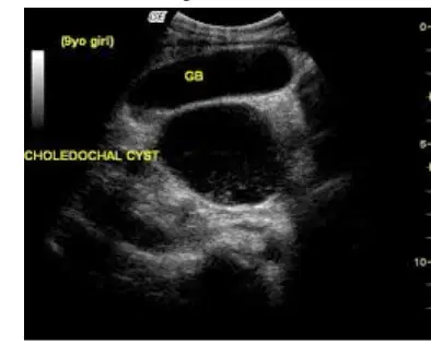
What is the ultrasound sign associated with choledochal cysts?
string of beads with type 5
The ultrasound image shows multiple gallstones within the gallbladder.
The ultrasound image shows a thickened gallbladder wall with pericholecystic fluid.
The ultrasound image shows a dilated pancreatic duct with no cystic structure.
Which liver pathology is associated with oral contraceptive use?
Liver cell adenoma and focal nodular hyperplasia
Which lab value is most associated with bile duct obstruction?
When the main portal vein is dilated, what pathology should you be suspicious of?
Portal hypertension
Cholecystitis
Renal artery stenosis
Pancreatitis
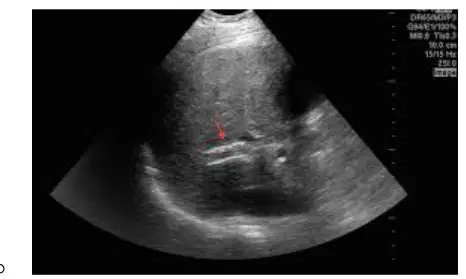
A TIPS shunt on ultrasound appears as which of the following?
A hyperechoic tubular structure connecting the portal and hepatic veins
A cystic lesion in the liver parenchyma
A solid mass with irregular borders
A hypoechoic area in the gallbladder wall
What is hepatic candidiasis?
Liver complication in HIV or other immunocompromised patients
A type of lung infection caused by bacteria
A genetic disorder affecting the kidneys
A neurological disorder causing seizures
What do the acinar cells of the pancreas do?
Secrete digestive enzymes (exocrine)
Produce insulin (endocrine)
Store bile
Filter blood
In a sagittal view of the pancreas, which blood vessel should you see open fully in a horizontal plane?
Splenic vein
Superior mesenteric vein
Portal vein
Inferior vena cava
What do the Islets of Langerhans cells of the pancreas do?
Allows hormones to be distributed throughout the body to stimulate other organs or functional units (endocrine)
Produce digestive enzymes for the breakdown of food (exocrine)
Store bile for fat emulsification
Absorb nutrients from digested food
How is the biliary tree affected by a pancreatic head mass?
Obstructing flow through CBD
Causing dilation of the pancreatic duct only
Increasing bile production
Reducing blood supply to the liver
What are the other names of the pancreatic ducts?
Wirsung - main
Santorini - accessory
Oddi - sphincter
Luschka - minor
What is the most common variant of the GB (gallbladder)?
Junctional fold (Hartmann's pouch) - body and infundibulum
Phrygian cap - fundus
Septate gallbladder
Double gallbladder
What are the causes of GB wall thickening? Bile duct wall thickening?
Obstruction causes back up of bile, inflammation, infection, then thickened walls
Hyperthyroidism
Diabetes Insipidus
Asthma
Describe the Murphy sign.
Rebound tenderness over the GB area
Pain relieved by eating fatty foods
Jaundice with pruritus
Palpable mass in the left lower quadrant
Describe the WES sign.
Wall echo shadow or double arc shadow sign; 2 parallel curved echogenic lines separated by thin anechoic space with distal shadowing; Contract GB containing stones
Well-defined hypoechoic mass in the liver with central scar
Multiple cystic lesions in the kidney with thin septations
Thickened gallbladder wall with pericholecystic fluid
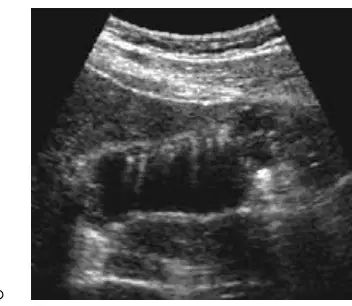
What is adenomyomatosis? What does it look like on ultrasound?
Benign proliferation of GB wall tissue; Comet tail
Malignant tumor of the pancreas; Hypoechoic mass
Inflammation of the liver; Increased echogenicity
Gallstone formation; Acoustic shadowing
Where would you find a Klatskin tumor?
Bifurcation where the left and right hepatic ducts join to form the common hepatic duct
Head of the pancreas
Lower esophagus
Ileocecal valve
Where is the proximal and distal most portions of the biliary tree?
Proximal: intrahepatic bile ducts; Distal: Distal CBD near the Ampulla of Vater
Proximal: gallbladder; Distal: hepatic ducts
Proximal: cystic duct; Distal: common hepatic duct
Proximal: pancreatic duct; Distal: intrahepatic bile ducts
Describe the ultrasound appearance of cholelithiasis.
Echogenic material within bile duct, shadowing, can be mobile or nonmobile; Dilated bile ducts.
Anechoic cystic structure with thin walls and posterior enhancement.
Homogeneous hypoechoic mass with increased vascularity.
Irregular hyperechoic lesion with no shadowing and no mobility.
What is a Phrygian cap?
Fold between fundus and body of gallbladder.
Congenital absence of the gallbladder.
A type of liver cyst.
A fold in the duodenum.
What is a thickened substance that is found in the GB which may create a fluid/fluid level appearance?
Sludge.
Gallstones.
Bile duct stricture.
Polyp.
What are the valves of Heister and where would you find them?
Found within cystic duct; Keep duct from kinking.
Located in the hepatic artery; Regulate blood flow to liver.
Present in the common bile duct; Prevent backflow of bile.
Found in the pancreatic duct; Control enzyme release.
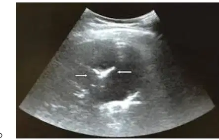
The ultrasound appearance of pneumobilia is best described as:
Echogenic foci with dirty shadowing and reverberation artifacts in the biliary tree
Anechoic tubular structures within the liver
Homogeneous hypoechoic areas in the gallbladder wall
Multiple cystic lesions in the liver parenchyma
Describe white and red pulp of the spleen.
Red- more abundant than white, consists of branching venous sinuses, contains lymphocytes, macrophages, and RBC. White- round masses are scattered through red pulp, surrounds an arteriole, composed of malpighian corpuscles and lymphatic follicles, and large numbers do not contain RBC.
Red- less abundant than white, contains only lymphocytes, no venous sinuses. White- forms the outer capsule of spleen, contains only RBC.
Red- forms the capsule of spleen, contains only connective tissue. White- found only in the center, contains only plasma cells.
Red- contains only arteries and veins, no lymphatic tissue. White- consists of adipose tissue and fibroblasts.
What is an ectopic spleen called?
Wandering spleen, aberrant spleen, and floating spleen.
Accessory spleen, horseshoe spleen, and splenunculus.
Splenomegaly, hypersplenism, and asplenia.
Polysplenia, hyposplenia, and splenic infarct.
Describe a pancreatic phlegmon.
An inflammatory process that spreads along fascial pathways.
A benign tumor of the pancreas.
A cystic lesion filled with pancreatic enzymes.
A congenital abnormality of the pancreatic duct.
Which pathologies would you see the starry sky sign with?
Pneumocystis carinii
Tuberculosis
Hepatitis
Emphysema
What is an accessory spleen?
Variant; More common and can be found throughout body, usually in hilum of spleen (30% of patients).
Congenital absence of the spleen.
A malignant tumor of the spleen.
A cystic lesion found only in the liver.
What is another term for elevated white blood cells?
Leukocytosis.
Anemia.
Thrombocytopenia.
Erythrocytosis.
What might be the cause of a splenic infarct?
Emboli usually originates in heart (ventricular fibrillation).
Vitamin B12 deficiency.
Gallstones in the bile duct.
Chronic pancreatitis.
How could splenic torsion happen?
Wandering or ectopic spleen (migrates); Due to lacking normal peritoneal attachments.
Excessive iron accumulation in the spleen.
Direct trauma to the spleen only.
Congenital absence of the spleen.
Describe an angiosarcoma.
Rare, large; Malignant; Anemia usually present, mets to liver; US appearance similar to hemangioma
Common, small; Benign; No anemia, no metastasis; US appearance always cystic
Always benign; Associated with high WBC count; Never metastasizes; US appearance is hyperechoic
Frequently found in children; Always causes jaundice; US appearance is calcified
What is the greater omentum?
a large apron-like fold of visceral peritoneum that hangs down from the greater curvature of the stomach and the transverse colon
a muscle that separates the thoracic and abdominal cavities
a ligament connecting the liver to the diaphragm
a membrane covering the lungs
Which ultrasound signs might be seen with biliary obstruction?
Antler or stellate branching, parallel channel sign, shotgun sign, and too many tubes sign
Double bubble sign, target sign, and coffee bean sign
String of pearls sign, whirlpool sign, and crescent sign
Snowstorm appearance, comet tail artifact, and ring-down artifact
What is the difference between a thoracentesis and a paracentesis?
Thoracentesis involves removing fluid from the pleural space (around the lungs), while paracentesis removes fluid from the abdominal cavity (ascites)
Thoracentesis is used to treat kidney stones, while paracentesis is used to treat gallstones
Thoracentesis removes air from the stomach, while paracentesis removes air from the lungs
Thoracentesis and paracentesis are both procedures to remove blood clots from arteries
What is cirrhosis?
Cirrhosis is a diffuse process in which liver parenchyma is replaced by fibrous and abnormal nodules. It is irreversible and progressive
Cirrhosis is an acute infection of the liver caused by a bacterial pathogen, leading to rapid liver enlargement and high fever.
Cirrhosis is a benign tumor of the liver that is usually asymptomatic and does not affect liver function.
Cirrhosis is a temporary inflammation of the liver that resolves without any long-term effects.
What is the most common form of glycogen storage disease?
Von Gierke’s disease
Pompe disease
Cori disease
Andersen disease
Which ligaments attach the spleen to the stomach and the left kidney?
Gastrosplenic attaches the spleen to the greater curvature of the stomach; Splenorenal to left kidney
Falciform attaches the spleen to the diaphragm; Hepatogastric to left kidney
Round ligament attaches the spleen to the pancreas; Splenocolic to left kidney
Lienorenal attaches the spleen to the liver; Gastrophrenic to left kidney
What is Budd Chiari syndrome?
Segmental obstruction of hepatic portion of IVC and dilated hepatic veins
Budd Chiari syndrome is a condition caused by obstruction of the renal arteries.
Budd Chiari syndrome is a condition caused by infection of the gallbladder.
Budd Chiari syndrome is a condition caused by inflammation of the pancreas.
Where is the appendix usually found on the body? (Fill in the blank)
Right lower quadrant/ right iliac-Abdominal wall under McBurney’s point- between umbilicus and anterior iliac crest
Left upper quadrant- below the rib cage
Right upper quadrant- near the liver
Left lower quadrant- near the sigmoid colon
Causes of fatty infiltration
obesity, chronic ETHO abuse, hyperlipidemia
diabetes, excess corticosteroids, pregnancy
sever hepatitis, glycogen storage disease, CF, and pharmaceutical
epigastric pain, gas, jaundice
weight loss, nausea, and vomiting

