
Font size
 Worksheets
OBGYN WK 1 Pt 2
Total questions: 103
Worksheet time: 2hrs 42mins
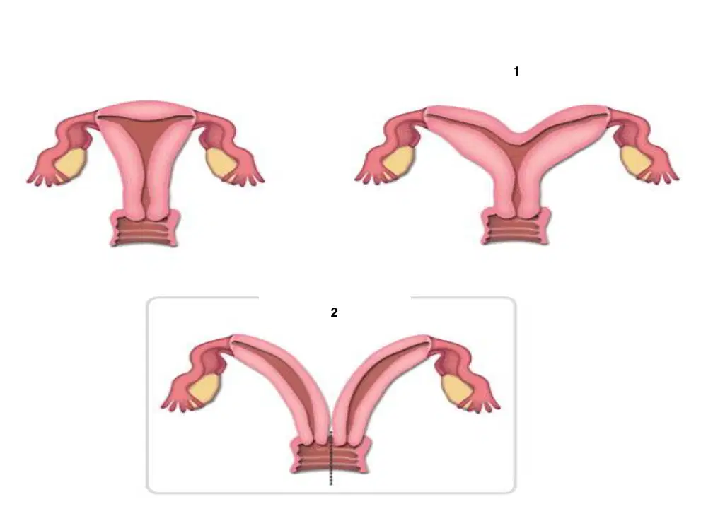
1
(a)
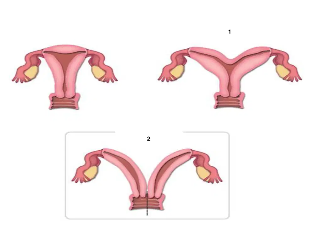
2
(a)
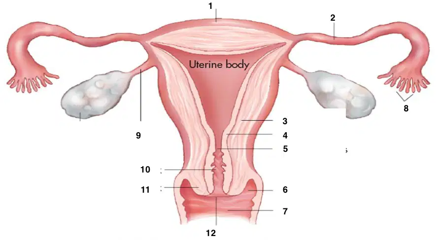
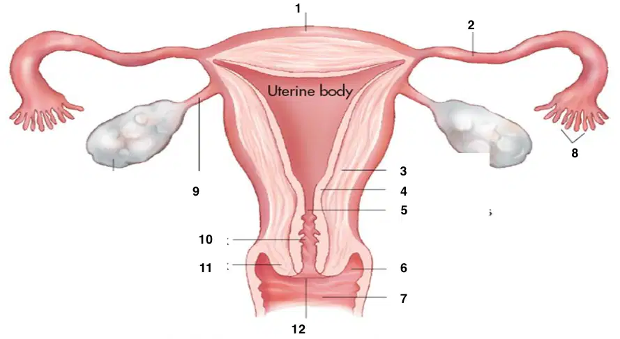
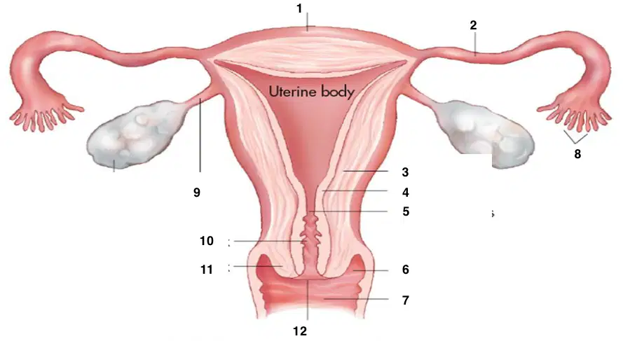
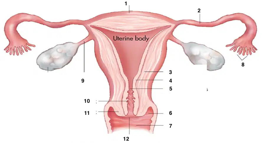
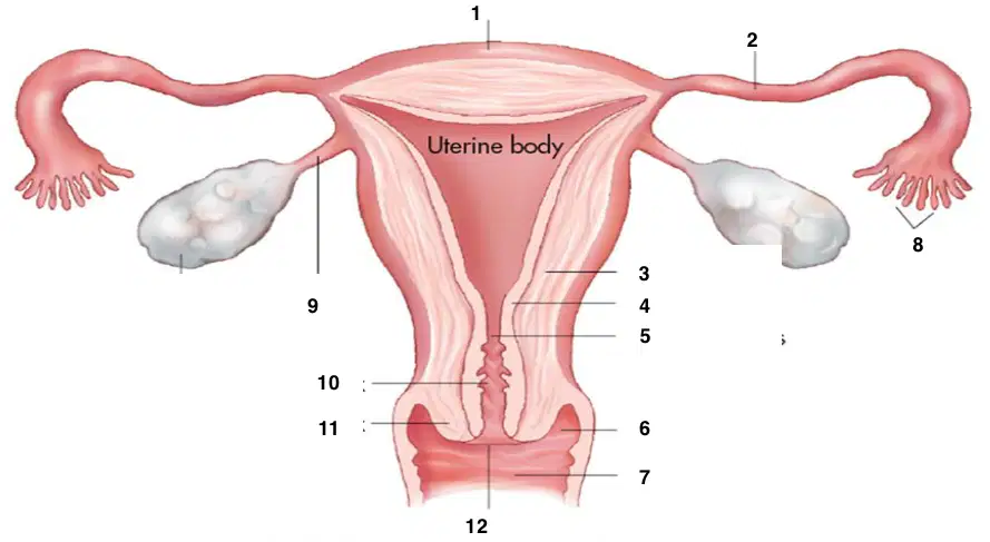
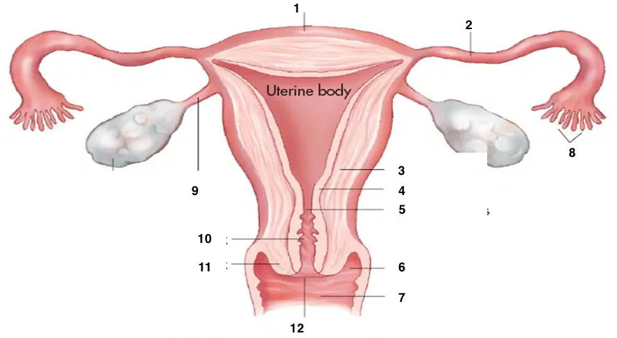
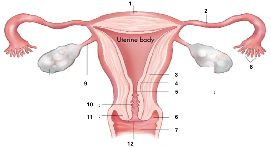
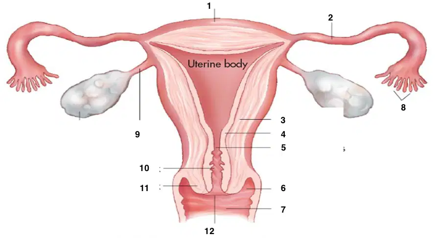
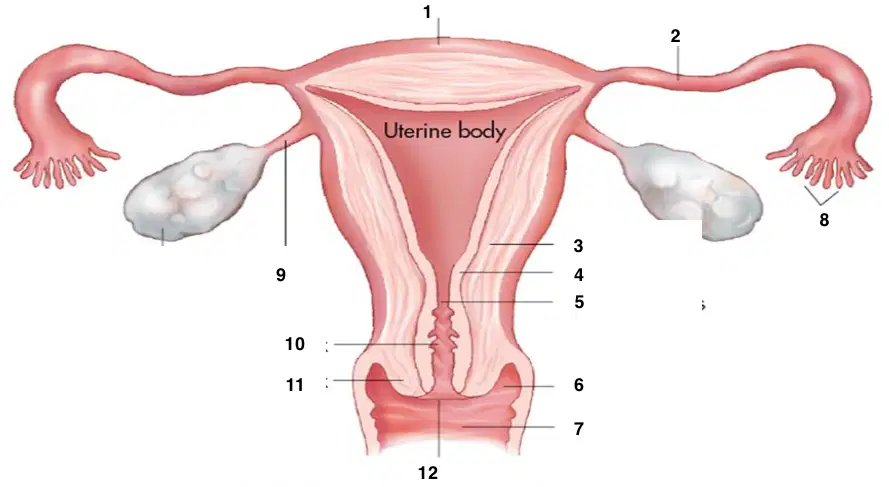
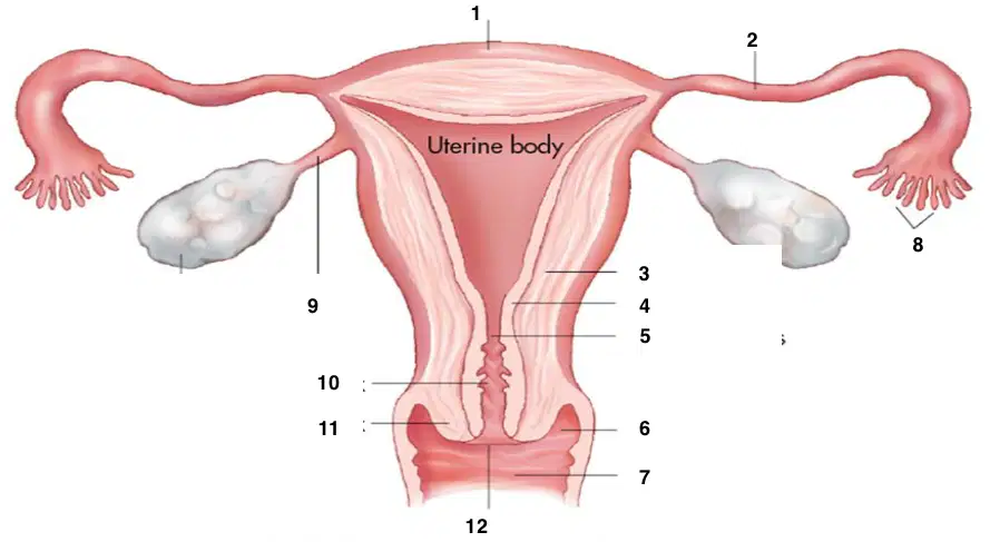
Widest and most superior part of the uterus
(a)
Largest portion of the uterus; between the fundus and cervix
(a)
Perimetrium
Myometrium
Endometrium
(a)
where the fallopian tubes enter the uterine cavity
(a)
Centrally located
(a)
Lower, cylindrical part of the uterus
(a)
Serous outer layer of uterus; serosa
(a)
Muscular middle layer of uterus composed of thick, smooth muscle supported by connective tissue
(a)
Inner mucous membrane, glandular portion of uterine body
(a)
Which of the following are layers of the endometrium
Perimetrium
Myometrium
Superficial or functional (zone functionalis)
Deep basal (zona basalis)
1
(a)
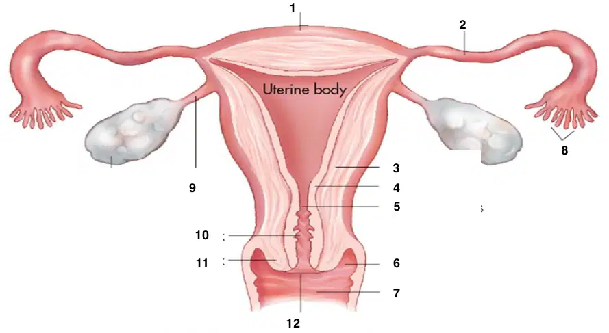
2
(a)
3
(a)
4
(a)
5
(a)
6
(a)
7
(a)
8
(a)
9
(a)
10
(a)
11
(a)
12
(a)
The (a) drapes over the uterine fundus and body to divide the pelvis into
anterior (A) and posterior (P) sections
The peritoneum extends (a) from the uterus, forming the broad ligaments and creating the mesosalpinx as it folds over the fallopian tubes
The (a) is another fold of the peritoneum, which forms
posterior to the broad ligament as it folds over the ovary
Lateral aspect of uterus to pelvic sidewall
(a)
Posterior fold of broad ligament; encloses ovary
(a)
Upper fold of broad ligament; encloses fallopian tube
(a)
Fundus to anterior pelvic sidewalls; holds uterus forward
(a)
Extend across pelvic floor; firmly supports cervix
(a)
Extend from uterine isthmus downward, along side rectum to sactum; firmly supports cervix
(a)
Extends from lateral aspect of ovary to pelvic sidewall
(a)
Extends medially from ovary to uterine cornua
(a)
Broad, round, cardinal, uterosacral, suspensory, and ovarian ligaments are all (a)
Average position of the uterus
(a)
Cervical canal at 90-degrees with vaginal canal
(a)
Uterine body and fundus bent forward upon the cervix
(a)
Cervical canal at an angle less than 90-degrees
with the vaginal canal
(a)
Uterine body and fundus bent backward upon the cervix
(a)
Entire uterus to the pelvic right side
(a)
Entire uterus to the left pelvic side
(a)
entire uterus tipped forward
(a)
(body and fundus
folded anteriorly toward the
cervix)
(a)
(body and fundus folded posteriorly upon the
cervix)
(a)
(entire uterus tilted backward with fundus and body folded posteriorly upon the cervix)
(a)
(entire uterus tilted backward)
(a)
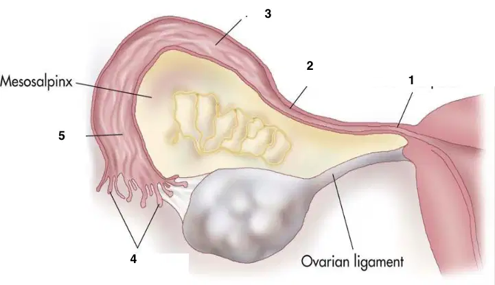
Funnel-shaped lateral tube that projects beyond the broad ligament to overlie the ovaries
(a)
Fingerlike projectors draped over the ovary
(a)
Widest part of the tube, where fertilization occurs
(a)
Hardest part; lies just lateral to the uterus
(a)
Pierces the uterine wall at the cornua
(a)

1
(a)
2
(a)
3
(a)

4
(a)

5
(a)
Almond shaped structures
Posterior to the uterus at the level of the cornua
(a)
3 cm long in a menarcheal female
(a)
12 cm; blood is supplied by ovarian arteries and veins
(a)
Suspended in the mesoovarium
(a)
The ovaries are (a) to the external iliac vessels
The ovaries are (a) to the internal iliac vessels and ureter
Dual blood supply: receives from ovarian artery and uterine artery
(a)
Blood drained by ovarian vein into IVC on the Right and into renal vein on the Left
(a)
It surrounds central medulla & consists primarily of Follicles (in varying stages of development)
(a)
It is covered by layer of dense connective tissue, Tunica albuginea,
surrounded by germinal epithelium)
(a)
Composed of connective tissue containing blood, nerves, lymphatic
vessels, and some smooth muscle at region of hilum
(a)

A
(a)

B
(a)

This image shows a (a)
The ovaries produce:
Reproductive cell, ovum
Testosterone
Periods
Fallopian tubes
Which two hormones are secreted by the ovaries
Testosterone
Progesterone
Estrogen
Lume
Secreted by the follicles
(a)
(a) is secreted by the corpus luteum
Each female ovary contains (a) oocytes at the time of birth
from the inferior/medial pole of the ovary to the uterine cornua
(a)
from the infundibulum of the fallopian tube and the lateral aspect of the ovary to the lateral pelvic wall
(a)
Variable postion ovaries
(a) to the internal iliac artery and vein
Variable position ovaries
(a) to the external iliac artery and vein
Variable position ovaries
Ellipsoid shape with long axis oriented (a)
Location highly variable as ligaments loosen, especially after pregnancy
(a)
medial psoas border
(a)
medial and posterior to the arteries
(a)
posterior to ureters and ovaries
(a)
Posterior to arteries
(a)
between the layers of the broad ligaments, lateral to
the uterus
(a)
Arcuate arteries
Radial arteries
Spiral and straight arteries
(a)
branch laterally off the aorta, run within the suspensory
ligaments, and anastomose with the uterine arteries
(a)
Right vein drains into the IVC directly, whereas the Left vein drains
into the left renal vein
(a)
Arclike arteries that encircle uterus in outer third of myometrium
(a)
Branches of arcuate arteries that extend from myometrium to base of
endometrium
(a)
Branches of radial arteries that
supply zona basalis &
functionalis of endometrium
(a)
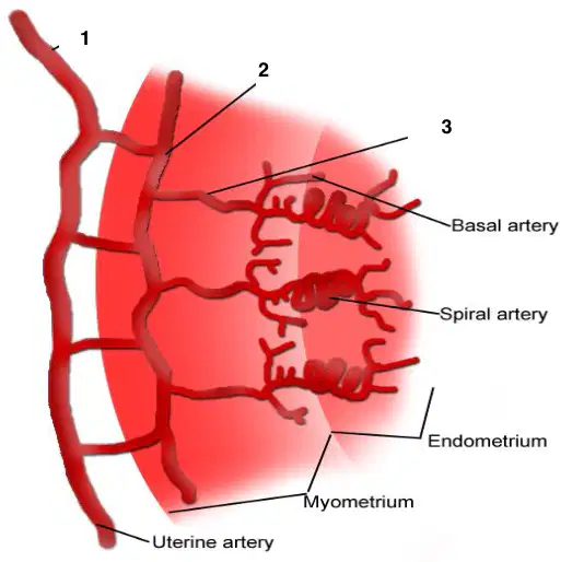
1
(a)
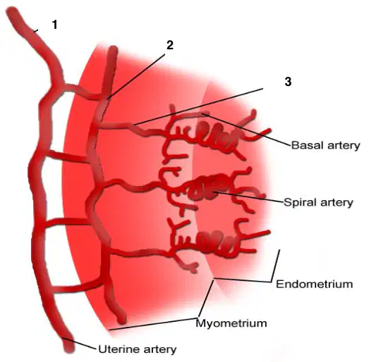
2
(a)
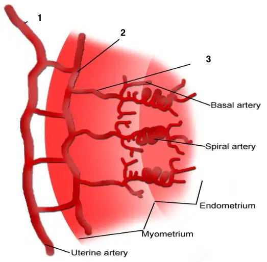
3
(a)
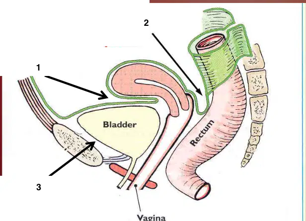
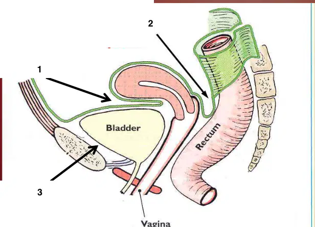
anterior cul- de-sac; anterior to the fundus between the uterus and bladder
(a)
posterior cul-de-sac; posterior to the uterine body and cervix, between the uterus and rectum
(a)
space of Retzius; between bladder and symphysis
pubis
(a)
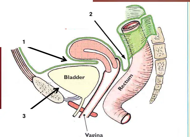
1
(a)
2
(a)
3
(a)

