
 Worksheets
Quality Assurance in Ultrasound Systems
Total questions: 119
Worksheet time: 2hrs 50mins
Routine, periodic evaluation of the ultrasound system to guarantee optimal image quality
(a)
Assessment of system components
(a)
Repairs
(a)
Preventative maintenance
(a)
Record keeping
(a)
Ensure proper operation of units
(a)
Detect gradual changes and avoid minimizing downtime
(a)
Reduce number of non-diagnostic exams
(a)
Reduce repeated exams
(a)
AIUM 100 mm Test Object
(a)
Tissue equivalent phantom
(a)
Doppler phantom
(a)
The beam profile device is also called a
(a)
Assessment under known, defined conditions
(a)
Constant instrument setting
(a)
Phantoms with measurable feature
(a)
Image in identical environment
(a)
(a) quality assurance is unbiased
(a) quality assurance is not influenced by individual experiences or beliefs
(a) quality assurance is factual and repeatable
(a) is influenced by individuals experiences or beliefs
(a) quality assurance is unverifiable and vary from person to person
(a) quality assurance is affected by opinions, beliefs, and assumptions
How many times a year should quality assurance be performed in an ultrasound lab?
1
2
3
4
What properties of AIUm 100 test object makes a good evaluation test?
Depth correction
Attenuation
Identical speed of sound to soft tissue
For an AIUM 100 test object, the speed of sound is (a) to that of soft tissue
What is one drawback of the AIUM 100 test object?
Speed of sound
It does not have the same attenuation as soft tissue so grayscale cannot be evaluated
Poor LATA
Poor LARRD
Can the AIUM test object evaluate gray scale?
Yes
No
Which of the following does the AIUM 100 test object evaluate?
Axial resolution
Lateral resolution
Dead zone
Electronic caliper accuracy
Attenuation
What condition must be true for the AIUM 100 test object to evaluate axial resolution?
When the pins in the object are perpendicular to the sound beams main axis
By scanning the pins located at the top of the test object
By comparing distances between the display with the actual distances
When the pins in the test object are parallel to sound beams main axis
Regarding the Lateral resolution aspect of the AIUM 100 test, the pins in the object are (a) to the sound beams main axis
How is the electronic caliper accuracy evaluated?
By comparing the distances between reflections on the display with the actual distances in the test object
When the pins in the test object are parallel to the sound beam's main axis
When the pins in the test object are perpendicular to the sound beam's main axis.
By scanning the pins located as the top of the test object, very close to the transducer
How is the dead zone evaluated?
When the pins in the test object are parallel to the sound beam's main
When the pins in the test object are perpendicular to the sound beam's main axis
By comparing the distances between reflections on the display with the actual distances in the test object
By scanning the pins located as the top of the test object, very close to the transducer
This phantom has similar features to soft tissue
(a)
What is the speed of sound in a tissue equivalent phantom?
1340 m/s
500 m/s
1540 m/s
1450 m/s
Speed of sound, attenuation, scattering characteristics, and echogenicity are all features of the (a)
Which of the following can properly evaluate gray scale, tissue textures, and multi focus?
AIUM 100 Test object
Tissue equivalent phantom
Doppler phantom
FAST phantom
What is the difference between the AIUM 100 Test object and Tissue equivalent phantom?
AIUM 100 test can not evaluate grayscale but the tissue equivalent object can
AIUM 100 test can evaluate grayscale but tissue equivalent object can not
Neither can evaluate grayscale
Both can evaluate grayscale
Evaluate gray-scale, tissue textures, multi-focus and adjustable phased array transducers.
(a)
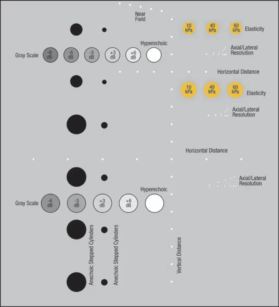
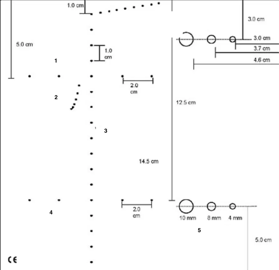
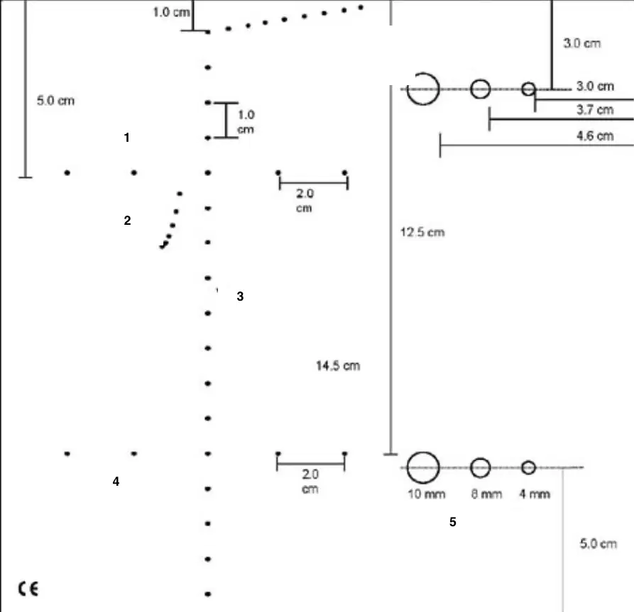
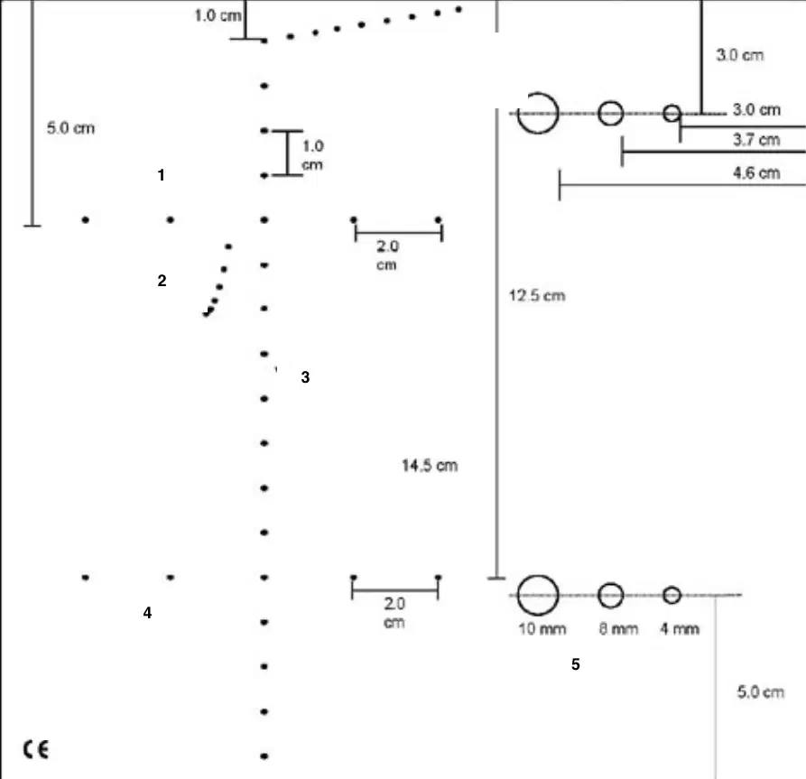
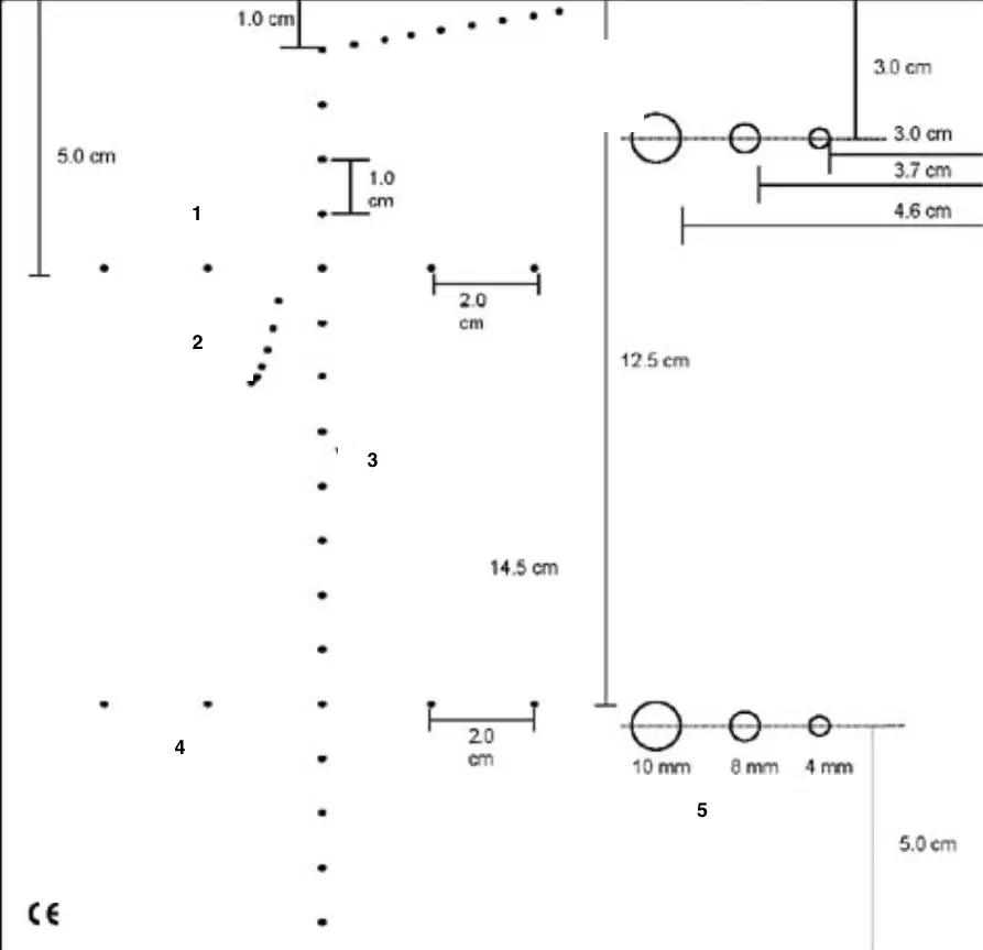
This diagram shows a (a) phantom
Depth calibration, lateral distance, axial resolution
(a)
Lateral resolution, lesion detection are (a)
Flow phantoms, circulation pump, and tissue equivalent phantoms are all apart of a (a)
Vibrating strings and moving belts are also apart of the (a)
The (a) evaluates pulsed doppler, CW doppler, color doppler, and power modeD
The (a) phantom assesses elevational resolution
Diffuse scattering plane at an angle to the incident beam
(a)
The slice thickness phantom is a (a) phantom
Regarding slice thickness phantoms , (a) slices decrease spatial resolution
With a slice thickness phantom, cystic structures may appear (a) with echoes

Which phantom is present in this image?
Tissue equivalent phantom
Doppler phantom
slice thickness phantom
AIUM 100 test object
What structures are contained in a normal sensitivity phantom?
Pins
solid masses
amplification
cystic structures
With normal sensitivity performance measurements the following are (a) displayed: pins, solid masses, and cystic structures
Output, TGC, and amplification adjusted establish (a)
Performance measurements for (a) include: output power, amplification, set to maximum practical levels, and measures the maximum depth
Sensitivity is the ability of the system to display (a) echoes
Should sensitivity vary from one exam to the next?
Yes
NO
The dead zone is an area (a) to the transducer that results in inaccurate images?
The dead zone is originated by ___ and ___ (from transmitting to receiving)
(a)
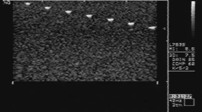
Dead zone is the (a) series of pins in a test object
Shallowest depth at which uniform tissue texture appears, with Tissue Equivalent.
(a)
Which type of transducers result in a thinner dead zone?
Higher frequency
Lower frequency
Broken crystal, detached backing material, longer pulse duration =
(a)
When using a stand off pad, where does the dead zone occur?
First layer of the pins
Within the stand off pad
Deepest zone
Shortest zone
If you were to fill in a glove with water and put that on a patient, when you scan where would the dead zone occur?
Deepest layer
Within the water filled glove
Shallowest layer of skin
Highest intensity
• Narrowest beam
• Best lateral resolution
(a)
(a) must be carefully evaluated
Smallest distance at which two pins parallel to the sound beam are displayed as two different echoes
(a)
Scanning sets of successively closed spaced pins in the phantom
(a)
Minimum distance at which two side-by-side pins are displayed as two distinct images
(a)
Measure width of reflections on the display created by point targets in the phantom
(a)
Scanner sensitivity and reliability has (a) and many instruments include internal diagnostic testing start-up mechanisms which identify improper electronic operation
Penetration, distance accuracy, image uniformity, accuracy of image display, and accuracy of image recording
(a)
Top surface.
•Pins at the top of test object.
(a)
Distance from transducer to an area in the near field covered by reverberation
(a)
Top surface.
•Rod group in the middle (separated 1, 2, 3, 4, 5 mm) in oblique plane
(a)
The smallest separation of the two closest rods seen
(a)
Side surface of the object.
•Scanning same rod for axial resolution
(a)
The smallest separation of the two closest rods seen
(a)
Vertical and horizontal accuracy of the electronic calipers.
•Scanning vertical and horizontal equidistant rods from the top (each rod separated 20 mm)
(a)
Measure the distance between the rods and compare to the actual distance
(a)
Measures the weakest echo signal.
•Determined by the gain setting, with no TGC
(a)
The bottom rod of the group of six vertical equidistant rods is used
(a)
Dead zone: Evaluate by the (a) surface of the object and scanning the pins located at the top of the object
It documents the distance from the face of the transducer to an area within the near field which cannot be used to image due to reverberation artifact or transducer design
(a)
Vertical and horizontal accuracy
(a)
Vertical and horizontal accuracy. Calibration by scanning vertical and horizontal equidistant rods from the top
(a)
Assessment of depth and horizontal accuracy involves comparing the known distance between rods with
the distance indicated by the calipers on the image
(a)
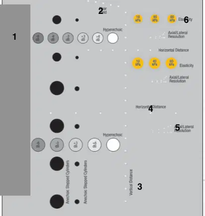
1
(a)
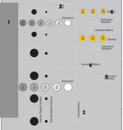
2
(a)
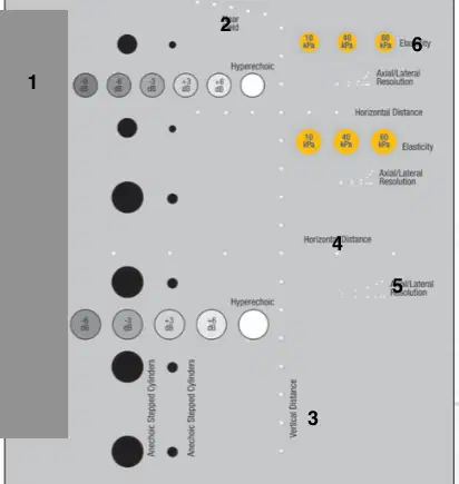
3
(a)
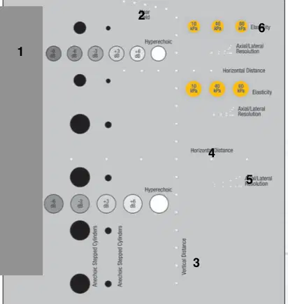
4
(a)
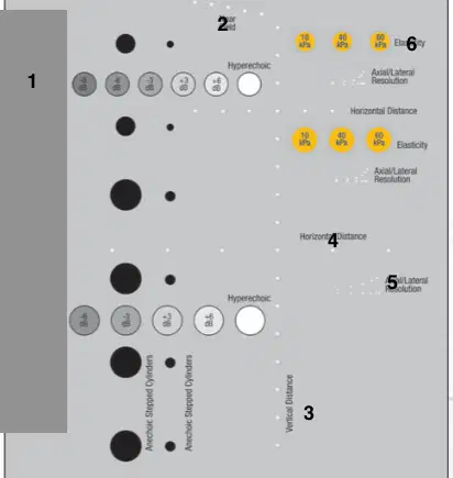
5
(a)
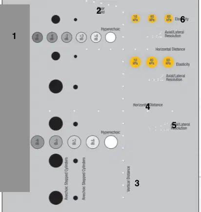
6
(a)
Use pins parallel to the sound bea
Top surface of the object
Rod group in the middle
Measure the two pins with space and are closest together
(a)
Pins perpendicular to the sound beam
Top surface of the phantom
Use middle rods closest together and off set to the side
Measure the set of pins closest side to side and visualized as two separate pins
(a)
Tissue Phantom: Use cylindrical objects within.
Inclined Plane Phantom: Two sections forming a flat interface oriented at 45 degrees
(a)
TGC characteristics: Measures system uniformity to display echoes of equal magnitude
(a)
System Sensitivity: Measures maximum depth of visualization.
Contrast Resolution: Gray scale targets
(a)
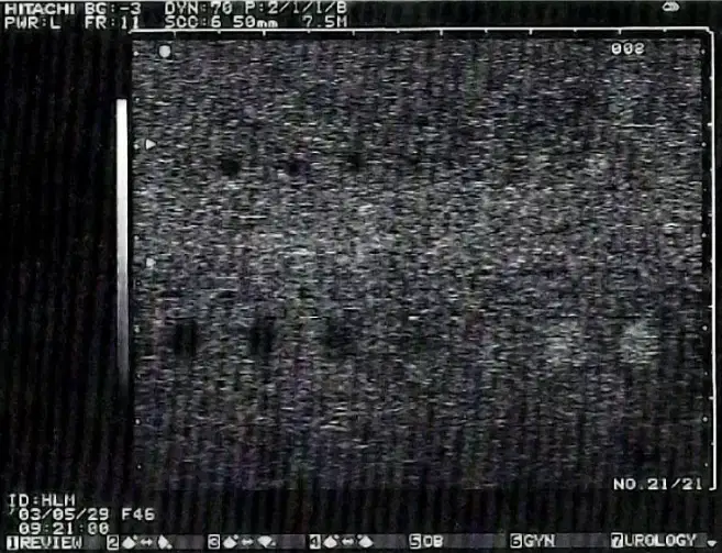
What is this used for?
(a)
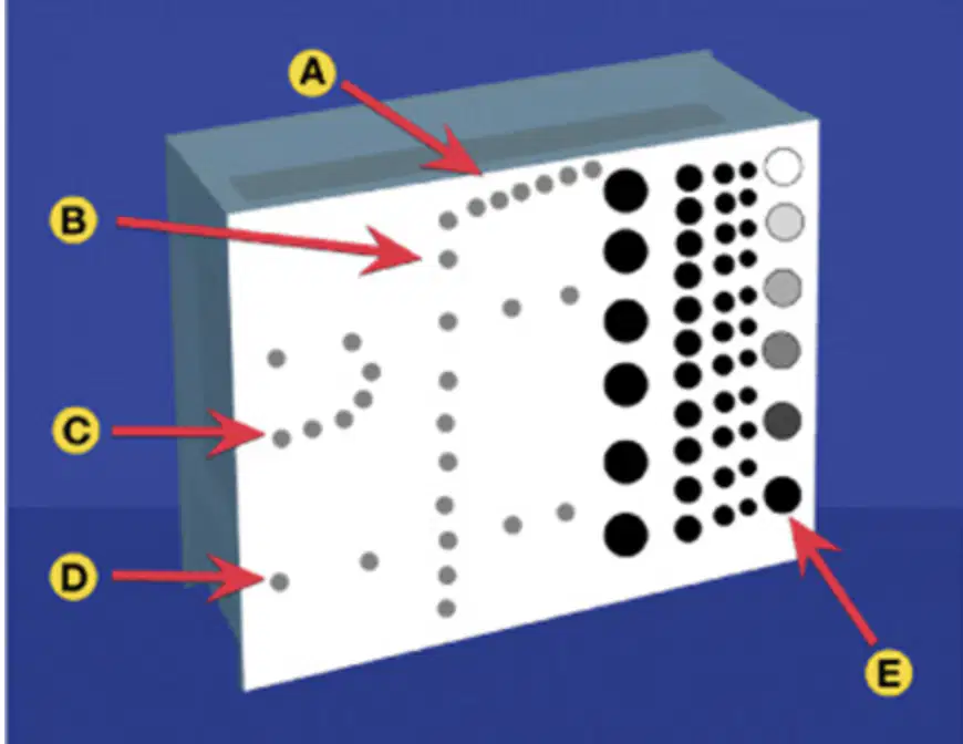
A
(a)
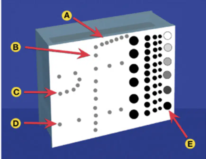
B
(a)
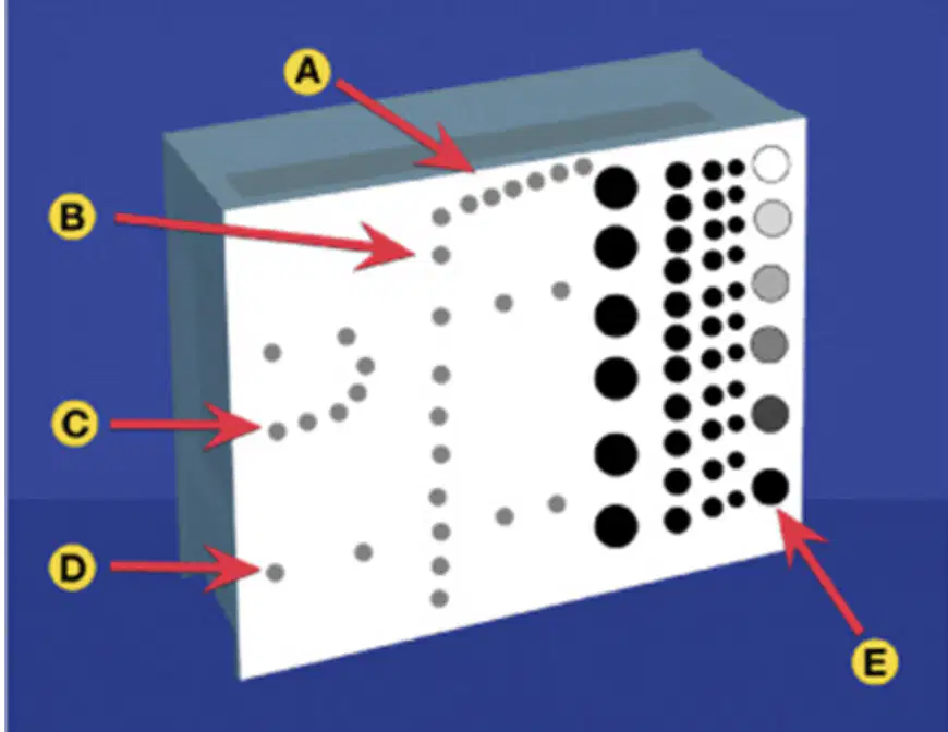
C
(a)
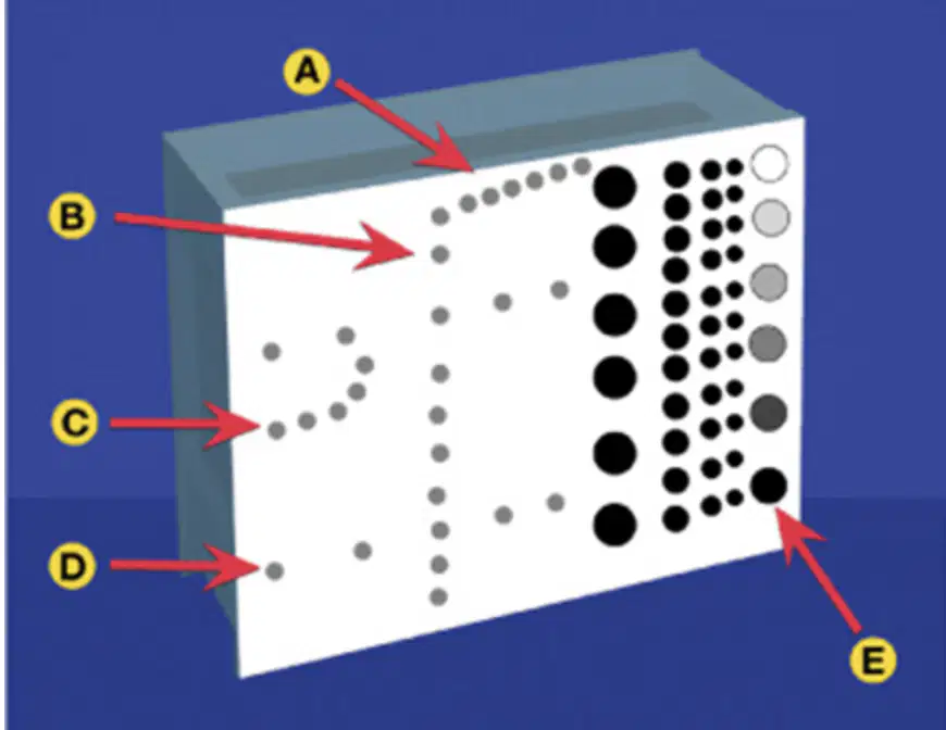
D
(a)
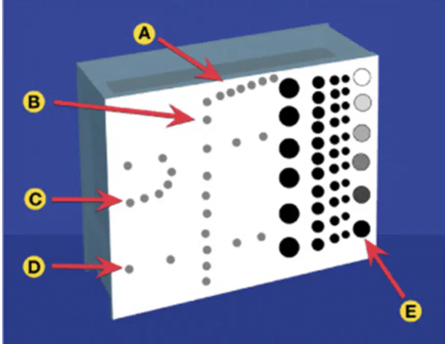
E
(a)
(a) are those at which all the pins, solid masses, and cystic structures in the test phantom are accurately displayed
(a) is evaluated with the output power and amplification of the system set to maximum practical levels
Does TEP assess Doppler?
No
Yes
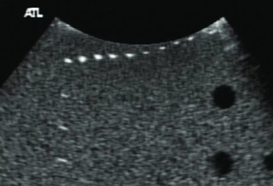
The dots at the top of this image =
(a)
The dead zone is assessed with the (a) series of pins
Which Doppler characteristics can be assessed with a Doppler Phantom?
Pulsed
Continuous
Power
Normal
The (a) results from transducer ringing and the time it takes to switch from the transmit to receive mode
Which of the following would NOT result in an increasingly deeper dead zone?
Shorter pulse duration
Cracked crystal
Detached backing material
Longer pulse duration
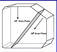
This image shows the (a) phantom
1
(a)
2
(a)
3
(a)
4
(a)
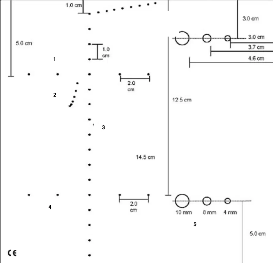
5
(a)

