
 Worksheets
Chapter 08: Cervical and Thoracic Spine
Total questions: 80
Worksheet time: 1hrs 20mins
On average, how many separate bones make up the adult vertebral column?
33
28
26
31
The spinal cord tapers off to a point distally at the vertebral level of:
lower L5.
L2-3 intervertebral space.
L1-L2.
L4-5 intervertebral space.
Which of the following is the first compensatory spinal curvature to develop in the young child?
Thoracic
Cervical
Sacral
Lumbar
Scoliosis is defined as an abnormal or exaggerated (a) _____ curvature.
Concave
Convex
Lateral
"Swayback" Lumbar
Kyphosis is defined as a(n):
normal lumbar curvature.
normal convex sacral curvature.
normal compensatory curve of thoracic spine.
abnormal thoracic curvature with increased convexity.
The bony structures connected directly to the vertebral body are the:
(a)
Fill in the blank: The bony projections on vertebrae that serve as points of articulation with adjacent vertebrae are called _________.
articular processes
spinous processes
transverse foramina
vertebral bodies
The most posterior aspect of a typical vertebra is the:
pedicle.
spinous process.
lamina.
body.
The joints between articular processes of vertebra are termed_____joints.
zygapophyseal
fibrous
intervertebral
synarthrodial
Which aspect of the intervertebral disk is composed of semigelatinous material?
Nucleus pulposus
Annulus fibrosus
Conus medullaris
Cauda equina
Which of the following statements is true?
All adult vertebrae are separated by intervertebral disks.
All thoracic vertebrae have at least one facet for rib articulation.
The intervertebral disks are tightly bound to the vertebral bodies to prevent movement within the vertebral column.
All of the above are true statements.
Which of the following statements is true for a typical adult vertebra?
The transverse processes extend laterally from the junction of the pedicles and laminae.
The transverse processes are present only on thoracic vertebrae containing rib connections.
The spinous process extends posteriorly from the body of the vertebra.
All of the above are true statements.
Which of the following features makes the cervical vertebra unique as compared with other vertebrae of the spine?
Transverse foramina and double (bifid) tips on spinous processes
Overlapping vertebral bodies
Presence of zygapophyseal joints
All of the above
Where is the articular pillar located on a cervical vertebra?
Between the superior and inferior articular processes
Between the pedicle and the body
Between the spinous process and the lamina
Nowhere
Which term best defines or describes the vertebral body of C1?
The smallest of all vertebral bodies
A column of bone supported by an intervertebral disk
A large bony mass
There is no vertebral body at C1.
Which of the following thoracic vertebra(e) possess no facets for costotransverse joints?
T9-10
T11-12
T1
None of the above
The zygapophyseal joints for the typical cervical vertebra lie at an angle of ___ in relation to the midsagittal plane.
45°
60°
70° to 75°
90°
The most prominent aspect of the thyroid cartilage corresponds to the vertebral level of:
C5.
C1-2.
The zygapophyseal joints for the thoracic spine lie at an angle of _____ in relation to the midsagittal plane.
45°
60°
70° to 75°
90°
T2-3 intervertebral disk space is found at the level of the:
jugular notch.
sternal angle.
xiphoid process.
2 inches (5 cm) below the jugular notch.
The gonion corresponds to the vertebral level of:
C1.
C4-5.
C3.
C7.
The xiphoid process corresponds to the vertebral level of:
T8.
T 9-10.
T7.
T12.
Along with increasing the source image receptor distance (SID), what other factor(s) will improve spatial resolution for lateral and oblique projections of the cervical spine?
Increasing object image receptor distance (OID)
Using a small focal spot
Using higher kV, lower mAs
Using a breathing technique
Which of the following pathologic conditions is defined as “a condition of the spine characterized by rigidity of a vertebral joint”?
Spondylitis
Ankylosing spondylitis
Spondylosis
Scheuermann disease
Which of the following clinical conditions will require a decrease in manual technical factors?
Advanced osteoporosis
Severe scoliosis
Moderate lordosis
Scheuermann disease
Which factor is most important to open up the intervertebral joint spaces for a lateral thoracic spine projection?
Use sufficiently high kV.
Collimate the spine region to reduce scatter.
Keep vertebral column parallel to tabletop.
Angle the central ray (CR) 5° to 10° caudad.
Which position or projection of the cervical spine will best demonstrate the zygapophyseal joint spaces between C1 and C2?
Anteroposterior (AP) axial
Lateral
45° posterior or anterior oblique
AP open mouth
Along with the upper incisors, what other bony landmark must be aligned for the AP open-mouth projection?
Base of skull
Tip of mandible
Inferior margin of lower incisors
Thyroid cartilage
How much CR angulation is required for the AP axial projection for the cervical spine?
CR is perpendicular to the image receptor.
5° to 10° cephalad
15° to 20° cephalad
15° to 20° caudad
Why are the anterior oblique projections (right anterior oblique [RAO]/left anterior oblique [LAO]) preferred over the posterior oblique projections of the cervical spine?
To make the patient more comfortable
To reduce the thyroid dose
To prevent overlap of the mandible over the spine
To place the zygapophyseal joints closer to the image receptor
Which of the following positions will demonstrate the left intervertebral foramina of the cervical spine?
RAO
Left posterior oblique (LPO)
Lateral
LAO
Which of the following projections will best demonstrate the zygapophyseal joints of the cervical spine?
AP axial
Posterior oblique
Lateral
Anterior oblique
Which of the following is NOT a correct evaluation criterion for the AP axial C spine projection?
C3 to T2 vertebral bodies should be visualized.
Spinous processes are seen to be equal distances from the vertebra body lateral borders.
Center of the collimation field is at C4.
All of the above are correct criteria.
Which of the following factors does not apply to a lateral projection of the cervical spine?
Use a 72-inch (183 cm) SID.
Suspend respiration upon full inspiration.
Perform horizontal-beam lateral projection if trauma is suspected.
Center CR to thyroid cartilage.
The chin is extended for a lateral projection of the cervical spine to:
open up the C1-2 joint space.
demonstrate the articular pillars.
open up the intervertebral joint space.
prevent superimposition of the mandible upon the spine.
The PA projection (Judd method) is intended to demonstrate the atlantoaxial joints between C1 and C2.
True
False
The posterior cervical oblique projections demonstrate the intervertebral foramina and pedicles on the side closest to the image receptor.
True
False
The AP axial-vertebral arch projection may be performed to better demonstrate the:
C1 and C1-2 atlantoaxial joint spaces.
articular pillars of C4-7.
open intervertebral disk spaces of C3-T1.
dens within the foramen magnum.
What type of CR angle is recommended when performing the AP axial C spine projection erect?
15° cephalad
20° cephalad
15° caudad
25° cephalad
What type of CR angle is required for posterior oblique (left posterior oblique [LPO]/right posterior oblique [RPO]) positions of the cervical spine?
15° caudad
5° to 10° cephalad
None; the CR is perpendicular to the image receptor (IR).
Which of the following factors will enhance the visibility of the vertebral bodies during a lateral projection of the thoracic spine?
Use a high kV.
Increase SID.
Use a breathing technique.
Flex the spine.
Which of the following positions will best demonstrate the left zygapophyseal joints of the thoracic spine?
LPO
RPO
RAO
Lateral
How much rotation of the body is required for an LAO projection of the thoracic spine (from the plane of the table)?
20°
45°
50° to 60°
70°
Which of the following projections will project the dens within the shadow of the foramen magnum?
AP open mouth
AP projection (Fuchs method)
Twining method
None of the above
A radiograph of an AP open-mouth projection of the cervical spine reveals that the zygapophyseal joints are not symmetric. No fracture or subluxation is present. Which one of the following positioning errors most likely led to this radiographic outcome?
Excessive CR angulation
Insufficient CR angulation
Rotation of the spine
Excessive flexion of the head
A radiograph of an AP axial projection of the cervical spine reveals that the intervertebral joints are not open. The following analog factors were used for this projection: 5° cephalad angle, 40-inch (100 cm) SID, grid, slight extension of the skull, and the CR centered to the thyroid cartilage. Which of the following modifications should be made during the repeat exposure?
Keep the CR perpendicular to the image receptor.
Increase the SID to 72 inches (183 cm).
Center the CR to the gonion.
Increase the CR angulation.
A radiograph of an AP thoracic spine projection reveals that the upper thoracic spine is overexposed. The lower vertebrae have acceptable density and contrast. Which one of the following modifications will improve the visibility of the upper thoracic spine?
Place the upper spine under the cathode side of the tube.
Use a compensating (wedge) filter.
Increase the kV to decrease the contrast of the upper spine.
Use a breathing technique.
A radiograph of a lateral thoracic spine reveals that there is excessive density along the posterior aspect of the spine. Even with good collimation, the scatter radiation reaching the image receptor obscures the spinous processes. What can the technologist do to improve the visibility of the posterior elements of the spine?
Use higher-speed screens and film.
Increase the kV, lower the mAs.
Use a higher-ratio grid.
Place a lead mat on the tabletop just posterior to the patient.
A radiograph of an AP open-mouth projection reveals that the base of the skull is superimposed over the dens. What positioning error led to this radiographic outcome?
Excessive flexion of the skull
Excessive extension of the skull
Excessive CR angulation
Rotation of the skull
A radiograph of a lateral projection of the cervical spine reveals that the ramus of the mandible is superimposed over the spine. What could the technologist have done to prevent this?
Increase the extension of the skull.
Increase the flexion of the skull.
Angle the CR 3° to 5° caudad.
Have the patient hold 5 to 10 lb in each hand.
A radiograph of an RAO of the cervical spine reveals that the lower intervertebral foramina are not open. The upper vertebral foramina are well visualized. Which positioning error most likely lead to this radiographic outcome?
Excessive CR angulation
Insufficient CR angulation
Insufficient rotation of the upper body
Incorrect CR placement
A patient comes to the radiology department for a cervical spine routine. The study is ordered for nontraumatic reasons. The AP open-mouth projection reveals that the base of the skull and upper incisors are superimposed, but they are obscuring the upper portion of the dens. On the repeat exposure, what should the technologist do to demonstrate the entire dens?
Increase the flexion of the skull.
Increase the extension of the skull.
Perform the Fuchs or Judd method.
Perform the AP chewing projection.
A patient enters the emergency department (ED) because of a motor vehicle accident (MVA). The patient is on a backboard and wearing a cervical collar. The technologist is concerned about the artifacts that the collar will project on the spine during the horizontal beam lateral projection. What should the technologist do in this situation?
Leave the collar on during the exposure.
Remove the cervical collar but be careful not to move the patient’s head.
Ask the patient to remove the collar herself (for liability reasons).
Ask the ED physician to remove the collar and to hold the patient’s head while the technologist is initiating the exposure.
A lateral projection of the thoracic spine reveals that the upper aspect, which is a primary area of interest on this patient, is obscured by the patient’s shoulders. Which of the following changes will best demonstrate this region of the spine?
Use a compensating (wedge) filter and repeat the exposure with increased kV.
Angle the CR 10° to 15° cephalad.
Perform the projection with the patient erect and holding weights in hand during exposure.
Perform a cervicothoracic (swimmer’s) lateral position.
A patient enters the ED with a cervical spine injury as a result of a fall. The initial horizontal beam lateral projection reveals no subluxation or fracture. The ED physician is concerned about a whiplash injury. Which of the following routines would be most helpful in diagnosing this type of injury?
Patient in the swimmer’s lateral position
Oblique projections of the cervical spine
Patient in hyperflexion and hyperextension lateral positions
AP Ottonello method
A patient comes to radiology for a thoracic spine routine. The patient has a history of arthritis of the spine. The radiologist requests that additional projections be taken to demonstrate the zygapophyseal joints. What positions and/or projections would be ideal to demonstrate these structures?
Pillar view projection of interest region
Swimmer’s lateral projection
Spot AP and lateral projections of interest region
70° oblique projections
An RPO position of the cervical spine requires a 45° oblique of the body with a 15° caudad CR angle.
True
False
Another term for a breathing technique during exposure is the orthostatic technique.
True
False
The anterior oblique thoracic spine demonstrates the upside zygapophyseal joints.
True
False
For an average-size adult, which of the following would receive the highest skin dose?
AP “wagging jaw” cervical spine, 3-second exposure time
AP thoracic spine
Lateral cervical spine at 72-inch (183 cm) SID
Lateral thoracic spine
The thyroid dose for a posterior cervical oblique is_____greater than that of the anterior oblique.
two times
one fourth
more than 10 times
None of these; there is no significant difference with proper collimation.
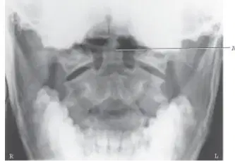
This radiographic image represents which of the following projections and/or positions?
AP Fuchs method
AP pillar projection
AP axial C spine projection
AP open-mouth projection
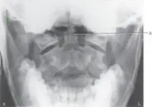
A critique of the image demonstrates which repeatable error?
Mislabeling of axes
Incorrect scale usage
Omission of units
Inconsistent data representation
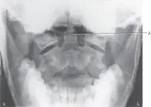
The error demonstrated on this radiograph is:
Excessive flexion of the skull
Excessive extension of the skull
Mouth not open far enough
None of the above are repeatable errors.
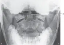
The structure labeled A, which should be well demonstrated on this projection, is the:
pedicle.
lamina.
Odontoid process
inferior articular process.
A digital (CR) image taken of a lateral thoracic spine projection demonstrates poor visibility of the spine. The image was taken on a full 14 × 17-inch (35 × 43 cm) IR. The following factors were used during the exposure: 80 kV, 80 mAs, 40-inch (102 cm) SID, and collimation to the size of the IR. Which one of the following modifications will result in a more diagnostic image?
Decrease kV.
Decrease mAs.
Collimate to spine.
Increase SID.
Which of the following projections would provide assessment of cervical spine for anteroposterior mobility?
Cervicothoracic projection
AP axial-vertebral arch projection
Judd or Fuchs projection
Hyperflexion and hyperextension projections
Which of the following projections will best demonstrate a compression fracture of the thoracic spine?
AP projection
Lateral projection
Posterior oblique positions
Anterior oblique positions
Which imaging modality will best demonstrate herniated nucleus pulposus in the cervical spine?
Computed tomography (CT)
Hyperflexion and hyperextension lateral projections
Magnetic resonance imaging (MRI)
Nuclear medicine
A patient enters the ED due to an MVA. He is on a backboard and in a cervical collar. The initial lateral cervical spine projection demonstrates C1 to C6. The patient has broad and thick shoulders. Because the hospital is in a rural setting, no CT scanner is available. Which of the following modifications would best demonstrate the lower cervical spine?
Repeat the exposure and increase kV.
Repeat the exposure but ask the patient to stand up and do the lateral projection erect.
Repeat the exposure but have a student pull down on the arms during the exposure.
Perform the horizontal beam cervicothoracic (swimmer’s) lateral position.
A patient comes to radiology for a follow-up study for a clay shoveler’s fracture. Which of the following projections will best demonstrate the extent of this fracture?
AP cervical spine
Lateral thoracic spine
Lateral cervical spine
AP axial-vertebral arch projection
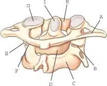
The structure anterior arch of the atlas is labeled ______.
H
A
C
F
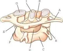
Stress from a severe whiplash type of injury may cause a fracture of the structure labeled ______.
I
A
C
F
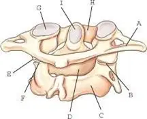
The structure transverse process is labeled ______.
C
A
F
H
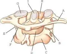
The body of C2 is labeled _____
D
A
B
C
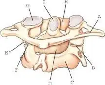
The superior articular facet is labeled _____
G
A
C
E
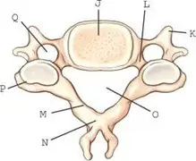
The structure transverse foramen is labeled _______.
Q
P
R
S
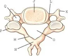
The part vertebral foramen is labeled _______.
O
A
B
C
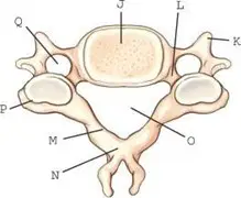
The structure pedicle is labeled _______.
L
A
C
F
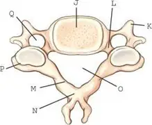
The structure lamina is labeled _____
M
A
C
F

