
 Worksheets
Radiographic Periodontal Diagnosis
Total questions: 51
Worksheet time: 26mins
Periodontal disease is defined as:
an inflammatory condition affecting the tissues surrounding the teeth
a viral infection of the oral cavity
a genetic disorder causing tooth loss
a condition affecting only the tongue
The clinical manifestations of periodontal disease include:
Bleeding gums, tooth mobility, gum recession, and bad breath
Tooth whitening, increased saliva, and strong enamel
Frequent headaches, blurred vision, and ear pain
Nasal congestion, sore throat, and cough
Radiographic manifestations of periodontal disease include which of the following?
Loss of alveolar bone height
Increased radiopacity of the periodontal ligament space
Thickening of the lamina dura
Presence of impacted teeth
A radiographic examination alone is enough to diagnose periodontal disease.
True
False
Sometimes
Only in severe cases
What is an EARLY radiographic indicator of periodontal bone loss in POSTERIOR teeth.
Curving of the alveolar crest and widened crestal PDL space
Furcation bone loss
Severe loss of bone height
Loosening teeth
Which of the following is a local irritating factor that may be visualized in radiographs and increases the risk of periodontal disease?
Calculus
Plaque
Gingivitis
Gingival recession
Poorly contoured or overextended ______ can be detected by radiographs, which could be a risk factor for periodontal disease. Fill in the blank.
restorations
implants
braces
crowns
Radiographs help in assessing root length and morphology and ______-to-root ratio. Fill in the blank.
crown
apex
pulp
enamel
Open interproximal contacts, which may be sites for food impaction, can be identified using ______. Fill in the blank.
radiographs
explorers
mouth mirrors
periodontal probes
Radiographs provide information about the position of the maxillary sinus in relation to a ______ deformity. Fill in the blank.
periodontal
skeletal
muscular
dental
Radiographs can help identify missing, supernumerary, impacted, and ______ teeth. Fill in the blank.
tipped
erupted
decayed
loose
What type of imaging provides the highest spatial resolution?
Intraoral imaging
Panoramic imaging
Computed tomography (CT)
Magnetic resonance imaging (MRI)
What is the primary modality in detecting radiographic signs of periodontitis?
BW (Bitewing)
OPG (Orthopantomogram)
Lateral Cephalogram
CBCT (Cone Beam CT)
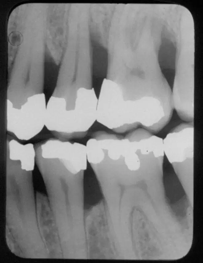
In intraoral imaging, what is the primary reason for vertical Bitewings rather that horizontal Bitewings?
To help diagnose interproximal caries
To help diagnose root caries
Both A and B
To help characterize severe periodontal bone loss
What technique is ideal to avoid distortion in PA (Periapical) intraoral imaging?
Parallel technique
Bisecting angle technique
SLOB technique
Occlusal technique
Fill in the blank: Intraoral radiography provides a 2-dimensional view of ______ structures. Bony defects overlapped by higher bony walls may be hidden.
3-D
soft tissue
vascular
muscular
Fill in the blank: Intraoral radiographs show _____ bone destruction compared to what is actually present.
more
less
equal
Fill in the blank: Intraoral radiographs provide very limited information about the depth of ______ tissue pockets.
soft
hard
dense
calcified
Fill in the blank: Bone level measurement from the CEJ is not valid where there is ______ or passive eruption.
supraeruption
attrition
abrasion
caries
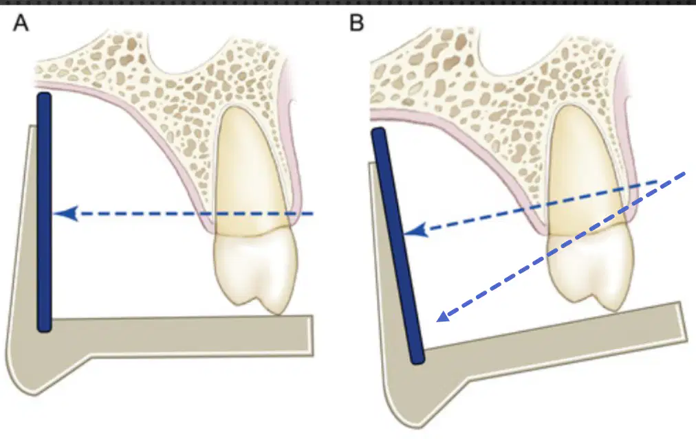
In a maxillary periapical, the beam is oriented from above, where does this project the buccal plate of bone?
Apically
Coronally
Mesially
Distally
Which imaging modality can give a rough idea but is limited by distortion and poor image detail?
Panoramic
CBCT
MRI
Ultrasound
CBCT is not recommended to be used routinely.
True
False
What can limit the usefulness of CBCT imaging?
Metal artifacts
High radiation dose
Short scan time
Low patient comfort
Some complex defects are better viewed _________
3-dimensionally
in black and white
with the naked eye
under low light
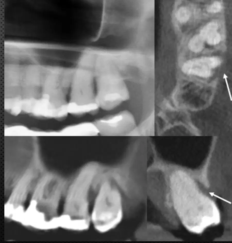
What does CBCT stand for in dental imaging?
Cone Beam Computed Tomography
Cranial Bone Computed Tomography
Cylindrical Beam Computed Tomography
Centralized Beam Computed Tomography
Healthy periodontium: The alveolar crest cortex is continuous with which structure?
lamina dura
periodontal ligament
gingival sulcus
cementum
What is the height range of the crest from the CEJs in a healthy patient?
0.5 to 2.0 mm
2.5 to 4.0 mm
3.0 to 5.0 mm
1.5 to 3.0 mm
In posterior teeth, the crest is parallel to which adjacent structures?
CEJs
Root apices
Pulp chambers
Occlusal surfaces
The periodontal ligament (PDL) is often wider at which portion?
cervical portion
apical portion
middle third
furcation area
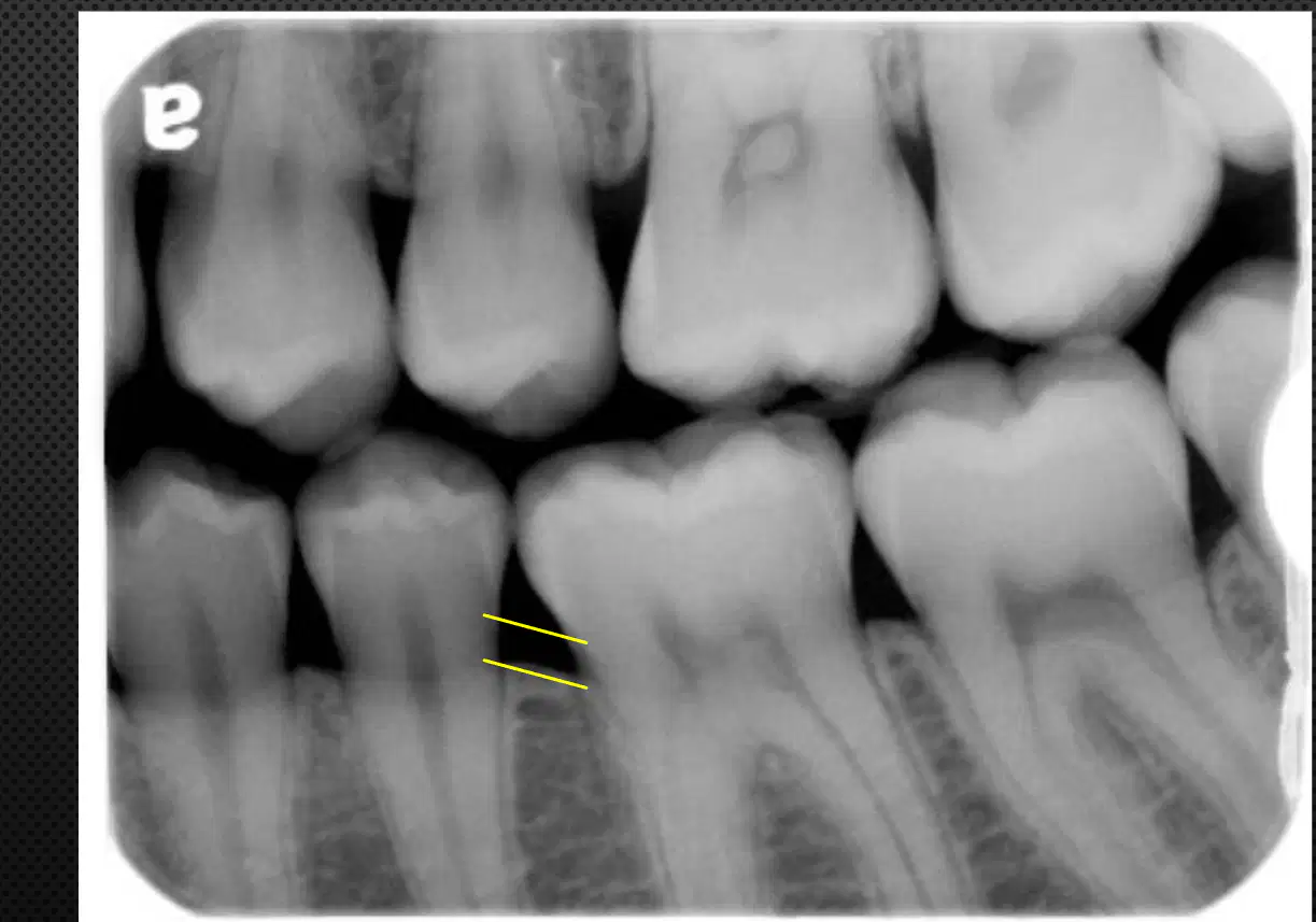
Describe the radiographic appearance of the periodontal tissues between teeth #19 and 20.
Normal crestal bone and periodontal ligament appearance
Rounded corners of crestal bone/ periodontal ligament indicating early periodontal bone loss
Severe, horizontal periodontal bone loss
Localized, vertical periodontal bone loss adjacent to tooth #19
In health, between anterior teeth, the crest is:
Flat and poorly defined
Pointed and well-defined
Rounded and irregular
Absent
A well-corticated crest indicates the absence of ________ periodontal disease.
ongoing
chronic
acute
localized
Lack of cortication always indicates disease.
True
False
Acute lesions in periodontal disease tend to be:
Sclerotic (radiopaque)
Radiolucent
Calcified
Transparent
Chronic lesions in periodontal disease tend to be:
Radiolucent
Sclerotic (radiopaque)
Transparent
Brittle
Which of the following is an early change in morphology associated with periodontal disease?
Localized erosion of the alveolar bone crest
Increased bone density
Formation of new bone
Thickening of the lamina dura
Fill in the blank: Early changes in morphology in posterior teeth include loss of sharp angle between the ______ and lamina dura.
alveolar crest
root
enamel
pulp
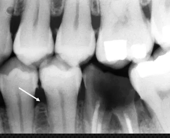
Significant attachment loss must occur 6-8 months before any radiologic bone loss is evident in periodontal disease.
True
False
What are the changes observed in the anterior region with periodontal disease?
A) Blunting of alveolar crest and loss of bone height
B) Increase in bone height
C) No change in alveolar crest
D) Formation of new bone
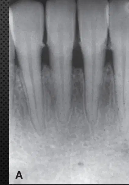
Describe the periodontal status of the radiograph.
Loss of periodontal bone height
Rounding, or blunting of the alveolar crest
Calculus can be visualized
All of the above
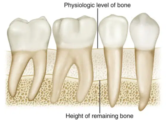
Refer to the diagram included. What does the 'physiologic level of bone' represent in the context of dental anatomy?
The normal height of bone supporting the teeth
The amount of bone lost due to disease
The height of the tooth crown
The thickness of the gum tissue
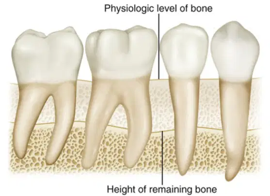
In the diagram included, the 'height of remaining bone' refers to the amount of bone that is still present after ________ has occurred.
severe, horizontal periodontal bone loss
mild, horizontal periodontal bone loss
bone growth
gum healing
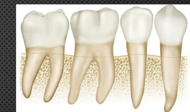
Horizontal bone loss occurs in a plane (a) to the CEJs of the teeth.
Not parallel
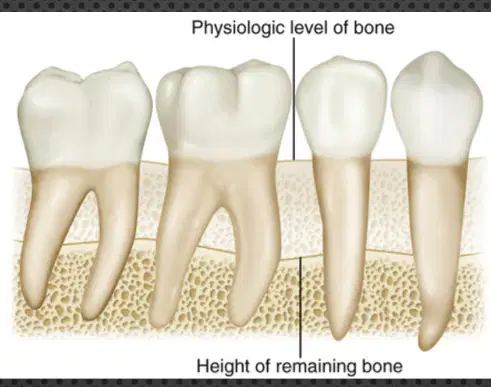
Vertical bone loss occurs in a plane (a) to the CEJs of the teeth.
Which of the following is a pattern of bone loss?
Horizontal
Vertical
Both A and B
None of the above
Fill in the blank: Localized bone loss is limited to less than ___% of all sites.
30
50
70
90
Fill in the blank: Generalized bone loss involves more than ___% of sites.
30
10
50
70
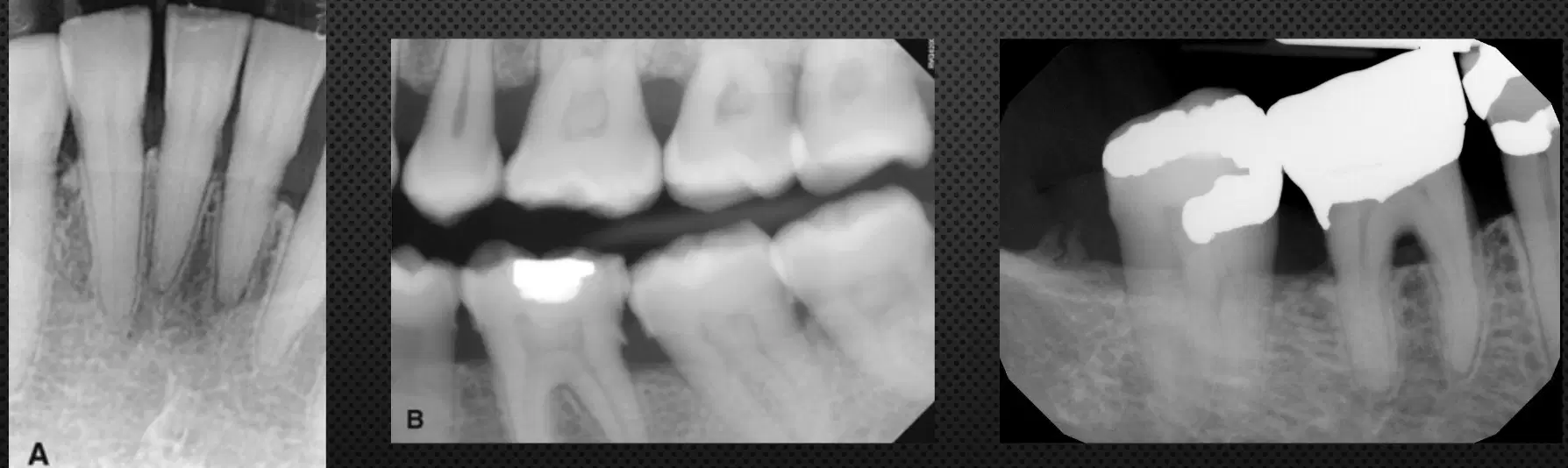
Horizontal bone loss is seen in which regions as shown in the images?
Anterior region
Posterior region
Both anterior and posterior regions
Neither region

In the included images, more severe horizontal bone loss is seen in the image on the right with _______ involvement.
furcation
crown
apical
gingival

In the provided radiograph, what does the abnormal widening of the PDL space (arrow) indicate?
Horizontal bone defect
Vertical bone defect
Normal bone structure
Tooth decay
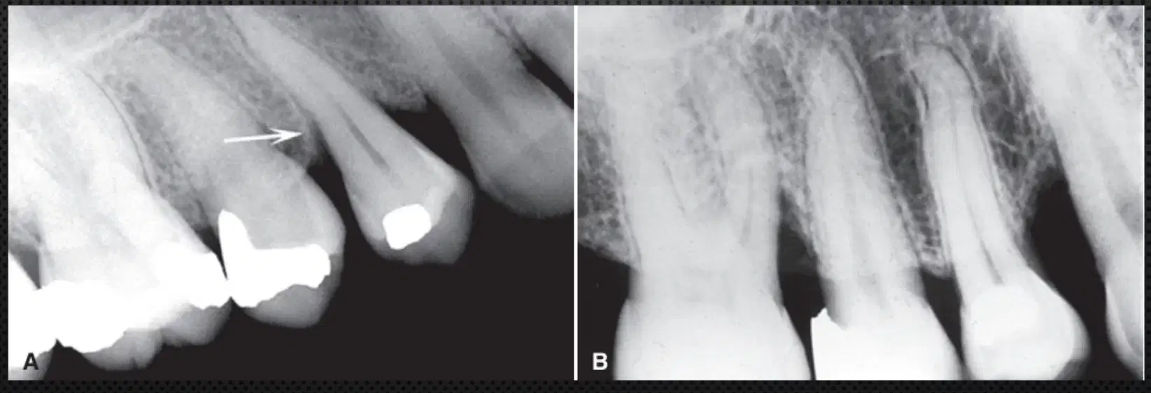
In these maxillary periapical images, which surfaces are affected by more severe vertical defects?
Mesial surface of the first molar and distal surface of the canine in image B
Distal surface of the first molar and mesial surface of the canine in image A
Occlusal surface of the first molar and canine in image A
Lingual surface of the first molar and canine in image B

