
 Worksheets
Oral Radiology Quality, Infection, Prescribing, and Legal
Total questions: 50
Worksheet time: 25mins
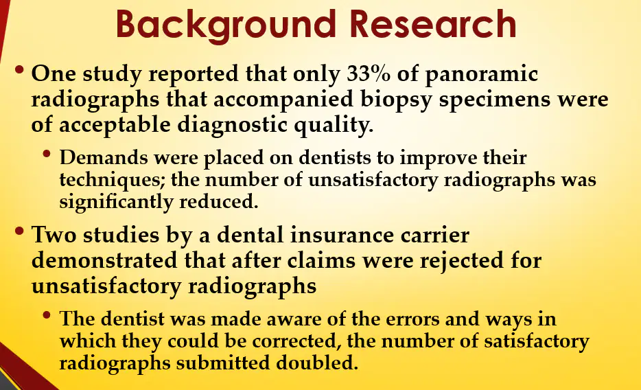
After claims were rejected for unsatisfactory radiographs, what happened to the number of satisfactory radiographs submitted by dentists?
It decreased.
It stayed the same.
It doubled.
It was not affected.
According to the ADA protocol for dental radiographic examinations, what should be developed and implemented in each dental health care setting?
Quality assurance protocols
Patient referral forms
Financial audit procedures
Marketing strategies
According to the ADA protocol for dental radiographic examinations, who should survey all X-ray units upon their placement and resurvey the equipment every four years?
A qualified expert
The dental assistant
The dentist
The receptionist
According to the ADA statement on Recommendations for Dental Radiographic Examinations, written policies and procedures for the safe use of ionizing radiation in dental practice should include which of the following?
Criteria for patient selection
Frequency of exposing radiographs on patients
Retaking radiographs consistent with current, accepted dental practice
All of the above

According to Table 16.1 White and Pharaoh's Schedule of Radiographic Quality Assurance Protocols, how often should an office "Enter findings in retake log"?
Daily
Weekly
Monthly
Yearly

According to Table 16.1 White and Pharaoh's Schedule of Radiographic Quality Assurance Protocols, how often should an office "inspect and calibrate the x-ray unit"?
Daily
Weekly
Monthly
Yearly
Which of the following is a quality control test for dental X-ray machines?
Half-value layer (HVL)
Blood glucose
Pulse oximetry
Body temperature
Which of the following is a quality control test for dental X-ray machines?
Focal spot size
Blood cholesterol
Blood urea
Spirometry
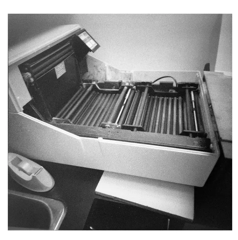
What is the recommended frequency for replenishing processing solutions in film-based radiography?
Daily
Weekly
Monthly
After every use
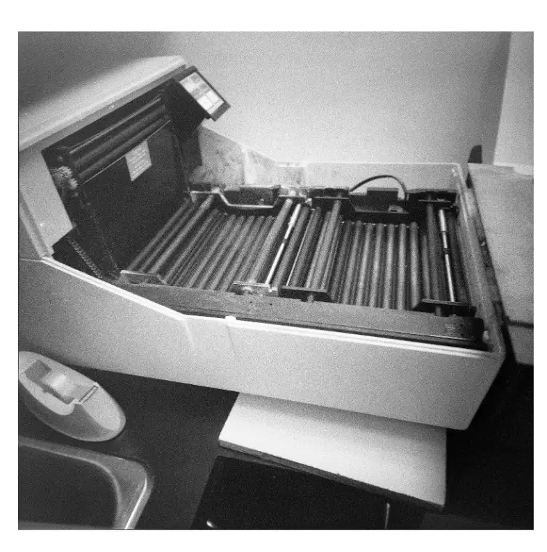
What is the recommended frequency for replacing processing solutions in film-based radiography?
Weekly
Monthly
Daily
Every six months
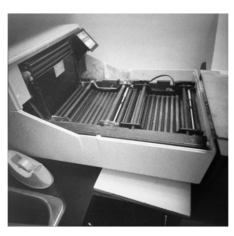
What is the recommended frequency for cleaning intensifying screens in film-based radiography?
Monthly
Weekly
Annually
Daily
What is the purpose of the Safelighting Test (Coin Test)?
To ensure that copper attenuates x-rays in your system
To ensure that safelighting exposes film over time
To ensure that safelighting does not expose film too much
To ensure the developing chemicals are still working correctly
What is the purpose of the Stepwedge Test?
To ensure that the x-ray tube is working
To measure the resolution of the imaging system
To ensure the processing chemicals are functioning properly
To test the composition of the stepwedge material
At the end of the day, what should be done in the Stepwedge Test to compare the results?
Expose another film beneath the stepwedge
Discard the chemicals
Use a different stepwedge
Skip the comparison
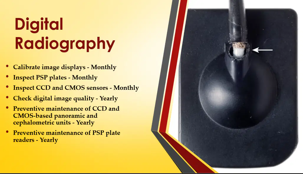
How often should PSP plates, CCD, and CMOS sensors be inspected in digital radiography?
Monthly
Annually
Weekly
Daily
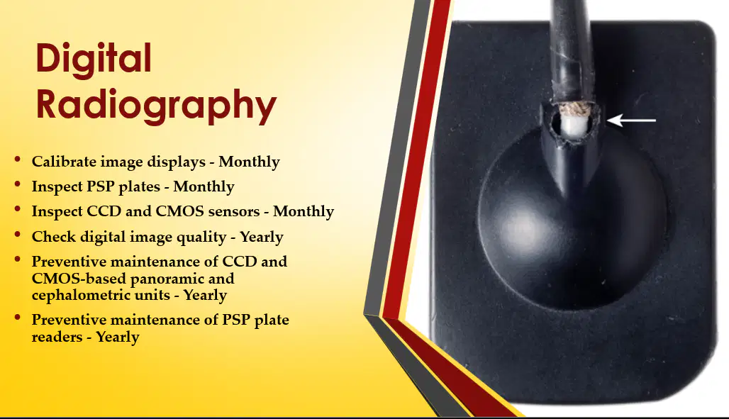
How often should digital image quality be checked in digital radiography?
Yearly
Monthly
Every 5 years
Weekly
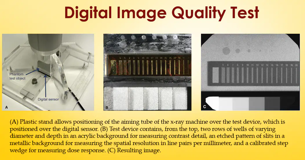
The resulting image from the Digital Image Quality Test shows a pattern used to measure _________?
spatial resolution in line pairs per millimeter
image contrast in grayscale levels
exposure time in milliseconds
color depth in bits
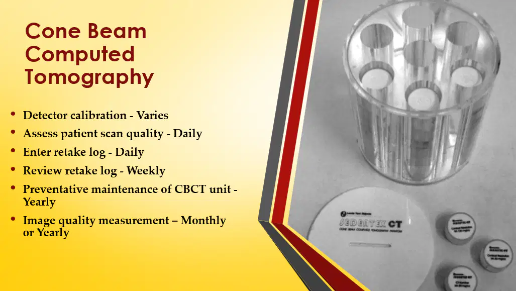
How often should patient scan quality be assessed in Cone Beam Computed Tomography?
Daily
Weekly
Monthly
Annually
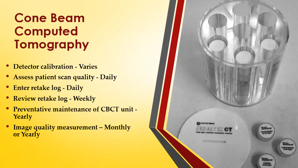
What is the recommended frequency for preventive maintenance of the CBCT unit?
Yearly
Monthly
Every 5 years
Weekly
What is subject contrast in X-ray imaging?
The black, white, and grey differences in the image caused by different degrees of attenuation as the X-ray beam is transmitted through different parts of the patient's tissues.
The amount of radiation absorbed by the X-ray machine during imaging.
The speed at which X-rays travel through the air before reaching the patient.
The color of the X-ray film after exposure to light.
Which of the following is a factor that subject contrast depends on?
Differences in tissue thickness and density
Patient's age
Room temperature
X-ray machine color
Fill in the blank: The X-ray beam used should be sufficiently ________ to pass through the patient to a varying degree, and interact differentially with the digital image receptor (solid-state or phosphor plate) to produce a well-contrasted black, white, and grey image.
penetrating
reflective
diffuse
opaque
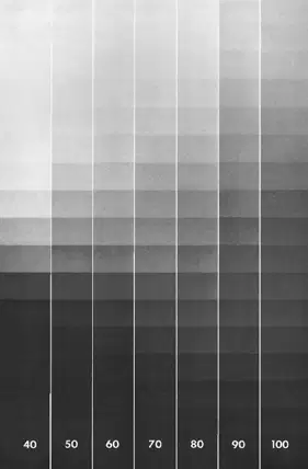
What does high contrast in an image mean?
Many shades of gray
A lot of black and white, with few shades of gray
Long grayscale
Low kVp
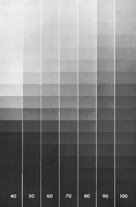
Contrast: High contrast has a lot of black and white, not many shades of gray, this is called a ______ contrast scale.
SHORT
LONG
MEDIUM
GRADUAL
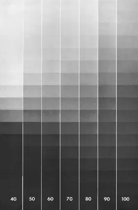
Contrast: Low contrast has many shades of gray, referred to as a ______ gray scale.
LONG
SHORT
MEDIUM
NARROW
Increasing kVp reduces contrast.
True
False
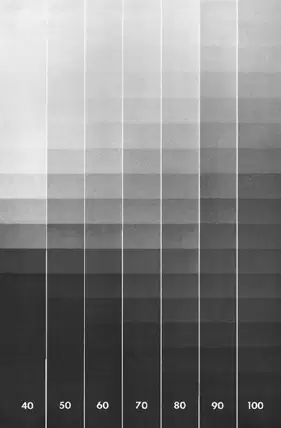
According to the diagram, at which kVp is the contrast highest?
40
60
80
100
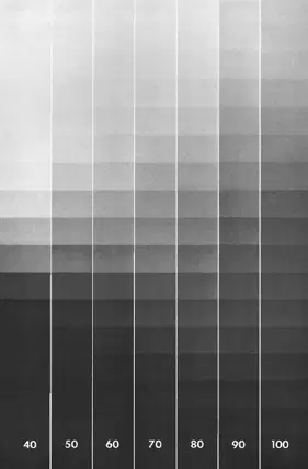
For dental radiography, kVp should be at ______.
60-80 (65)
30-40 (35)
90-110 (100)
120-140 (130)
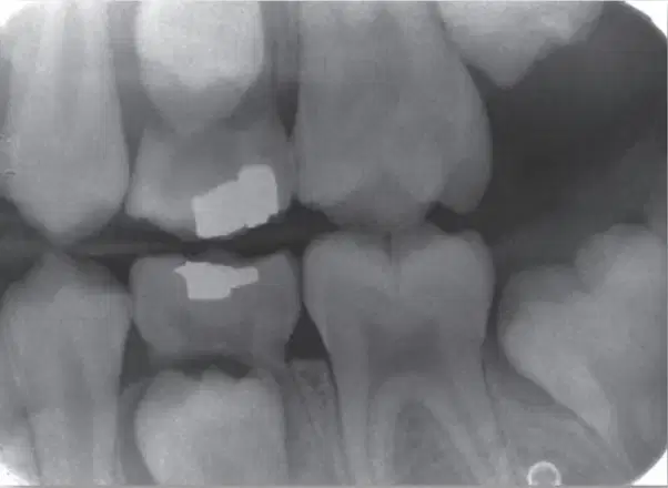
What is wrong with this radiograph?
Too light
Too dark
Too much contrast
Insufficient contrast
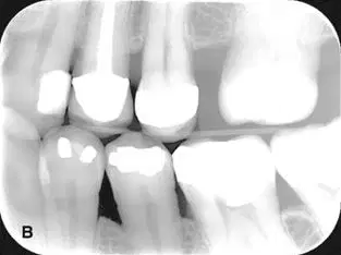
What is nearly wrong with this radiograph?
Too light
Too dark
Insufficient contrast
Nothing
Low contrast image can be caused by a litany of reasons. What is the most common cause of low contrast in digital images?
Overexposure
Underexposure
Sensor fog
Underdevelopment
What is the most common cause of image noise in digital images?
Overexposure
Underexposure
Patient movement
Music on too loud
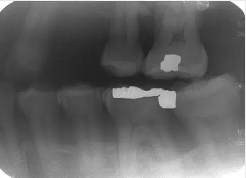
What is wrong with this radiographic?
Too light only
Too dark only
Insufficient contrast only
Both too dark and insufficient contrast
This is film fog. What causes film fog?
Improper safelighting
Light exposure
Overdevelopment
Deteriorated or expired film
Any of the above
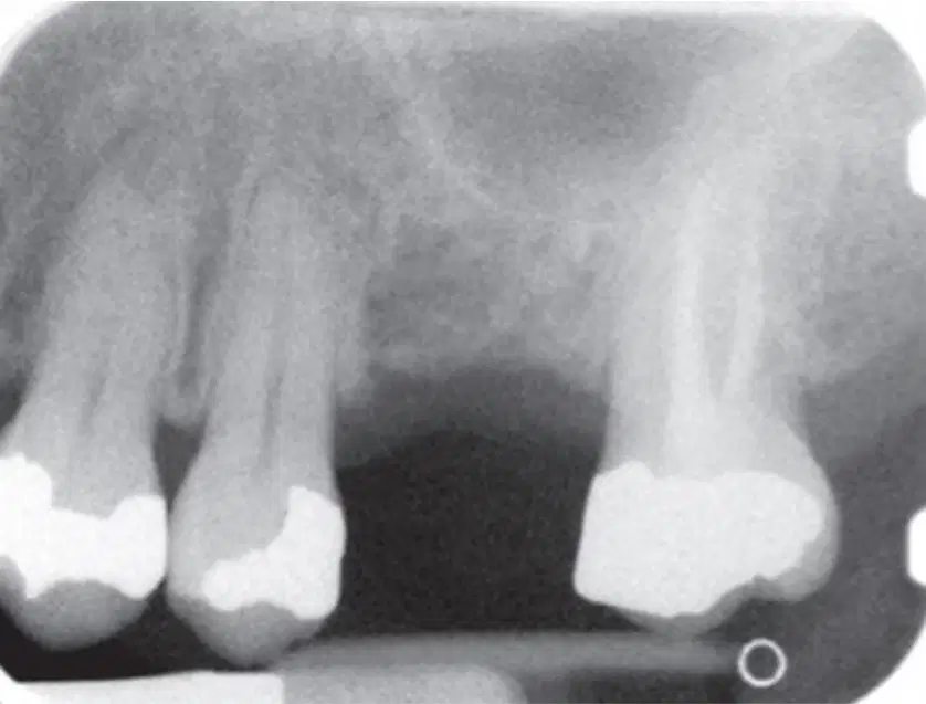
What is primarily wrong with this image?
Cone cut
Noise
Low contrast
Nothing
This is a radiographic image from a PSP plate. What is a possible cause of the light image?
Overexposure
Fed through the scanner backwards
Exposure to light after x-ray exposure
Exposure to light before x-ray exposure
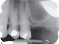
What type of sensors could generate the error you see in the top right corner of this radiograph?
Film and PSP
Film and Direct Digital (CCD or CMOS)
CCD or CMOS only
Any of the above
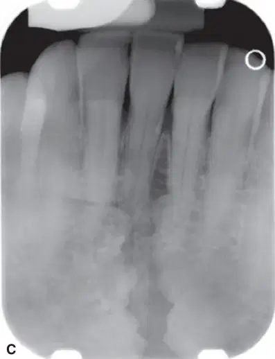
What type of sensors could generate the error you see in this radiograph?
Film and PSP
Film and Direct Digital (CCD or CMOS)
CCD or CMOS only
Any of the above
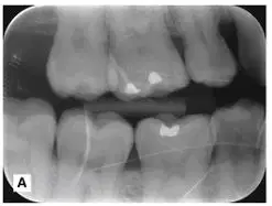
What is the likely cause of the problem seen in this radiograph?
Developer chemical splash
Light exposure
Scratched PSP plate
Software malfunction
What is appropriate infection control after using the Nomad on a patient?
Sterilize the Nomad
Disinfect the Nomad
Wipe the Nomad with paper towel only
Takethe Nomad home and wash it in your dishwasher, just make sure there are no meatloaf chunks on other dishes in the dishwasher the same time
You see a student exposing radiographs and touching a door handle between exposures. The door handle has no barrier. Is this appropriate Infection Control?
Yes
No
According to Dr. Mecham, everything in an operatory that might be touched during a procedure should
Be disinfected between patients
Have a clean protective barrier (tape, plastic bag) place over it prior to the procedure
Both A and B
When is it OK to use a digital sensor WITHOUT a plastic barrier?
When it's new
If it has been wiped down REAL GOOD
After it has been sterilized
Never ever
Prescribe radiographs: 5-year-old boy. Posterior teeth in contact. No evident caries.
No radiographs needed
Bitewings only
Bitewings and panel
FMX
Prescribe radiographs: 25-year-old woman. 6 month checkup for fractured incisor (crown). No caries on BW 6 months ago. Low caries risk. Fractured incisor tests vital.
No radiographs needed
Periapical incisor and bitewings
FMX
FMX, Pano, and CBCT
Prescribe radiographs: 45-year-old man 1 year return appointment. One year ago multiple amalgam restorations were placed, RCT #30. 5 mm buccal furcation pocket #3.
No radiographs needed
Bitewings only
Bitewings and periapicals of #3 and 30
FMX, Pano, and CBCT
Prescribe radiographs: Pregnant woman, 2nd trimester. Severe pain and swelling in right maxilla. Clinical observation shows large carious lesion on #2.
No radiographs due to pregnancy
Right molar BW only
Right molar PA only
Right molar BW and PA
A Dentist refers a patient to an imaging center to receive a CBCT for implant planning. A skull base tumor was visible on the CBCT but not diagnosed. Is the referring dentist liable for this mistake?
Yes
No
Unclear
For the dentist who didn't notice the skull base tumor on a CBCT. This is an example of:
Disclosure
Malpractice
Negligence
Informed consent

Dental images are the property of:
The dentist
The patient
The insurance company
The Oral Radiologist who taught you in dental school, IOW, all your image are belong to me

