
 Worksheets
Where do blood cells come from?
Total questions: 96
Worksheet time: 48mins
What is hematopoiesis?
The production and development of blood cells.
The breakdown of glucose in the liver.
The process of bone formation.
The filtration of waste in the kidneys.
Hematopoiesis begins with self-renewing ________ and ends with peripheral blood cells. Fill in the blank.
hematopoietic stem cells (HSC)
megakaryocytes
erythrocytes
lymphocytes
Where does hematopoiesis primarily occur in adults?
In the bone marrow
In the liver
In the spleen
In the thymus
Which of the following is NOT a lineage of blood cells?
Erythrocyte
Myeloid
Lymphocyte
Neuron
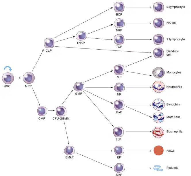
Which cell type is at the origin of all blood cells?
Hematopoietic stem cell (HSC)
Erythrocyte
Lymphocyte
Megakaryocyte
Hematopoiesis begins as early as the ________ after fertilization.
19th day
10th day
25th day
30th day
Production of erythrocytes occurs ________ (Fill in the blank)
in the bone marrow
in the liver
in the spleen
in the kidneys
Which of the following is NOT produced during embryonic hematopoiesis?
Erythrocytes
Macrophages
Megakaryocytes
Neurons
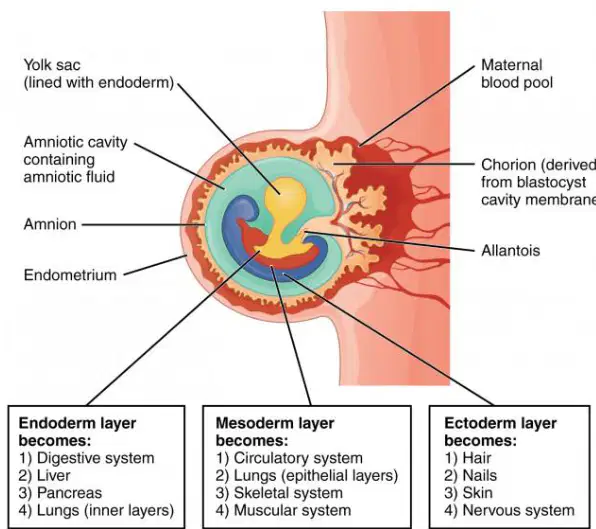
The endoderm layer becomes which of the following?
Circulatory system
Digestive system
Hair
Muscular system

The mesoderm layer becomes which of the following?
Skin
Liver
Circulatory system
Pancreas
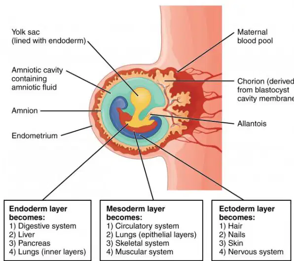
The ectoderm layer becomes which of the following?
Pancreas
Hair
Liver
Lungs
Primitive erythropoiesis _____ erythrocyte production in the yolk sac.
is
are
was
will be
Develop from clusters of cells in the mesenchyme called _____ (Fill in the blank)
blood islands
germ layers
somites
neural crests
Primitive erythrocytes _____
retain their nucleus
lack any cytoplasm
are multinucleated
contain hemoglobin only after maturation
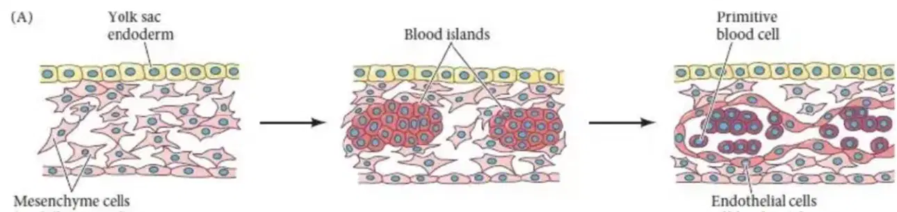
What is the name of the clusters of cells in the mesenchyme that develop into blood cells?
blood islands
lymph nodes
osteons
islets of Langerhans
At how many weeks of gestation does definitive erythropoiesis begin?
Four weeks of gestation
Six weeks of gestation
Eight weeks of gestation
Twelve weeks of gestation
Definitive erythropoiesis begins in which region?
Yolk sac
Aorta-gonads-mesonephros (AGM) region
Placenta
Liver
Erythrocytes arise from ________.
HSCs
Osteocytes
Chondrocytes
Neurons

According to the diagram, which region in the embryo is responsible for definitive erythropoiesis?
AGM region
Yolk sac
Liver
Placenta
AGM makes a wider range of hematopoietic cells than the yolk sac.
True
False
Around the third month of fetal development, the liver is the primary site of blood cell production. Spleen produces ____________.
lymphocyte
erythrocyte
platelet
plasma cell
Which organ is the primary site of blood cell production around the third month of fetal development?
Spleen
Liver
Bone marrow
Kidney
Which type of hemoglobin replaces embryonic hemoglobin during fetal development?
Hemoglobin A
Hemoglobin S
Hemoglobin F (Fetal)
Hemoglobin C
Myeloid lineage gives rise to which of the following?
B cells and T cells
Erythrocytes (RBCs), megakaryocytes/platelets, granulocytes, and monocytes
Only erythrocytes
Only lymphocytes
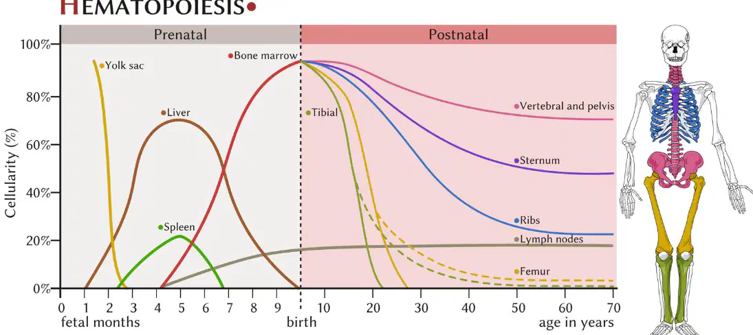
Around which month of gestation does the bone marrow become the primary site of hematopoiesis?
Third month
Fifth month
Seventh month
Ninth month
Bone marrow continues as the primary source of blood production after birth and during adult life.
True
False
Fill in the blank: Around the seventh month of gestation, ______ becomes the primary site of hematopoiesis.
bone marrow
liver
spleen
yolk sac
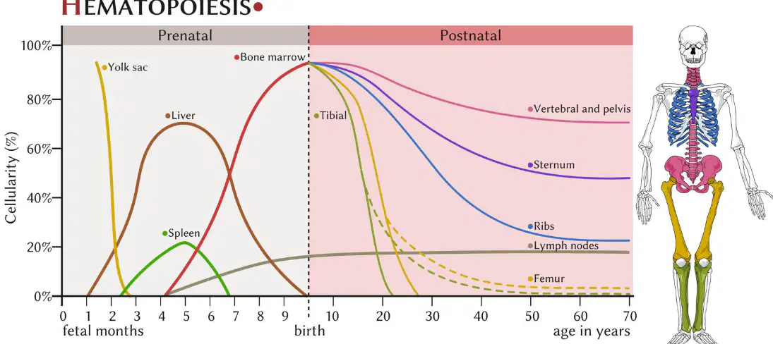
According to the graph, which organ is the main site of hematopoiesis during the early fetal months?
Liver
Yolk sac
Spleen
Bone marrow
Which of the following is NOT an organ or tissue involved in adult hematopoietic tissue?
Bone marrow (BM)
Spleen
Thymus
Kidney
Fill in the blank: The tissue and organs regulating the proliferation, maturation, and destruction of blood cells include ________, spleen, thymus, and lymph nodes + mucosa-associated lymphoid tissue (MALT).
Bone marrow (BM)
Liver
Pancreas
Kidney
Which two organs does the class focus on, according to the notes on the slide?
Bone marrow and Spleen
Thymus and Spleen
Bone marrow and Thymus
Spleen and Lymph nodes
Bone marrow (BM) is blood-forming tissue found between the ________ of spongy bone. Fill in the blank.
trabeculae
osteons
lamellae
canaliculi
Name the two major types of cells found in bone marrow. Fill in the blanks: 1) Hematopoietic cells 2) ________
Stromal cells
Epithelial cells
Neuronal cells
Muscle cells
What is the function of stromal cells in bone marrow?
Provide structure support and cytokines to regulate hematopoiesis.
Initiate muscle contraction in bone tissue.
Produce antibodies to fight infections directly.
Transport oxygen throughout the body.
In the diagram, which part of the bone is labeled as 'compact bone'?
The outer dense layer of the bone is labeled as 'compact bone'.
The spongy inner region of the bone is labeled as 'compact bone'.
The central cavity containing marrow is labeled as 'compact bone'.
The cartilage covering the bone ends is labeled as 'compact bone'.
The main function of the vascular compartment in bone marrow is:
to provide a pathway for the exchange of cells and nutrients between the blood and bone marrow
to store fat for energy reserves
to produce bone matrix and support bone structure
to regulate the immune response within the bone marrow
Fill in the blank: The site of __________ and entry into the bloodstream is the vascular compartment of bone marrow.
blood cell development
bone formation
fat storage
hormone secretion
Which of the following are included in the vascular compartment of bone marrow?
Arterioles
Capillaries
Sinusoids
All of the above
Hematopoietic cells in bone marrow include all blood cells in different stages of development.
True
False
The function of adipose tissue (fat cells) in the stromal cells of bone marrow is:
to store energy and provide support to the bone marrow microenvironment
to produce red blood cells
to form the hard structure of bone
to transport oxygen in the blood
Erythroblasts compose _______ of marrow cells and are produced near venous sinuses.
25-30%
5-10%
50-55%
70-75%
Erythroblasts develop in _______.
erythroblastic islands
lymph nodes
alveoli
glomeruli
Erythroblastic islands contain a central macrophage surrounded by _______ in different states of maturation.
erythroblasts
lymphocytes
megakaryocytes
neutrophils
Erythroblasts are the _______ version of RBC and detected in bone marrow.
immature
mature
inactive
fragmented
Macrophages have two major functions in bone marrow: 1. Phagocytosis: Ingest ________ of maturing erythroblasts.
nuclei
mitochondria
ribosomes
cytoplasm
Macrophages have two major functions in bone marrow: 2. Secretion of hematopoietic ________.
cytokines
enzymes
hormones
antibodies
What are cytokines?
Enzymes that digest food
Signal proteins of immune systems
Hormones that regulate blood sugar
Structural proteins in muscles
Granulocytes are produced in nests that are mainly seen close to the ________.
arterioles
veins
lymph nodes
capillaries
Why do neutrophils come across more in BM smears?
Because they are more abundant.
Because they are larger in size.
Because they stain darker.
Because they are less fragile.
Which of the following cell types is NOT labeled in the provided BM smear diagram?
Neutrophilic metamyelocyte
Promyelocyte
Erythrocyte
Myeloblast
BM—Smear: Nuclei are clumped ____________.
together
apart
randomly
individually
What type of cells are large, polyploid (DNA content > 2N) cells?
Megakaryocytes
Erythrocytes
Neutrophils
Lymphocytes
What do megakaryocytes produce after cytoplasmic fragmentation?
Platelets
Red blood cells
White blood cells
Plasma cells
Where are megakaryocytes located?
Near vascular sinus
Inside lymph nodes
Within alveoli
In the renal cortex
Identify the cell labeled in the diagram and describe its function.
The cell labeled in the diagram is a megakaryocyte. Its function is to produce platelets after cytoplasmic fragmentation.
The cell labeled in the diagram is an erythrocyte. Its function is to transport oxygen in the blood.
The cell labeled in the diagram is a lymphocyte. Its function is to produce antibodies for immune response.
The cell labeled in the diagram is a neutrophil. Its function is to engulf and destroy bacteria.
What are lymphocytes produced in?
Lymphoid aggregates near arterioles
Bone marrow sinusoids
Red blood cells
Platelets
Mature lymphocytes may aid in hematopoiesis by releasing _________.
cytokines
antibodies
hormones
enzymes
Plasma cells have a lot of ______ apparatus in them.
Golgi
Mitochondria
Lysosome
Endoplasmic reticulum
Plasma cells are B cells that produce _________.
antibodies
hormones
enzymes
neurotransmitters
What is attached to the nucleus in plasma cells?
Perinuclear halo
Mitochondria
Ribosome
Nucleolus
Identify the cell shown in the image labeled 'Plasma cell'.
Plasma cell
Neutrophil
Eosinophil
Monocyte
Adipocytes are cells whose cytoplasm is mostly replaced with a single _________.
fat vacuole
nucleus
mitochondrion
lysosome
Which of the following is a function of adipocytes in bone marrow?
Mechanically control the volume of BM
Produce red blood cells
Absorb nutrients
Secrete digestive enzymes
Adipocytes provide steroids and cytokines that influence hematopoiesis and maintain osseous bone integrity.
True
False
Identify the large, clear cells present in the bone marrow smear.
Adipocytes
Osteoclasts
Megakaryocytes
Plasma cells
BM—Composition: The BM region lining the ________________ of bone is called the endosteal compartment. (Fill in the blank)
inner surfaces
outer surfaces
marrow cavity
periosteum
BM—Composition: Hemopoietic cells in the endosteal compartment are mainly ________________.
dormant HSCs
mature erythrocytes
adipocytes
megakaryocytes
Which of the following are stromal cells found in the endosteal compartment?
Osteoclasts
Osteoblasts
Adipose cells
All of the above
What is the main function of the endosteal compartment in bone?
Site of bone remodeling
Production of red blood cells
Storage of fat
Attachment of muscles
What is the primary function of osteoblasts in a BM-Smear?
Destroy old bone
Form new bone
Transport oxygen
Produce antibodies
What is the maximum diameter of osteoblasts as mentioned in the BM-Smear slide?
10 mcM
20 mcM
30 mcM
50 mcM
Osteoblasts resemble plasma cells except that the perinuclear halo is ______ from the nuclear membrane.
detached
attached
continuous
fused
The cytoplasm of osteoblasts may be less ______ compared to plasma cells.
basophilic
acidophilic
eosinophilic
chromophilic
Osteoblasts are normally found ______ in a BM-Smear.
in groups
scattered singly
within sinusoids
lining blood vessels
Plasma cells are more purple-blueish compared to osteoclasts.
True
False
What is the primary function of osteoclasts?
Build bone
Breakdown bone
Transport oxygen
Produce hormones
Osteoclasts develop from which type of cells?
Osteoblasts
HSCs
Megakaryocytes
Erythrocytes
Which of the following best describes the morphology of osteoclasts?
Uninucleated cells
Multinucleated cells
Small round cells
Spindle-shaped cells
Osteoclasts are larger than osteoblasts, reaching up to ______ in diameter.
100 mcM
10 mcM
1 mm
500 nm
Osteoclasts resemble megakaryocytes except their nuclei are ______ and often contain nucleoli.
discrete
fused
lobulated
fragmented
Refer to the images of BM smears. Which cell type is shown in the images?
Osteoblasts
Osteoclasts
Erythrocytes
Lymphocytes
For a new blood cell to reach circulation, it needs to squeeze through which cells of the blood vessel?
Platelet cells
Endothelial cells
Red blood cells
Macrophages
BM—Blood Cell Egress: The process of a new blood cell reaching circulation is driven by ________.
cytokines
antibodies
platelets
hormones
The stroma in bone marrow is composed of reticular cells, collagenous fibers, and extracellular matrix.
True
False
BM—Blood Cell Egress: Which of the following is NOT labeled in the bone marrow diagram?
Stem cell
Platelets
Neuron
Macrophage
During the first four years of life, what are nearly all marrow cavities composed of?
Adipocytes (yellow marrow)
Hematopoietic cells (red marrow)
Compact bone
Cartilage
After four years of age, what happens to the red marrow in the shafts of long bones?
It is gradually replaced by adipocytes (yellow marrow).
It increases in volume and activity.
It turns into compact bone tissue.
It becomes the primary site of white blood cell destruction.
By age 25, hematopoiesis is limited to the marrow of which bones?
Skull, ribs, sternum, scapulae, clavicles, vertebrae, pelvis, upper half of sacrum, proximal end of long bones.
Femur, tibia, fibula, humerus, radius, ulna only.
Mandible, maxilla, nasal bones, zygomatic bones only.
Phalanges, metacarpals, metatarsals, tarsals only.
Red marrow continues to decrease with age.
True
False
In the diagram, which color represents red marrow and which color represents yellow marrow?
Red color represents red marrow and yellow color represents yellow marrow.
Yellow color represents red marrow and red color represents yellow marrow.
Both red and yellow colors represent red marrow.
Both red and yellow colors represent yellow marrow.
BM Hypoplasia: Hematopoietic tissue becomes ____________.
inactive
hyperactive
fibrotic
calcified
Which of the following is NOT a cause of BM Hypoplasia?
Autoimmune disorders
Chemicals
Toxins
Viral infections
Medications/drugs
In BM Hypoplasia, fat cells increase providing a cushion for the marrow.
True
False
You see excessive production of fat cells in BM Hypoplasia.
True
False
What is a key feature seen in BM Hypoplasia?
Excessive production of fat cells
Increased number of megakaryocytes
Hypercellular bone marrow
Increased erythropoiesis
What is BM Hyperplasia?
Non-cancerous increase in blood cells
Cancerous decrease in blood cells
Non-cancerous decrease in blood cells
Cancerous increase in blood cells

