NEW
Font size
 Worksheets
BIO2251-01N: Week 9.1 - Muscle Org
Total questions: 41
Worksheet time: 21mins
Which statement best defines the origin of a muscle?
The movable point of attachment where the action occurs
The immovable attachment of a muscle
The thickest part of a muscle belly
The connective tissue that links bone to bone
The nerve supply to a muscle
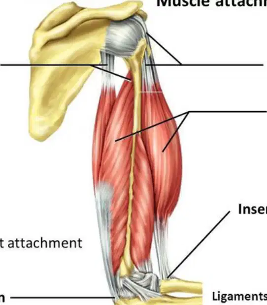
According to the diagram of the upper limb, where is the insertion located relative to the origins?
Superior and proximal to the origins
At the same location as the origins
Inferior and distal compared to the origins
Posterior to the scapula
Medial to the muscle bellies
Which pairing correctly matches each connective tissue with what it connects?
Ligaments: muscle to bone; Tendons: bone to bone
Ligaments: bone to bone; Tendons: muscle to bone
Ligaments: muscle to muscle; Tendons: bone to cartilage
Ligaments: muscle to cartilage; Tendons: bone to bone
Ligaments: bone to muscle; Tendons: bone to bone
A student wants to predict how a muscle’s action will influence movement at a joint. Based on the material, what is the most appropriate first step?
Measure the cross-sectional area of the muscle belly
Identify what structures the muscle is attached to
Determine the resting membrane potential of the muscle fibers
Estimate the fiber type composition (slow vs. fast)
Assess local blood supply to the muscle
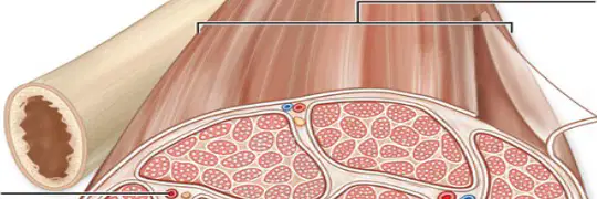
Which connective tissue layer encloses the entire skeletal muscle, as shown surrounding the outer surface in the diagram?
Epimysium
Perimysium
Endomysium
Deep fascia
Tendon
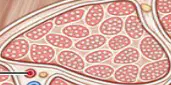
In the hierarchical organization of muscle, a fascicle is best described as which of the following?
A single contractile protein within a myofibril
A bundle of muscle fibers wrapped within the perimysium
The connective tissue sheath that surrounds the whole muscle
A tendon connecting muscle to bone
A capillary network within muscle tissue
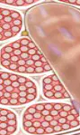
Which connective tissue directly surrounds individual muscle fibers within a fascicle?
Epimysium
Perimysium
Endomysium
Deep fascia
Sarcolemma
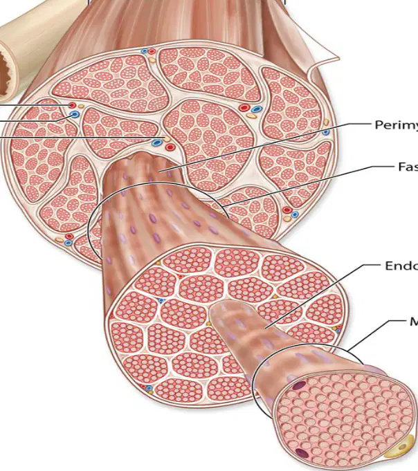
Identify the correct outer-to-inner sequence of coverings from the whole muscle to the individual fiber.
Endomysium → Perimysium → Epimysium
Perimysium → Epimysium → Endomysium
Epimysium → Perimysium → Endomysium
Deep fascia → Endomysium → Epimysium
Perimysium → Endomysium → Epimysium
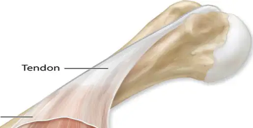
Which structure is shown transmitting force from muscle to bone and is continuous with the connective tissue layers?
Nerve
Artery
Tendon
Vein
Deep fascia
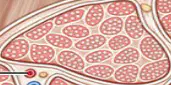
A researcher isolates a bundle of muscle fibers each wrapped by connective tissue. Which term best identifies both the bundle and its specific wrapping?
Fascicle and epimysium
Fascicle and perimysium
Muscle fiber and endomysium
Muscle fiber and perimysium
Fascicle and endomysium
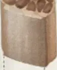
Which connective tissue layer surrounds an individual muscle fiber in skeletal muscle?
Epimysium
Perimysium
Endomysium
Deep fascia
Tendon
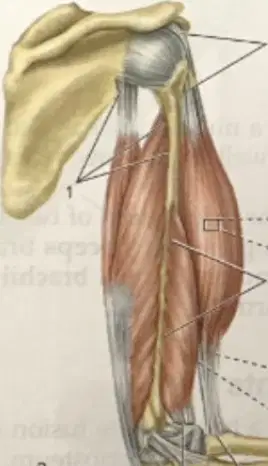
In the diagram of the posterior view of a laterally rotated arm, which structure represents the dense connective tissue that lies external to epimysium and separates muscles into functional groups?
Perimysium
Deep fascia
Endomysium
Epimysium
Tendon
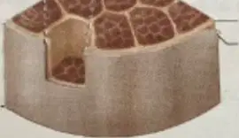
Which label would identify a bundle of muscle fibers within a muscle?
Fasciculus
Myofibril
Sarcomere
Aponeurosis
Endosteum
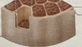
Which connective tissue layer surrounds a fasciculus?
Epimysium
Perimysium
Endomysium
Deep fascia
Reticular lamina
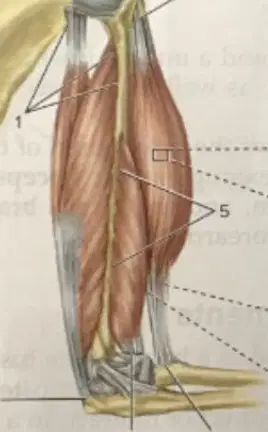
Which structure anchors muscle to bone at both proximal and distal attachments in the figure?
Ligament
Periosteum
Tendon
Epimysium
Endomysium
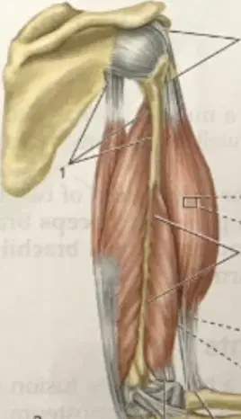
According to the figure, which muscle has a labeled distal attachment near the forearm?
Biceps brachii
Brachialis
Triceps brachii
Deltoid
Supraspinatus
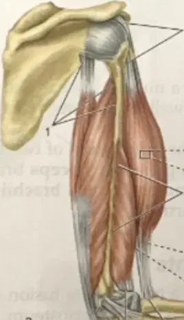
Which pair correctly matches the muscle with its labeled proximal attachment in the figure?
Biceps brachii — olecranon
Brachialis — coracoid process
Triceps brachii — humeral shaft (posterior)
Deltoid — ulna
Brachioradialis — scapular spine
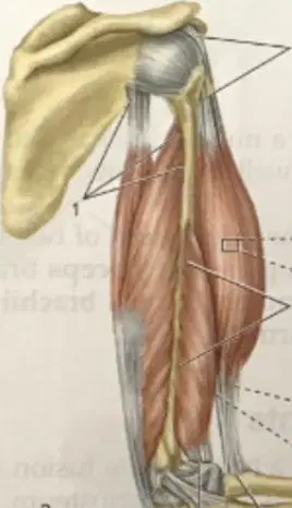
Which statement best distinguishes epimysium from deep fascia in the figure?
Epimysium surrounds individual fibers; deep fascia surrounds fascicles.
Epimysium surrounds the whole muscle; deep fascia lies external and groups muscles.
Both epimysium and deep fascia surround the whole muscle with no difference.
Epimysium is a tendon; deep fascia is a ligament.
Epimysium lies external to deep fascia.
In the microscopic inset (D), the small cylindrical units labeled as muscle fibers are enclosed by which layer?
Perimysium
Epimysium
Deep fascia
Endomysium
Periosteum
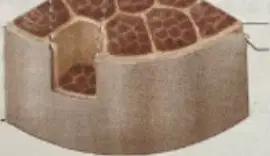
If a fasciculus is damaged but surrounding structures remain intact, which layers are most likely still continuous?
Endomysium and tendon
Perimysium and epimysium
Deep fascia and endomysium
Periosteum and endomysium
Sarcolemma and deep fascia
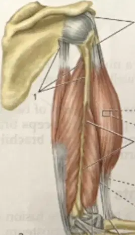
Which labeled arm muscle lies deep to biceps brachii and has both proximal and distal attachments indicated?
Brachialis
Triceps brachii
Deltoid
Brachioradialis
Coracobrachialis
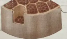
Considering the hierarchical organization depicted, which sequence correctly lists structures from outermost to innermost around a muscle fiber?
Deep fascia → Epimysium → Perimysium → Endomysium
Epimysium → Deep fascia → Perimysium → Endomysium
Deep fascia → Perimysium → Epimysium → Endomysium
Perimysium → Epimysium → Deep fascia → Endomysium
Endomysium → Perimysium → Epimysium → Deep fascia
Which statement best defines the neuromuscular junction?
A gap junction that electrically connects two muscle fibers
A chemical synapse between a motor neuron and a muscle fiber
A mechanical connection between bone and tendon
An electrical synapse between two interneurons
Which structure releases neurotransmitter via exocytosis at the neuromuscular junction?
Myofibril
Sarcoplasmic reticulum
Synaptic vesicle
T tubule
Which membrane of the muscle cell receives the neurotransmitter at the neuromuscular junction?
Sarcolemma
Myofibril
Axon of neuron
Sarcoplasmic reticulum
What is the functional role of the synaptic cleft in the neuromuscular junction?
It stores calcium for muscle contraction
It is the extracellular space where neurotransmitter diffuses from neuron to muscle
It generates action potentials in the axon hillock
It forms the T tubule network inside the muscle fiber
Which pathway correctly traces the signal from neuron to muscle contraction initiation?
Axon of motor neuron → Synaptic vesicle exocytosis → Sarcolemma depolarization → T tubule conduction
Myofibril contraction → T tubule depolarization → Axon potential → Synaptic vesicle endocytosis
Sarcoplasmic reticulum release → Axon depolarization → Synaptic cleft diffusion → Myelin formation
T tubule conduction → Axon depolarization → Synaptic vesicle exocytosis → Sarcolemma hyperpolarization
Which structure in the muscle fiber directly surrounds myofibrils and stores calcium?
Sarcoplasmic reticulum
Sarcolemma
Synaptic cleft
Terminal bouton
The terminal bouton is best described as which part of the motor neuron?
The initial segment of the axon where action potentials begin
The swollen end of the axon that contains synaptic vesicles
The dendritic spine receiving input from other neurons
The node of Ranvier between myelin segments
What is the primary function of T tubules in skeletal muscle fibers?
Store ATP for contraction
Transmit action potentials from the sarcolemma into the fiber’s interior
House synaptic vesicles for neurotransmitter release
Degrade neurotransmitter in the synaptic cleft
Which pairing correctly matches structure with location at the neuromuscular junction?
Myofibrils — inside the axon terminal
Synaptic vesicles — within the terminal bouton
Sarcoplasmic reticulum — outside the muscle fiber
Mitochondria — only in the neuron, never in muscle
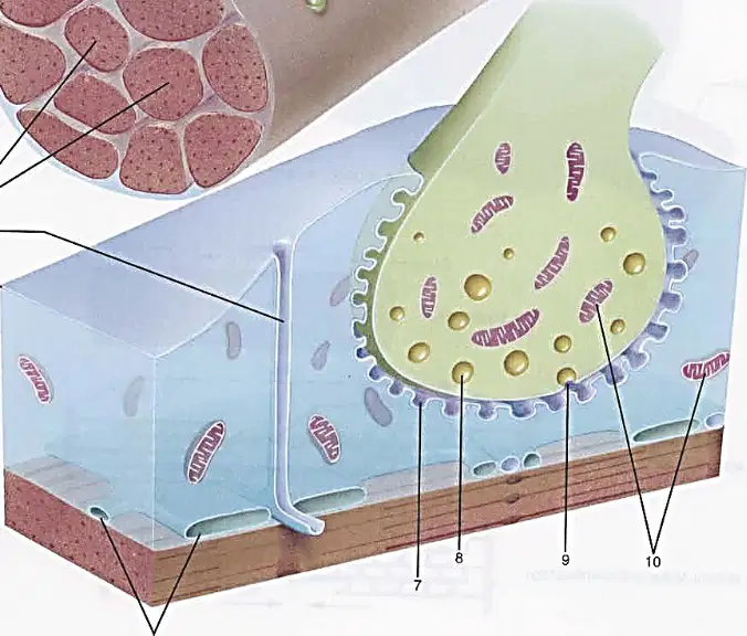
In the labeled diagram of a neuromuscular junction, which component is most directly responsible for providing energy for synaptic vesicle cycling and ion pumping?
Mitochondria
Myofibrils
Synaptic cleft
Sarcolemma
Which statement best describes the A band in a sarcomere?
Light region containing thin filaments (mostly actin)
Dark region containing thick filaments (mostly myosin)
Region that disappears during contraction
Area composed only of Z lines
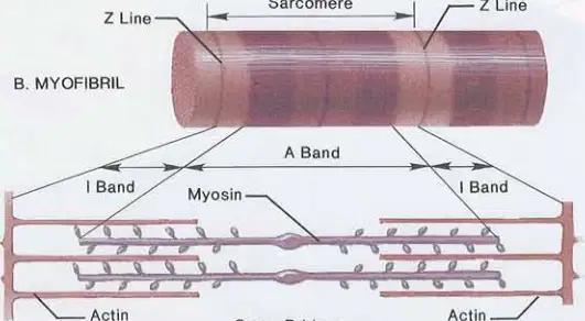
In the myofibril diagram of a resting muscle fiber, which labeled region primarily contains actin?
A band
Z line
I band
M line
At rest, the myosin heads are in which configuration according to the material?
Bound rigidly to actin forming cross-bridges
Detached and in the cocked, extended position
Folded against the thick filament and inactive
Partially hydrolyzed ADP-free state
During contraction, what immediate event allows force generation between filaments?
Actin depolymerizes to shorten the sarcomere
Myosin heads bind to actin, forming cross-bridges and pivot
Z lines dissolve to reduce sarcomere length
Mitochondria release calcium to stiffen myosin
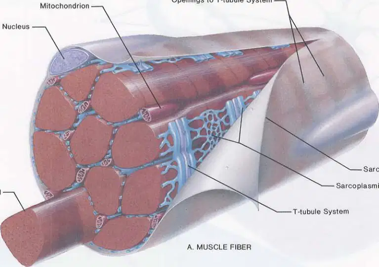
Which sequence best represents the hierarchy from whole structure to contractile proteins as depicted?
Sarcomere → Muscle fiber → Myofibril → Actin/Myosin
Muscle fiber → Myofibril → Sarcomere → Actin/Myosin
Myofibril → Muscle fiber → Sarcomere → Actin/Myosin
Muscle fiber → Sarcomere → Myofibril → Actin/Myosin
What happens to the I band during contraction based on the sliding interaction of actin and myosin?
It widens as myosin slides outward
It narrows as thin filaments slide toward the center
It becomes darker due to added myosin
It shifts to the center of the sarcomere
Which component defines the boundaries of a sarcomere?
A bands
Z lines
I bands
T-tubules
In the context of cross-bridge cycling, which action directly follows myosin binding to actin according to the description?
Myosin head pivots forward and back, producing movement
Actin detaches from the Z line
Thick filaments move past the Z line
Myosin heads return to the cocked position without movement