
 Worksheets
DFT-EMREE-PULMONOLOGY🫁-18-10-2025-STUDYWITHMAXEMO
Total questions: 10
Worksheet time: 8mins
74 Y/O M, a chronic smoker, c/o persistent cough & SOB x3 months. Hx of recent weight loss. O/E, he appears cachectic. Vitals: BP 155/95 mmHg, HR 102/min. Noteworthy findings include hyperpigmentation of the oral mucosa, palmar creases, and soles. Ix reveal: Glucose 190 mg/dL, K+ 2.9 mEq/L. A chest X-ray shows a right-sided hilar mass. What is the most likely Dx?
Cushing's disease
Addison's disease
Conn's syndrome
Ectopic ACTH syndrome
Bronchial carcinoid tumor
A 26-year-old male with a four-year history of heavy smoking presents to the emergency department with an acute onset of severe, sharp left-sided chest pain and progressive shortness of breath. He has no history of trauma or known underlying lung disease. On examination, he is tachypneic. Auscultation reveals markedly diminished air entry over the entire left hemithorax. Percussion of the left chest is hyperresonant. A chest radiograph confirms a large left-sided primary spontaneous pneumothorax. The underlying pathophysiology of this patient's condition is most commonly associated with which of the following findings?
Rupture of subpleural apical blebs
Necrotizing pneumonia leading to a bronchopleural fistula
Rupture of large bullae secondary to emphysematous changes
Fracture of a rib penetrating the visceral pleura
Iatrogenic injury during central venous catheter placement
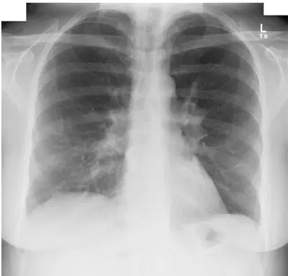
A 35-year-old male presents with a 2-month history of progressive shortness of breath and a dry cough. He also complains of pain in both ankles and notes the recent appearance of tender, reddish nodules on his shins. He is afebrile. His chest X-ray is provided, demonstrating prominent bilateral hilar and right paratracheal lymphadenopathy. What is the most likely diagnosis?
Tuberculosis
Hodgkin's lymphoma
Sarcoidosis
Histoplasmosis
Silicosis
A child diagnosed with moderate persistent bronchial asthma, presents to the clinic in Sharjah with a two-week history of a progressively hoarse voice. The patient's asthma symptoms have been well-controlled since starting a new maintenance therapy which includes a high-dose inhaled corticosteroid. There is no associated fever, sore throat, or cough. Examination of the oropharynx is unremarkable. What is the most likely cause of this patient's hoarseness?
Acute viral laryngitis
Laryngeal candidiasis
Vocal cord nodules from chronic coughing
Laryngopharyngeal reflux
Spasmodic dysphonia
A patient c/o intermittent wheeze and cough. Symptoms occur 4-5 days per week, and the patient is awakened by cough approximately twice a week. A SABA (short-acting beta-agonist) is used almost daily for relief. The patient also has a Hx of eczema. Based on the frequency of symptoms, what is the most accurate classification of this patient's asthma?
Intermittent
Mild Persistent
Moderate Persistent
Severe Persistent
65 Y/O M presents to the ED with a 1-day Hx of sudden-onset high fever, shaking chills, and a productive cough with rust-colored sputum. The patient c/o sharp right-sided chest pain that worsens with inspiration. O/E: T 39.5°C, HR 110/min, RR 24/min. Auscultation reveals crackles and bronchial breath sounds over the R lower lobe. CXR shows dense consolidation in the R lower lobe. What is the most appropriate initial antibiotic Rx for this inpatient?
Amoxicillin
Vancomycin + Piperacillin-tazobactam
Ceftriaxone + Azithromycin
Doxycycline
Oseltamivir
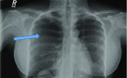
On a chest X-ray, the key diagnostic finding that confirms a pneumothorax is the visualization of the ________ line, which represents the edge of the collapsed lung.
mediastinal
diaphragmatic
costophrenic
visceral pleural
A patient presents with chronic sinusitis, coughing up blood (hemoptysis), and signs of kidney failure. This classic triad of symptoms is most characteristic of which disease?
Goodpasture's Syndrome
Sarcoidosis
Granulomatosis with Polyangiitis
Systemic Lupus Erythematosus
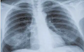
A patient presents with fever and cough. A chest X-ray is performed. What is the primary abnormality seen on this chest X-ray?
Normal chest
Right lower lobe consolidation
Left-sided pneumothorax
Enlarged heart
A 32-year-old female with no smoking history presents with recurrent spontaneous pneumothorax. A high-resolution computed tomography scan shows diffuse, uniformly distributed, thin-walled pulmonary cysts. What is the most likely diagnosis?
Langerhans Cell Histiocytosis
Lymphangioleiomyomatosis
Birt-Hogg-Dubé Syndrome
Centrilobular Emphysema

