
 Worksheets
BSC250L Terminology and Concepts – Vocabulary Assessment
Total questions: 143
Worksheet time: 2hrs 31mins
Nervous tissue: Which structures should you be able to identify?
Neurons
Neuroglial cells
Intercalated discs
Chondrocytes

Your collar bone is scientifically called
clavicle
sternum
phalanges
ulna

A lower arm bone that is on the pinky side
clavicle
radius
phalanges
ulna
Your collar bone is scientifically called
clavicle
sternum
phalanges
ulna
A lower arm bone that is on the pinky side
clavicle
radius
phalanges
ulna

A lower arm bone that is on the thumb side
clavicle
radius
phalanges
ulna

Upper arm bone is scientifically called
sternum
humerus
femur
biceps

Fingers are scientifically called
upper phalanges
tibia
cranium
fingers

Lower leg bone in the back is scientifically called
fibula
femur
sternum
humerus

Strongest and longest bone in the body
femur
sternum
humerus
cranium

Toes are known as your
feet
radius
lower phalanges
tibia

Your spine or backbone is scientifically called
vertebrae
spine
sternum
femur

Bone in your chest that protects your heart
cranium
ulna
femur
sternum

Lower leg bone in front. Known as your shin bone
tibia
radius
ulna
fibula

What bone is in red?
carpals
humerus
radius
ulna

What bone is shown in red?
clavicle
humerus
radius
ulna

What bone is shown in red?
clavicle
humerus
radius
ulna

What bone is shown in red?
clavicle
humerus
scapula
ulna

What bone is shown in red?
clavicle
humerus
scapula
ulna

What bone is shown in red?
pelvis
ribs
sternum
vertebrae

What bone is shown in red?
femur
fibula
patella
tibia

What bone is shown in red?
femur
fibula
patella
tibia

What bone is shown in red?
femur
fibula
patella
tibia

What bone is shown in red?
femur
fibula
patella
tibia
The ends of your fingers and toes are called
carpals
mandibles
phalanges
tarsals

What structure is shown?
mandible
patella
pelvis
vertebrae
Your skull is known as your
carpals
cranium
mandible
tarsals

What bones are shown in red?
carpals
metacarpals
metatarsals
tarsals

What bones are shown in green?
carpals
metacarpals
metatarsals
tarsals

What bone is shown in red?
carpal
metacarpal
metatarsal
tarsal

What bones are shown in green?
carpals
metacarpals
metatarsals
tarsals

What bones are shown in red?
pelvis
ribs
sternum
vertebrae

What bones are shown in blue?
humerus
radius
ribs
vertebrae

What bone is shown?
cranium
mandible
metacarpals
tibia

Identify the highlighted bone
Pelvis
Sacrum
Coccyx
Phalanges

Identify the highlighted bone
Pelvis
Sacrum
Radius
Tarsals

Identify the highlighted bone
Vertebrae
Coccyx
Scapula
Sacrum

Identify the highlighted bone
Humerus
Tibia
Femur
Patella

Identify the highlighted bone
Radius
Humerus
Femur
Tibia

Identify the highlighted bone
Tibia
Fibula
Femur
Scapula

Identify the highlighted bone
Maxilla
Skull
Femur
Sternum
Which of the following is NOT an example of a long bone?
forearm bone
wrist bone
femur
thigh bone
Either end of a long bone.
Diaphysis
Periosteum
Spongy Bone
Epiphyses
Shaft of a long bone.
Epiphysis
Compact Bone
Sternum
Diaphysis
Dense connective tissue covering the surface of a bone.
Trabeculae
Articuler cartilage
Periosteum
Spongy tissue
Hyaline cartilage that covers the ends of bones in synovial joints.
Endosteum
Periosteum
Articular cartilage
Synovial cartilage
Cavity in the diaphysis that contain red or yellow marrow.
Medullary Cavity
Marrow cavity
Nerve cavity
All of the above
Soft connective tissue found in the spaces in bones
Trabeculae
Osteon
Osteocyte
Marrow


What is does the picture show?
An osteon
A hair follicle
Hyaline cartilage
A flat bone


What is the name for the thin, spiderlike membrane in the middle of the bone.
Lamellae
Medullary Cavity


Which letter is labeling the lacunae?
a
b
c
d
e

What is shown in letter A?
canaliculi
osteocyte
lacuna
osteon
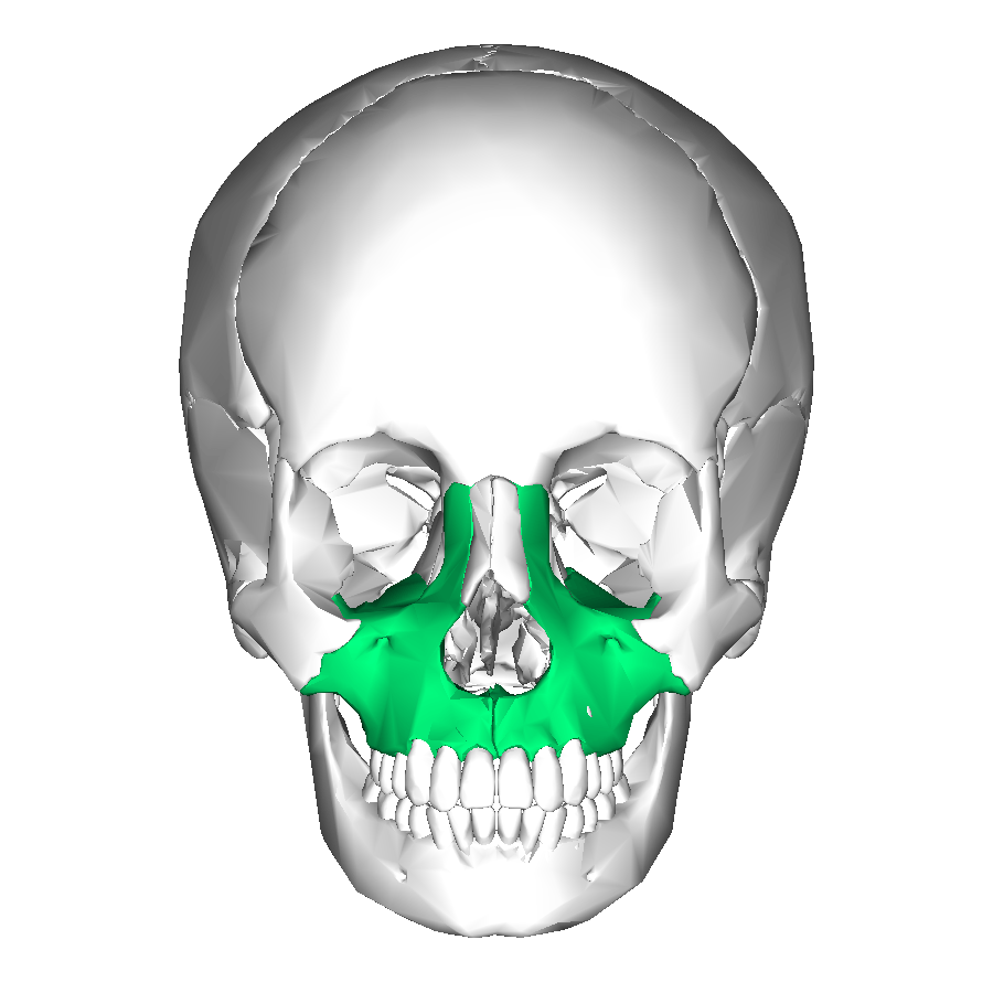


sagittal
squamous
coronal
lambdoid


The bone indicated by #5 is the
parietal bone
temporal bone
spheniod bone
occipital bone


What name is given to the scapula labelled A?
glenoid cavity
acromial process
coracoid process
infraspinous fossus

What name is given to the scapula labelled B?
glenoid cavity
acromial process
coracoid process
infraspinous fossus

What name is given to the scapula labeled C?
glenoid cavity
acromial process
coracoid process
infraspinous fossus

The red depression area at the back end of the humerus is called the
olecranon fossa
olecranon process
trochlear notch
coracoid process

The red area at the end of the humerus and ulna articulation is
acromion process
olecranon process
foramen magnum
coracoid process

Name this bone highlighted in green.
xyphoid process
coccyx
sacrum
suprasternal notch


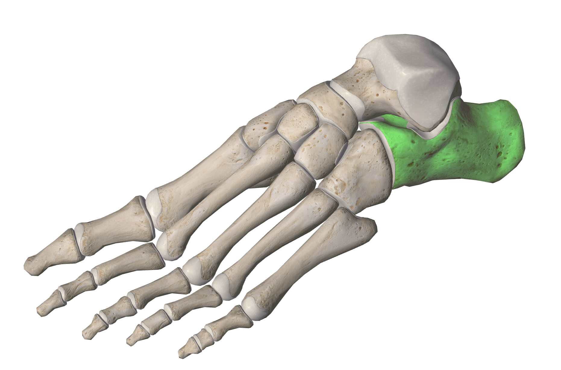

Which bone is highlighted in the picture?
Thoracic Vertebrae
Lumbar Vertebrae
Sacrum
Coccyx

What are the bones that make up the spine?
vertebrae
phalanges
metacarpals
metatarsals


5
ulna
coccyx
pelvic guide
tarsals
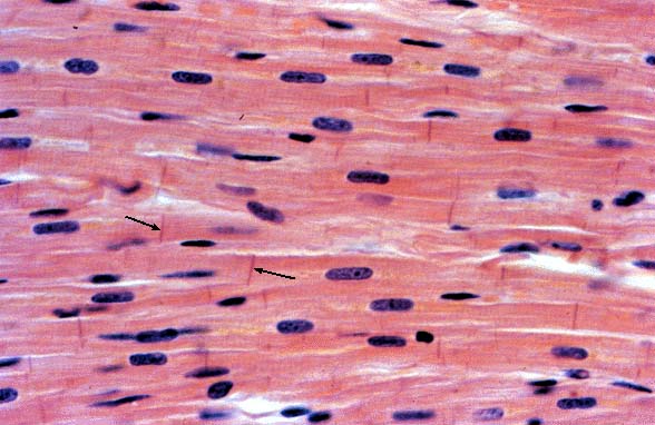

Identify this bone
Hyoid
Cervical Vertebra
Coccyx
Sternum
Lumbar Vertebra

Identify this bone
Thoracic Vertebra
Cervical Vertebra
Axis
Atlas
Lumbar Vertebra
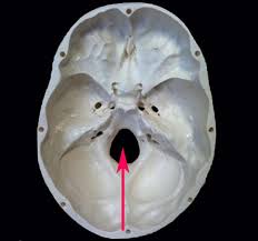
What SURFACE MARKING is this?
Vertebral Foramen
Occipital Condyles
External Auditory Meatus


coronal suture
squamous suture
lambdoid suture
sagittal suture

temporal foramen
mental foramen
external auditory meatus
zygomatic facial foramen

sagittal suture
coronal suture
lambdoid suture
squamous suture

nasal
ethmoid
sphenoid
lacrimal

nasal
lacrimal
ethmoid
sphenoid

nasal
lacrimal
ethmoid
sphenoid

Which bones are described as the lowermost scroll-shaped bones on the sidewalls of the nasal cavity?
Middle nasal concha
Inferior nasal conchae
Maxilla
Middle nasal conchae

Which bone forms part of the base of the skull and parts of the floor and sides of the orbit?
Sphenoid bone
Mandible
Frontal bone
Parietal bone


Which is bone 5
inferior nasal conche
nasal
vomer
lacrimal

Coronal Suture
Sagittal Suture
Lambdoidal Suture
Squamous Suture
Name the bone marking shaded in green.
Occipital condyles
Styloid process
Mental foramen
Foramen magnum

What is number 23?
Vomer
Coccyx
Radius
Fibula

What is number 30?
Zygomatic
Occipital
Mandible
Sphenoid
What is number 31?
Palatine
Occipital
Ethmoid
Calcaneus
What is number 32?
Maxillary
Parietal
Frontal
Atlas

What is number 33?
Incus
Malleus
Stapes
Manubrium
What is number 34?
Malleus
Incus
Stapes
Body
What is number 35?
Stapes
Incus
Malleus
Phalanges

What is number 36?
Hyoid
Mandible
Mastoid
Nasal Conchae
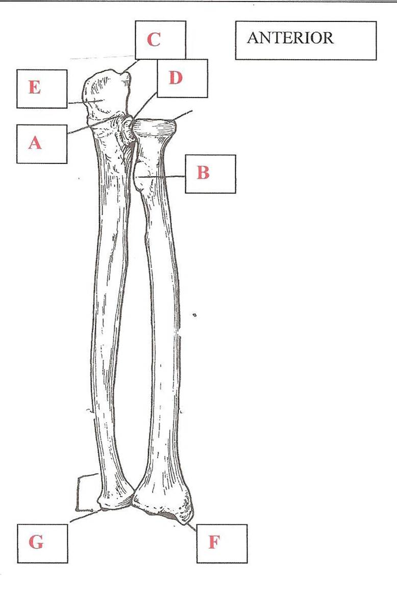
Label C (anterior view of the left forearm)
Semilunar Notch
Coronoid process
Radial tuberosity
Olecranon process

Label D (anterior view of the left arm)
Medial Malleolus
Lateral Malleolus
Medial condyle
Lateral condyle

Label A (anterior view of the left arm)
Medial epicondyle
Lateral epicondyle
Capitulum
Coronoid fossa
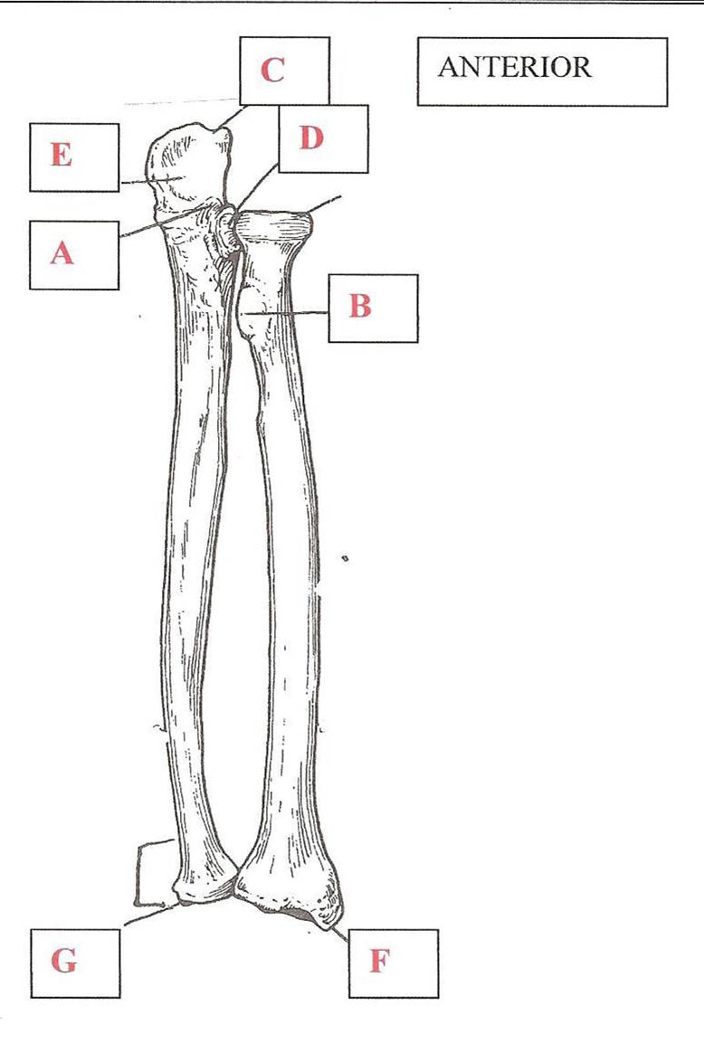
Label A (anterior view of the left forearm)
Semilunar Notch
Coronoid process
Radial tuberosity
Olecranon process
Calcaneus

What is the bone labeled 1?
ischium
pubis
ilium
acetabulum

What bone is labeled 2 and is shaded green?
ischium
pubis
ilium
acetabulum

What feature is labeled 6?
obturator foramen
acetabulum
greater sciatic notch
pubis symphysis

The iliac crest is at which number?
7
9
4
5

What feature is pointed out by arrow 8?
acetabulum
pubis symphysis
iliac spine
obturator foramen
Label A (anterior view of the left arm)
Medial epicondyle
Lateral epicondyle
Capitulum
Coronoid fossa

