
 Worksheets
Chapter 12
Total questions: 119
Worksheet time: 2hrs 59mins
The liver is located primarily in the of the abdomen.
LLQ
RLQ
RUQ
LUQ
The liver performs more than 100 different functions.
True
False
The liver is divided into major and minor lobes.
Three
Four
Two
Six
The liver secretes approximately mL of bile per day.
100 to 250
500 to 700
800 to 1000
12000 to 1500
The main function of bile is to:
break down cholesterol.
emulsify fats.
begin the digestion of proteins.
begin the digestion of complex sugars.
Which aspect of the gallbladder is located most posterior within the abdomen?
Fundus
Body
Neck
Apex
The average adult gallbladder is__cm long and_____cm wide.
7 to 10; 3
4 to 6; 2
12 to 15; 5 to 6
2 to 3; 1
What is the primary purpose of the membranous folds located within the cystic duct?
Produces cholecystokinin
Prevents gallstones from entering the gallbladder
Provides blood supply to the gallbladder
Prevents distention or collapse of the cystic duct
Where is bile formed?
Gallbladder
Duodenal mucosa
Liver
Pancreas
Which of the following functions are performed by the gallbladder?
Storage of bile
Concentration of bile
Contraction and release of bile
All of the above
What is a primary function of cholecystokinin?
Serves as an enzyme to break down certain food nutrients
Stimulates the production of bile
Stimulates the gallbladder to contract
Inhibits the formation of gallstones
Where is cholecystokinin produced?
Duodenal mucosa
Liver
Gallbladder
Pancreas
What is an older term for the pancreatic duct?
Duct of Wirsung
Hepatopancreatic duct
Duct of Langerhans
Ampulla of Vater
In 80% of all individuals, the common bile duct and the pancreatic duct unite before entering the duodenum.
True
False
What is an older term for the hepatopancreatic sphincter?
Duodenal papilla
Sphincter of Vater
Duodenal sphincter
Sphincter of Oddi
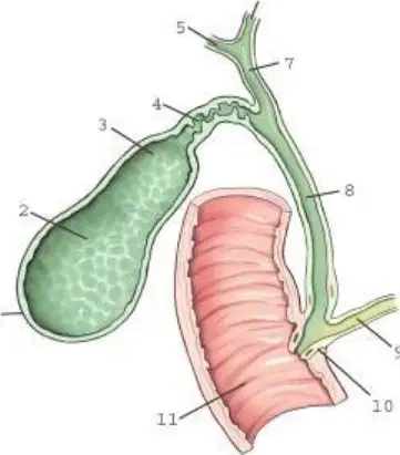
Which aspect of the gallbladder is labeled 1?
Neck
Fundus
Body
Apex
Which biliary structure is labeled 4?
Cystic duct
Common bile duct
Common hepatic duct
Right hepatic duct
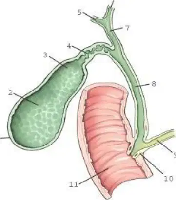
Which structure is labeled 6?
Cystic duct
Pancreatic duct
Right hepatic duct
Left hepatic duct
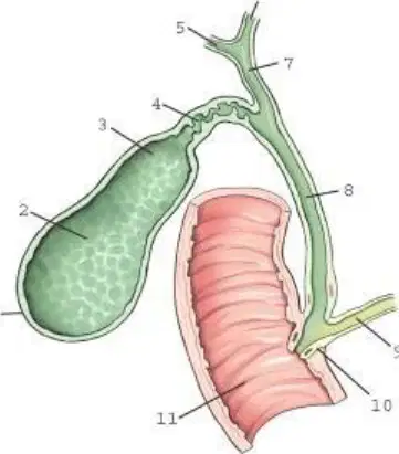
Which structure is labeled 9?
Pancreatic duct
Common bile duct
Cystic duct
Common hepatic duct
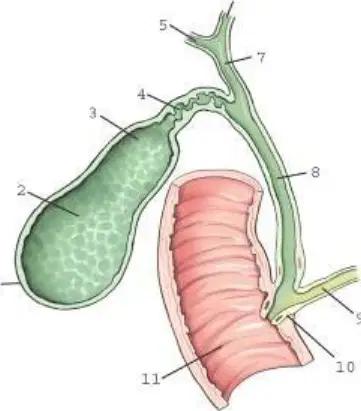
The region of the biliary system labeled 10 is:
Common bile duct
Cystic duct
Gallbladder
Hepatic duct
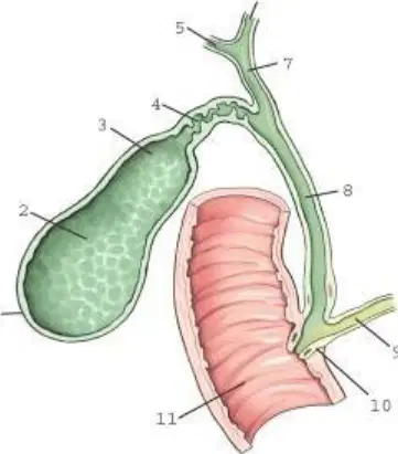
Which structure is labeled as number 11?
Duct of Vater
Hepatopancreatic ampulla
Duct of Wirsung
Hepatopancreatic sphincter
"Chole-" is a prefix for terms pertaining to the:
gallbladder.
ducts.
bile.
liver.
Radiographic examination of the biliary ducts only is termed:
cholecystography.
cholangiography.
cholelithiasis.
cholecystocholangiography.
Which of the following structures is not considered to be an accessory organ of digestion?
Liver
Pancreas
Salivary glands
Kidneys
Saliva contains certain enzymes to begin the digestion of:
starch.
minerals.
proteins.
lipids.
The act of swallowing is termed:
mastication.
digestion.
deglutition.
aphasia.
Which of the following structures is not one of the salivary glands?
Parotid
Sublingual
Submandibular
All of the above are salivary glands.
The esophagus is located___to the larynx.
anterior
posterior
inferior
proximal
The aortic arch and the create a normal indentation seen along the lateral border of the esophagus.
Pulmonary Artery
Left Primary Bronchus
Pulmonary Veins
Superior Vena Cava
Which two forces or processes propel food down the esophagus?
Peristalsis and gravity
Peristalsis and rhythmic segmentation
Gravity and deglutition
Deglutition and mastication
The opening between the stomach and esophagus is termed the:
cardiac antrum.
cardiac notch.
esophagogastric junction.
esophagocardiac junction.
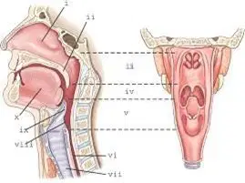
Part ii refers to the:
soft palate.
tonsils.
Hard Palate
Epiglotis
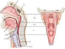
Part iv refers to the:
laryngopharynx.
nasopharynx.
oropharynx.
posterior oral cavity.
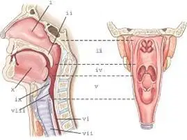
Part ix refers to the:
soft palate.
tonsils.
uvula.
epiglottis.
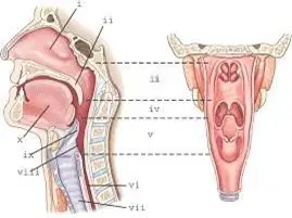
Refer to the diagram of the head and neck region. Which part is labeled as 'i'?
nasopharynx.
laryngopharynx.
oropharynx.
nasal cavity.
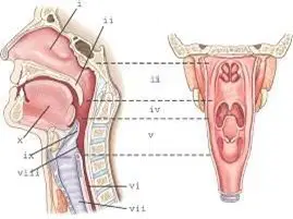
Refer to the diagram of the head and neck region. Part vii refers to the:
esophagus.
trachea.
spinal cord.
larynx.
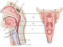
What is labeled as part III?
nasal sinus.
maxillary sinus.
nasal cavity.
upper oral cavity.
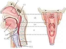
Refer to the diagram. Part V refers to the:
nasopharynx.
larynx.
oropharynx.
laryngopharynx.
The dilated portion of the distal esophagus is termed the:
cardiac antrum.
cardiac notch.
fundus.
incisura cardiaca.
Which of the following is not a main subdivision of the stomach?
Fundus
Cardium
Pylorus
Body
Which term describes the outer, lateral border of the stomach?
Lesser curvature
Incisura angularis
Gastric border
Greater curvature
What is the term for the longitudinal mucosal folds found within the stomach?
Gastric canal
Valvulae conniventes
Haustra
Rugae
Toward which aspect(s) of the stomach will barium gravitate with the patient in a prone position?
Body and pylorus
Fundus
Cardia
Greater curvature
Which aspect of the stomach is attached to the duodenum?
Fundus
Body
Pylorus
Cardiac
Which specific part of the pancreas is adjacent to the C-loop of the duodenum?
Tail
Body
Head
Neck
Which division of the duodenum contains the duodenal bulb or cap?
First (superior)
Third (horizontal)
Second (descending)
Fourth (ascending)
What structure indicates the junction between the duodenum and jejunum?
Duodenal cap
Suspensory muscle of the duodenum
Valvulae conniventes
C-loop of the duodenum
Which part of the stomach is referred to as the body and pylorus?
Body and pylorus
Fundus
Cardiac
Fundus and body
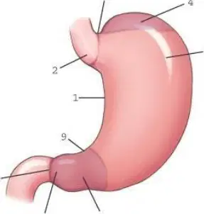
Which part of the stomach is labeled 6?
Body
Pyloric portion
Pyloric antrum
Pyloric canal
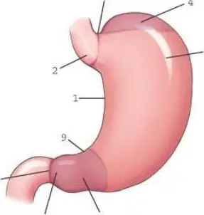
Which part of the stomach is labeled 2?
Esophagogastric junction
Incisura angularis
Incisura cardiaca
Cardiac antrum
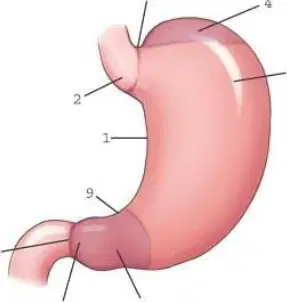
The part of the stomach labeled 9 is:
Cardia
Fundus
Body
Pylorus
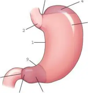
Which part of the stomach is labeled 3?
Cardiac notch
Angular notch
Pyloric orifice
Incisura cardiaca
Once food enters the stomach and is mixed with gastric secretions, it is termed:
bile.
lipids.
chyme.
biologic catalysts.
Which of the following substances are not digested chemically?
a. Minerals
b. Carbohydrates
c. Proteins
d. Lipids
Enzymes that aid in chemical digestion are classified as:
lipids.
biologic catalysts.
digestive acids.
Lipids (fats) are broken down to fatty acids and glycerol as they are digested and absorbed by the:
stomach and small bowel.
stomach only.
small bowel only.
None of the above (are not digested).
Which aspect of the gastrointestinal (GI) tract is primarily responsible for the absorption of digestive end products along with water, vitamins, and minerals?
Stomach
Small intestine
Large intestine
Pancreas
A high and transverse stomach would be found in a(n)____patient.
hyposthenic
asthenic
sthenic
hypersthenic
A stomach with the duodenal bulb at the level of L1-2 would be found in a(n)____patient.
hyposthenic
asthenic
sthenic
hypersthenic
What is the classification of barium sulfate as a contrast media?
Radiopaque
Radiolucent
Isodense
Negative contrast media
What type of solution is formed when barium is mixed with water?
Water-soluble solution
Isotonic solution
Hypotonic solution
Colloidal suspension
Sodium bicarbonate is often used as a negative contrast medium during an upper GI series.
True
False
Swallowed air can be used as a negative contrast medium during an upper GI series.
True
False
Which of the following statements is true about barium sulfate?
It should be discarded if not used within an hour after mixing.
The patient may become ill if it is not prepared with sterile water.
It rarely produces an allergic reaction.
It is soluble in water.
Which of the following clinical indications would mandate the use of an oral, water-soluble contrast agent?
Patient with esophageal reflux
Patient with a bezoar
Patient with a possible perforated bowel
Patient with a possible peptic ulcer
What type of contrast media is ideal for demonstrating a diverticulum within the stomach?
Double-contrast barium/negative contrast agent
Single-contrast barium sulfate
Water soluble
Radiolucent
Which of the following patient care concerns would prevent the use of an oral, water-soluble contrast medium?
The patient has a possible ulcer.
The patient is sensitive to iodine.
The patient has esophageal reflux.
The patient has dysphasia.
Digital fluoroscopy does not require the use of an image intensifier.
True
False
No cassettes are required for imaging with a digital fluoroscopy unit.
True
False
Digital fluoroscopy has distinct advantages but produces a slightly higher patient dose as compared with conventional fluoroscopy.
True
False
With digital fluoroscopy edge enhancement, brightness and contrast can be manipulated both during the procedure and also at a later time.
True
False
Which of the following is not one of the cardinal principles of radiation protection?
Time
Distance
Intensity
Shielding
Which one of the following cardinal principles of radiation protection is most effective in reducing the dose to the technologist during fluoroscopy?
Time
Distance
Intensity
Shielding
Which of the following devices reduces scatter exposure during fluoroscopy from the fluoroscopy tube?
Compression paddle
Bucky tray
Bucky slot cover
Lead gloves
Protective aprons worn during fluoroscopy must possess at least mm lead equivalency.
0.25
0.5
0.75
1
Which of the following conditions involves dilated veins in the distal aspect of the esophagus, which in some cases can lead to internal bleeding?
Esophageal Varices
Esophageal Reflux
Esophageal Hypertension
Esophageal thrombosis
A large outpouching of the proximal esophagus above the upper esophageal sphincter is termed:
Barrett esophagus.
esophageal varices.
esophageal reflux.
Zenker diverticulum.
Achalasia is generally defined as:
difficulty in swallowing.
a common malignancy of the esophagus.
motor disorder of the esophagus.
large outpouching of the esophagus.
Which of the following definitions would describe a bezoar?
Inflammation of the gastric lining
Outpouching of the stomach wall
Mass of undigested material
Stomach neoplasm
Duodenal ulcers are rarely malignant.
True
False
Only 5% of all ulcers lead to bowel perforation.
True
Flase
Research suggests that peptic ulcers may be caused by:
Smoking
Bacteria
Alcohol
Antibiotics
Gastritis is defined as inflammation of the:
Pancreas
Large intestine
Small Intestine
Stomach
Which of the following conditions is an example of GERD?
Esophageal varices
Gastric carcinoma
Esophageal reflux
Bezoar
What is the most common radiographic procedure performed to diagnose GERD?
Endoscopy
Esophagogram
Upper GI series
CT
Hypertrophic pyloric stenosis (HPS) is the most common form of intestinal obstruction found in infants.
True
False
Which of the imaging modalities or procedures is most effective and preferred in diagnosing HPS?
Nuclear medicine
CT
Upper GI series
Ultrasound
What is a potential risk associated with the use of water-soluble contrast agents, especially for geriatric patients?
Bowel obstruction
Cardiac arrest
Dehydration
Shock
When using computed radiography (CR), inadequate kV or mAs will produce a image.
Dark
Light
Blurry
Mottled
Which of the following technical factors will best enhance a digital image taken during an upper GI series?
Collimation
Low kV techniques
Long SID
Use of compensation (wedge) filter
Patient preparation for an esophagram includes NPO before the procedure.
4 to 6 hours
30 minutes to 1 hour
4 to 6 hours and no gum chewing or smoking
None of the above; patient preparation is not needed as long as an upper GI series is not scheduled to follow.
Most esophagograms begin with the patient:
recumbent-supine.
erect.
recumbent-prone.
in a left lateral decubitus position.
Why would a patient undergo Valsalva maneuver during an esophagogram?
To demonstrate possible esophageal varices
To demonstrate possible esophageal reflux
To demonstrate possible esophagitis
To demonstrate a possible bezoar
Which of the following procedures will not demonstrate possible esophageal reflux?
Toe-touch maneuver
Water test
Reverse Trendelenburg method
Compression technique
Which of the following positions is not normally considered to be part of the esophagogram basic routine?
Posteroanterior (PA)
Left lateral
Right anterior oblique (RAO)
Anteroposterior (AP)
Patient preparation for an adult upper GI series includes:
nothing; no patient preparation is required.
NPO 4 hours before the procedure.
NPO 8 hours before the procedure.
NPO 24 hours before the procedure.
Which of the following kV ranges should be used for an upper GI series using barium sulfate (single contrast study)?
80 to 90 kV
90 to 100 kV
100 to 125 kV
125 to 140 kV
1. Which of the following imaging modalities is an alternative to an esophagogram for detecting esophageal varices?
a. Sonography
CT
a. Nuclear medicine
a. MRI
Which of the following imaging modalities is ideal for demonstrating signs of Barrett esophagus?
Nuclear medicine
MRI
Sonography
CT
How much barium is typically given to a 3- to 10-year-old child during an upper GI series?
4 to 6 ounces
14 to 16 ounces
6 to 12 ounces
1 cup
Gastric emptying studies are performed using:
intraesophageal sonography.
radionuclides.
MRI.
CT.
CR centering for an esophagogram should be to the vertebral level of:
the sternal angle.
T3 or T4
T5 or T6
T7
How much obliquity is required for the RAO position for the esophagus?
10 to 15
20 to 30
35 to 40
45 to 60
The RAO position of the esophagus is preferred over the left anterior oblique (LAO) because it:
is a more comfortable position for the patient.
increases the visibility of the esophagus between the vertebrae and heart.
reduces thyroid exposure to the patient.
is easier for the patient to hold the cup of barium in his or her left hand.
What type of breathing instructions should be given to the patient during an esophagogram using a thin barium mixture?
Suspended respiration while continuing to swallow
Suspended inspiration after the last swallow
Shallow breathing and continued swallowing during exposure
Exposure immediately after last bolus is swallowed (patient will not be breathing immediately after swallow)
Other than the esophagogram, what other imaging modality is performed to diagnose Barrett’s esophagus?
Computed tomography
Nuclear medicine
Magnetic resonance
Sonography
Which of the following upper GI projections and/or positions will best demonstrate the pylorus and duodenal bulb in profile during a double-contrast study?
AP
LAO
PA
LPO
At what level should the CR and image receptor be centered for the RAO or PA upper GI projection and/or position on a sthenic-body type of patient?
Lower costal margin
L2
L3-4
Iliac crest
A radiograph taken during an upper GI series demonstrates poor visibility of the gastric mucosa. The following factors were used: 80 kV, 30 mAs, 1/40-second exposure time, and high-speed image receptors. Barium sulfate was used during the procedure. Which of the following factors needs to be modified during the repeat exposure?
Use of detail-speed screens (adjust mAs accordingly)
Shortened exposure time (increase mAs accordingly)
Use of a water-soluble contrast medium
Increased kV (decrease mAs as needed)
A PA radiograph taken on a hypersthenic patient during an upper GI series reveals that the pylorus and duodenal bulb are superimposed. Which of the following modifications will best eliminate the superimposition between these structures?
Roll the patient into a slight RAO position.
Angle the CR 35 to 45 cephalad.
Place a rolled towel or a filled compression paddle under the patient’s abdomen before exposure.
Increase the kV.
A patient comes to radiology for an upper GI series. Her clinical history indicates that there may be a tumor posterior to the stomach. Which one of the following projections and/or positions will best demonstrate this condition?
RAO
PA
Right Lateral
LPO
A pediatric patient enters the emergency department (ED) with a small, plastic object stuck in her esophagus. PA and lateral chest radiographs fail to demonstrate the foreign body. Which of the following procedures would be most effective in demonstrating the location of the object?
Upper GI series
Esophagogram with the water test
Esophagogram using very thick barium
Esophagogram using shredded cotton soaked in barium
A patient comes to radiology with a history of a gastric ulcer. Which of the following procedures would be most diagnostic to demonstrate this lesion?
Double-contrast upper GI series
Single-contrast barium sulfate upper GI series
Oral, water-soluble upper GI series
Carbon dioxide upper GI series
A patient comes to radiology for an esophagogram. The radiologist is concerned about the upper portion of the esophagus, near the level of T1, which did not visualize well on the previous routine projection-position esophagogram. Which of the following special projections and/or positions would be most helpful in demonstrating this region?
Soft tissue lateral
LAO
Swimmer’s lateral position
AP with 25 to 30 cephalad CR angle
One radiograph of an upper GI series needs repeating. The technologist is unsure which projection and/or position is seen on this radiograph. The fundus is filled with barium, and the pylorus and duodenal bulb are profiled and air filled. The patient was recumbent for all projections. Which projection and/or position needs to be repeated?
PA
LPO
RAO
Right Lateral
A patient comes to radiology for an upper GI series. The patient has a clinical history of hiatal hernia. Which of the following positions may be helpful in demonstrating this condition?
AP Trendelenburg position
Left lateral decubitus, AP projection
Dorsal decubitus, lateral projection
LAO with compression technique
A PA projection taken during an upper GI series performed on an infant reveals that the body and pylorus of the stomach are superimposed. What modification needs to be made during the repeat exposure to separate these two regions?
Perform the AP projection instead.
Angle the CR 20 to 25 cephalad.
Angle the CR 30 to 45 cephalad.
Use the AP Trendelenburg position.
During an upper GI series, the “halo” sign appears in the duodenum. Which of the following conditions will produce this radiographic sign?
Ulcer
Bezaor
Diverticulum
Tumor
A patient comes to radiology with a clinical history of HPS. Which of the following imaging modalities will best demonstrate this condition?
CT
Nuclear Medicine
Ultrasound
Upper GI Series

